Review
. 1992 Sep;12(5):423-71.
doi: 10.1002/med.2610120502.

Adenosine A1 and A2 receptors: structure--function relationships

Affiliations
Review

Adenosine A1 and A2 receptors: structure--function relationships

P J van Galen et al. Med Res Rev. 1992 Sep.
No abstract available


Figures

Figure 1
Figure 1
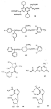
Basic structures of tricyclic nonxanthine antagonists.
Figure 2
Figure 2
Computer-generated models of the N6 region of (a) A1 and (b) A2 receptors. Shaded areas indicate presumed receptor boundaries. The N6 region of the A1 receptor is divided into subdomains (S1, S2, S3, A, B, C) where hydrophobic substitution may enhance affinity.
Figure 3
Figure 3
Model for the antagonist binding site of the A1 receptor. Indicated are areas of preferred electrostatic potential (+5 and -5 kcal/mol boundaries); a mtrogen atom at position 7 that is thought to act as a hydrogen bond acceptor; and areas where hydrophqbic substituents may enhance affinity.
Figure 4
Figure 4
Structures of some representative compounds (78–84) that enhance the actions of adenosine in an indirect manner.
Figure 5
Figure 5
(Overleaf) Alignment of the canine A1 and A2 receptors with the human β2- and α2A-adrenergic receptors, the rat Ml acetylcholine receptor, and bovine rhodopsin. Alignment was performed with the MACAW multiple sequence alignment program. Putative transmembrane sections (indicated by horizontal bars and roman numbers) were treated as blocks without allowing gaps. Nucleotide sequences were retrieved from the GenBank database; implied amino acid sequences were generated with the GeG Sequence Analysis Software Package.
Figure 5
Figure 5
(Overleaf) Alignment of the canine A1 and A2 receptors with the human β2- and α2A-adrenergic receptors, the rat Ml acetylcholine receptor, and bovine rhodopsin. Alignment was performed with the MACAW multiple sequence alignment program. Putative transmembrane sections (indicated by horizontal bars and roman numbers) were treated as blocks without allowing gaps. Nucleotide sequences were retrieved from the GenBank database; implied amino acid sequences were generated with the GeG Sequence Analysis Software Package.
Figure 6
Figure 6
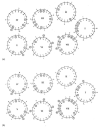
Helical wheel representations of the (a) A1 and (b) A2 receptors. Sequences are modeled as right-handed α-helices (Le., 3.6 residues per turn; thus consecutive residues appear at 100° intervals. Helices are viewed from the extracellular side; H 1 should be read clockwise, H II counterclockwise, and so on). The first and last residues of a helix are indicated by either i (in; Le., cytoplasmic) or 0 (out; Le., extracellular). The first 18 residues of a helix are indicated on the inside of the wheel, any remaining residues on the outside. The arrangement of the helices is based on the structure of bacteriorhodopsin, as determined by cryoelectron microscopy. Hydrophilic residues are encircled and shaded.
Figure 7
Figure 7
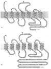
Membrane topology of the (a) A1 and (b) A2 receptors. The N terminus is located on the extracellular side, the C tenninus on the cytosolic side. Residues that are conserved in both adenosine receptor subtypes are shaded. Histidines that are putatively involved in ligand binding, and serines/threonines that potentially can be phosphorylated are shown in boldface. Potential sites for asparagine glycosylation and cysteine palmiloylation are also indicated.
Figure 8
Figure 8
A generalized map of the upper half of each transmembrane helix of the adenosine receptor, showing separate domains of hydrophobic and hydrophilic environment, and the location of the two histidine residues that are putatively involved in the ligand binding site. This model applies in both A1 and A2 receptors.

References

    1. Bruns RF. Nucleosid. Nucleotid. 1991;10:931.
    1. Daly J. w. J. Med Chem. 1982;25:197. - PubMed
    1. Jacobson KA, van Galen PJM, Williams M. J. Med. Chern. 1992;35:407. - PMC - PubMed
    1. Pantely GA, Bristow JD. Circulation. 1990;82:1854. - PubMed
    1. Eidelman O, Guay-Broder C, van Galen PJM, Jacobson KA, Fox C, Turner RJ, Cabantchik ZI, Pollard HB. Proc. Natl. Acad. Sci. USA. 1992;89:5562. - PMC - PubMed

LinkOut - more resources