

Maternal nutrient restriction alters gene expression in the ovine fetal heart
- PMID: 15133065
- PMCID: PMC1664914
- DOI: 10.1113/jphysiol.2004.061697
Maternal nutrient restriction alters gene expression in the ovine fetal heart
Abstract
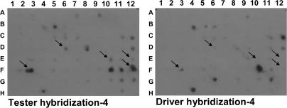
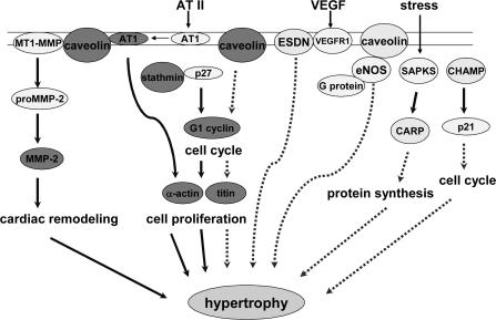
Adequate maternal nutrient supply is critical for normal fetal organogenesis. We previously demonstrated that a global 50% nutrient restriction during the first half of gestation causes compensatory growth of both the left and right ventricles of the fetal heart by day 78 of gestation. Thus, it was hypothesized that maternal nutrient restriction significantly altered gene expression in the fetal cardiac left ventricle (LV). Pregnant ewes were randomly grouped into control (100% national research council (NRC) requirements) or nutrient-restricted groups (50% NRC requirements) from day 28 to day 78 of gestation, at which time fetal LV were collected. Fetal LV mRNA was used to construct a suppression subtraction cDNA library from which 11 cDNA clones were found by differential dot blot hybridization and virtual Northern analysis to be up-regulated by maternal nutrient restriction: caveolin, stathmin, G-1 cyclin, alpha-actin, titin, cardiac ankyrin repeat protein (CARP), cardiac-specific RNA-helicase activated by MEF2C (CHAMP), endothelial and smooth muscle derived neuropilin (ESDN), prostatic binding protein, NADH dehydrogenase subunit 2, and an unknown protein. Six of these clones (cardiac alpha-actin, cyclin G1, stathmin, NADH dehydrogenase subunit 2, titin and prostatic binding protein) have been linked to cardiac hypertrophy in other species including humans. Of the remaining clones, caveolin, CARP and CHAMP have been shown to inhibit remodelling of hypertrophic tissue. Compensatory growth of fetal LV in response to maternal undernutrition is concluded to be associated with increased transcription of genes related to cardiac hypertrophy, compensatory growth or remodelling. Counter-regulatory gene transcription may be increased, in part, as a response to moderating the degree of cardiac remodelling. The short- and long-term consequences of these changes in fetal heart gene expression and induction of specific homeostatic mechanisms in response to maternal undernutrition remain to be determined.
Figures
Similar articles
-
Maternal nutrient restriction reduces concentrations of amino acids and polyamines in ovine maternal and fetal plasma and fetal fluids.Biol Reprod. 2004 Sep;71(3):901-8. doi: 10.1095/biolreprod.104.029645. Epub 2004 May 12. Biol Reprod. 2004. PMID: 15140798
-
Maternal nutrient restriction during early to mid gestation up-regulates cardiac insulin-like growth factor (IGF) receptors associated with enlarged ventricular size in fetal sheep.Growth Horm IGF Res. 2005 Aug;15(4):291-9. doi: 10.1016/j.ghir.2005.05.003. Growth Horm IGF Res. 2005. PMID: 16046163
-
Maternal nutrient restriction and the fetal left ventricle: decreased angiotensin receptor expression.Reprod Biol Endocrinol. 2005 Jul 14;3:27. doi: 10.1186/1477-7827-3-27. Reprod Biol Endocrinol. 2005. PMID: 16018810 Free PMC article.
-
Transcriptional regulation of the fetal cardiac gene program.J Pharmacol Sci. 2012;119(3):198-203. doi: 10.1254/jphs.12r04cp. Epub 2012 Jun 13. J Pharmacol Sci. 2012. PMID: 22786561 Review.
-
Expression profiling techniques for cardiac molecular phenotyping.Trends Cardiovasc Med. 2001 Aug;11(6):218-21. doi: 10.1016/s1050-1738(01)00110-4. Trends Cardiovasc Med. 2001. PMID: 11673051 Review.
Cited by
-
Gender differences in the expression of genes involved during cardiac development in offspring from dams on high fat diet.Mol Biol Rep. 2014 Nov;41(11):7209-16. doi: 10.1007/s11033-014-3605-8. Epub 2014 Jul 24. Mol Biol Rep. 2014. PMID: 25055976
-
Maternal Fructose Intake Exacerbates Cardiac Remodeling in Offspring with Ventricular Pressure Overload.Nutrients. 2021 Sep 18;13(9):3267. doi: 10.3390/nu13093267. Nutrients. 2021. PMID: 34579143 Free PMC article.
-
Transgenerational effects of prenatal nutrient restriction on cardiovascular and hypothalamic-pituitary-adrenal function.J Physiol. 2008 Apr 15;586(8):2217-29. doi: 10.1113/jphysiol.2007.147967. Epub 2008 Feb 21. J Physiol. 2008. PMID: 18292131 Free PMC article.
-
Developmental programming resulting from maternal obesity in mice: effects on myocardial ischaemia-reperfusion injury.Exp Physiol. 2009 Jul;94(7):805-14. doi: 10.1113/expphysiol.2009.047183. Epub 2009 Apr 24. Exp Physiol. 2009. PMID: 19395658 Free PMC article.
-
Sexual dimorphism in the fetal cardiac response to maternal nutrient restriction.J Mol Cell Cardiol. 2017 Jul;108:181-193. doi: 10.1016/j.yjmcc.2017.06.006. Epub 2017 Jun 19. J Mol Cell Cardiol. 2017. PMID: 28641979 Free PMC article.
References
-
- Adachi S, Ito H, Ohta Y, Tanaka M, Ishiyama S, Nagata M, et al. Distribution of mRNAs for natriuretic peptides in RV hypertrophy after pulmonary arterial banding. Am J Physiol. 1995;268:H162–H169. - PubMed
-
- Adachi S, Ito H, Tamamori M, Tanaka M, Marumo F, Hiroe M. Skeletal and smooth muscle alpha-actin mRNA in endomyocardial biopsy samples of dilated cardiomyopathy patients. Life Sci. 1998;63:1779–1791. - PubMed
-
- Aguilera A, Bajo MA, Rebollo F, Diez JJ, Diaz C, Paiva A, et al. Leptin as a marker of nutrition and cardiovascular risk in peritoneal dialysis patients. Adv Perit Dial. 2002;18:212–217. - PubMed
-
- Aihara Y, Kurabayashi M, Saito Y, Ohyama Y, Tanaka T, Takeda S, et al. Cardiac ankyrin repeat protein is a novel marker of cardiac hypertrophy: role of M-CAT element within the promoter. Hypertension. 2000;36:48–53. - PubMed
-
- Arnett DK. Genetic contributions to left ventricular hypertrophy. Curr Hypertens Rep. 2000;2:50–55. - PubMed
Publication types
MeSH terms
Grants and funding
LinkOut - more resources
Full Text Sources
Research Materials