. 2004 Jun;13(6):1466-75.
doi: 10.1110/ps.04648604. Epub 2004 May 7.

Accessibility of introduced cysteines in chemoreceptor transmembrane helices reveals boundaries interior to bracketing charged residues

Affiliations

Accessibility of introduced cysteines in chemoreceptor transmembrane helices reveals boundaries interior to bracketing charged residues

Thomas Boldog et al. Protein Sci. 2004 Jun.

Abstract

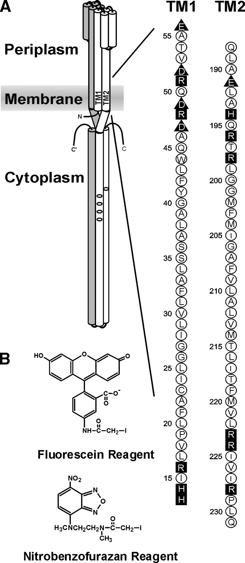
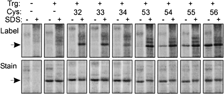
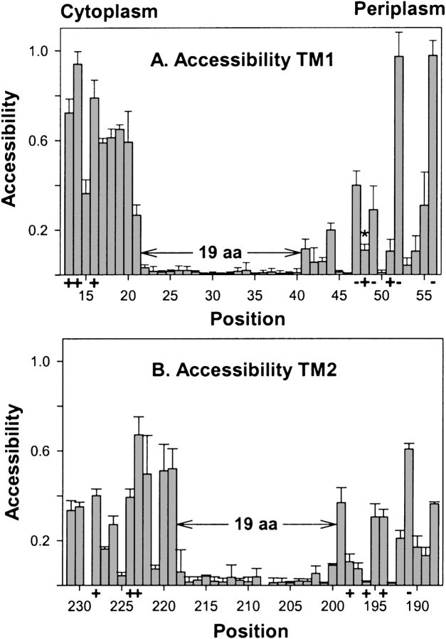
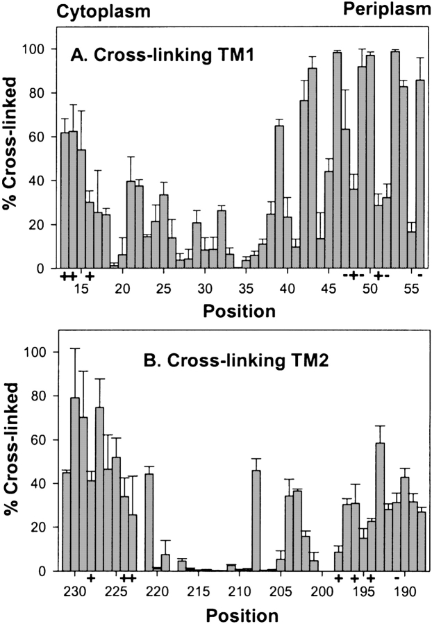
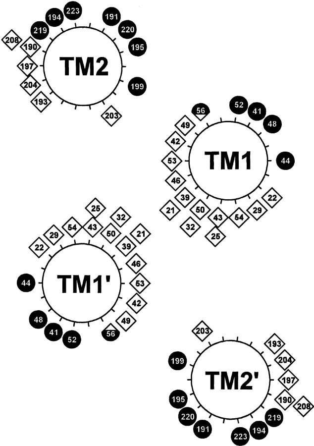
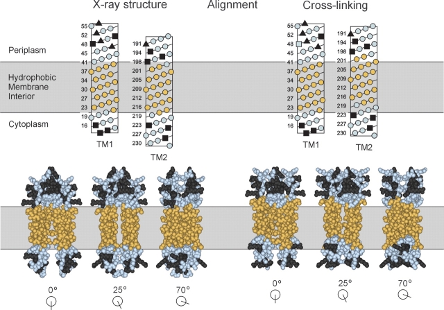
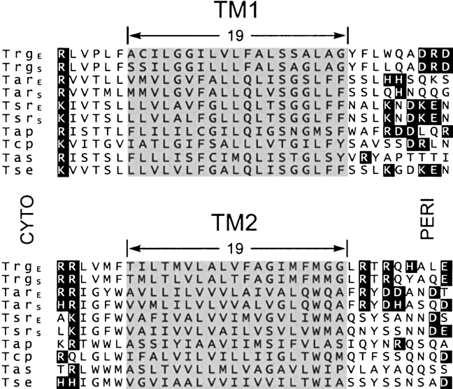
Two hydrophobic sequences, 24 and 30 residues long, identify the membrane-spanning segments of chemoreceptor Trg from Escherichia coli. As in other related chemoreceptors, these helical sequences are longer than the minimum necessary for an alpha-helix to span the hydrocarbon region of a biological membrane. Thus, the specific positioning of the segments relative to the hydrophobic part of the membrane cannot be deduced from sequence alone. With the aim of defining the positioning for Trg experimentally, we determined accessibility of a hydrophilic sulfhydryl reagent to cysteines introduced at each position within and immediately outside the two hydrophobic sequences. For both sequences, there was a specific region of uniformly low accessibility, bracketed by regions of substantial accessibility. The two low-accessibility regions were each 19 residues long and were in register in the three-dimensional organization of the transmembrane domain deduced from independent data. None of the four hydrophobic-hydrophilic boundaries for these two membrane-embedded sequences occurred at a charged residue. Instead, they were displaced one to seven residues internal to the charged side chains bracketing the extended hydrophobic sequences. Many hydrophobic sequences, known or predicted to be membrane-spanning, are longer than the minimum necessary helical length, but precise membrane boundaries are known for very few. The cysteine-accessibility approach provides an experimental strategy for determining those boundaries that could be widely applicable.


Figures

Figure 1.
Figure 1.
Chemoreceptor organization, the transmembrane regions of Trg, and the fluorescent sulfhydryl reagents. (A) (Left) A cartoon of the organization of a chemoreceptor homodimer. Amino (N) and carboxyl termini (C) are labeled, and one subunit is shaded. On the other subunit, the transmembrane regions TM1 and TM2 are labeled and the positions of methyl-accepting sites shown by ovals. The approximate position of the cytoplasmic membrane is shown as a shaded area. (Right) The amino acid sequences (single letter code) of the TM1 and TM2 regions of Trg. Neutral and hydrophobic residues are enclosed in open circles; charged residues, in dark symbols; negative charges, in triangles; and positive charges, in squares. (B) Structure of 5-IAF (Fluorescein Reagent) and N, N′-dimethyl-N-(iodoacetyl)-N′-(7-nitrobenz-2-oxa-1, 3-diazol-4-yl)ethyl-ene-diamine (Nitrobenzofurazan Reagent).
Figure 2.
Figure 2.
Labeling of cysteine-containing forms of Trg by a fluorescent sulfhydryl reagent. Trg lacking cysteine or containing a single cysteine at the indicated positions in the TM1 region was treated with 5-IAF in its native, membrane-embedded state (−) or in the presence of the denaturant and membrane solvent SDS (+), and separated from other proteins in the membrane vesicles by SDS-PAGE. The figure shows two fluorographic images of the same segments of SDS polyacrylamide gels, the upper ones generated by fluorescein fluorescence, and the lower ones, by the protein stain SYPRO red. The leftmost pairs of lanes show patterns for vesicles lacking Trg. The position of Trg, Mr ~60,000, is shown by the arrows.
Figure 3.
Figure 3.
Accessibility of introduced cysteines to 5-IAF. Experiments like those in Fig. 2 ▶ were performed to determine relative accessibility to 5-IAF of cysteine sulfhydryls at each of the indicated positions in membrane-embedded Trg. Mean values of accessibility (three independent membrane preparations) with standard deviations are plotted by residue number from cytoplasm to periplasm for the TM1 (A) and TM2 regions (B). Positions of charged residues in the wild-type protein are marked by “+” or “−” along the abscissa, and the sequence of 19 consecutive residues with minimal accessibility is marked by divergent arrows and labeled “19 aa.” Position 48 is marked in panel A by an asterisk (*) to indicate that the low accessibility is likely the result of bracketing negative charges restricting access by the negatively charged reagent (see text). Because of low receptor content, it was not possible to make reliable measurements for positions 208 and 229.
Figure 4.
Figure 4.
Oxidative cross-linking between introduced cysteines. Extent of oxidative cross-linking catalyzed by Cu(phenanthroline)3 was determined for cysteine pairs in homologous positions in the two subunits of the Trg homodimer using the same three membrane preparations used for experiments in Fig. 3 ▶. Mean values of percent cross-linking with std. dev. are plotted by residue number displayed from cytoplasm to periplasm for the TM1 (A) and TM2 regions (B). No data are shown for positions 199, 200, and 222 because of ambiguity in the results.
Figure 5.
Figure 5.
Distribution of maxima for accessibility and cross-linking. Local maxima for accessibility from Fig. 3 ▶ and for cross-linking from Fig. 4 ▶ are displayed as filled circles and open diamonds, respectively, on the helical wheel diagram in the model of the Trg transmembrane domain derived from comprehensive analysis of previous cross-linking data (Lee and Hazelbauer 1995). The local maximum for position 48, which is bracketed by two negatively charged residues, represents high accessibility to an uncharged sulfhydryl reagent, not the lower accessibility for a bulky, charged reagent plotted in Fig. 3A ▶ (see text). The hybrid symbol for position 56 reflects local maxima for both cross-linking and accessibility, created by a bulge in helical packing (see text).
Figure 6.
Figure 6.
Alignment of TM1 and TM2. Positions in TM1 and TM2 exhibiting minimal accessibility to 5-IAF (Fig. 3 ▶) are shown in gold, and positions outside this region are in blue. Charged residues, all of which are outside the region of minimal accessibility, are in dark gray. In the upper panel, sequences of the TM1 and TM2 regions are displayed in helical net diagrams and aligned along their long axes by two independent criteria. (Left) Extension of the homology model based on the X-ray structure of the periplasmic domain of chemoreceptor Tar (Milburn et al. 1991; Peach et al. 2002). (Right) Alignment by patterns of oxidative cross-linking between introduced cysteines (Lee et al. 1994; Hughson et al. 1997; Peach et al. 2002). The two alignments are placed relative to the membrane so that in each the minimally accessible positions in TM1 are all in the hydrophobic interior. The lower panel presents three space-filling views of the two alignments based on models of the entire transmembrane domain of Trg (Milburn et al. 1991; Peach et al. 2002). The models include TM1/TM1′ residues 13–56 and TM2/TM2′ residues 188–229.
Figure 7.
Figure 7.
Sequence alignments of TM1 and TM2 regions from enteric bacteria. The figure shows the TM1 and TM2 regions of a computer-generated alignment of the complete sequences of chemoreceptors Trg, Tar, and Tsr from E. coli and from Salmonella, Tap from E. coli, Tcp from Salmonella, and Tas and Tse from Enterobacter aerogenes. There are few residue identities in the regions shown. However, alignment in the TM2 region is the result of extension without gaps from the extensive stretch of residue identity among the sequences over much of the ~350-residue carboxyl terminal domain. Alignment in the TM1 region derives primarily from the correspondence of a cluster of positively charged residues on the amino-terminal side of the extended hydrophobic sequences. Charged residues are in dark boxes, and the segments identified as embedded in the hydrophobic environment for Trg are shaded for all sequences.

References

    1. Abramson, J., Smirnova, I., Kasho, V., Verner, G., Kaback, H.R., and Iwata, S. 2003. Structure and mechanism of the lactose permease of Escherichia coli. Science 301 610–615. - PubMed
    1. Altenbach, C., Greenhalgh, D.A., Khorana, H.G., and Hubbell, W.L. 1994. A collision gradient method to determine the immersion depth of nitroxides in lipid bilayers: Application to spin-labeled mutants of bacteriorhodopsin. Proc. Natl. Acad. Sci. 91 1667–1671. - PMC - PubMed
    1. Barnakov, A., Altenbach, C., Barnakova, L., Hubbell, W.L., and Hazelbauer, G.L. 2002. Site-directed spin labeling of a bacterial chemoreceptor reveals a dynamic, loosely packed transmembrane domain. Protein Sci. 11 1472–1481. - PMC - PubMed
    1. Bass, R.B. and Falke, J.J. 1998. Detection of a conserved α-helix in the kinase-docking region of the aspartate receptor by cysteine and disulfide scanning. J. Biol. Chem. 273 25006–25014. - PMC - PubMed
    1. Bass, R.B., Coleman, M.D., and Falke, J.J. 1999. Signaling domain of the aspartate receptor is a helical hairpin with a localized kinase docking surface: Cysteine and disulfide scanning studies. Biochemistry 38 9317–9327. - PMC - PubMed

Publication types

MeSH terms