

Structure of a novel c7-type three-heme cytochrome domain from a multidomain cytochrome c polymer
- PMID: 15133162
- PMCID: PMC2279975
- DOI: 10.1110/ps.04626204
Structure of a novel c7-type three-heme cytochrome domain from a multidomain cytochrome c polymer
Erratum in
- Protein Sci. 2009 Mar;18(3):675
Abstract
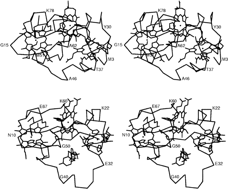
The structure of a novel c(7)-type cytochrome domain that has two bishistidine coordinated hemes and one heme with histidine, methionine coordination (where the sixth ligand is a methionine residue) was determined at 1.7 A resolution. This domain is a representative of domains that form three polymers encoded by the Geobacter sulfurreducens genome. Two of these polymers consist of four and one protein of nine c(7)-type domains with a total of 12 and 27 hemes, respectively. Four individual domains (termed A, B, C, and D) from one such multiheme cytochrome c (ORF03300) were cloned and expressed in Escherichia coli. The domain C produced diffraction quality crystals from 2.4 M sodium malonate (pH 7). The structure was solved by MAD method and refined to an R-factor of 19.5% and R-free of 21.8%. Unlike the two c(7) molecules with known structures, one from G. sulfurreducens (PpcA) and one from Desulfuromonas acetoxidans where all three hemes are bishistidine coordinated, this domain contains a heme which is coordinated by a methionine and a histidine residue. As a result, the corresponding heme could have a higher potential than the other two hemes. The apparent midpoint reduction potential, E(app), of domain C is -105 mV, 50 mV higher than that of PpcA.
Figures


Similar articles
-
Family of cytochrome c7-type proteins from Geobacter sulfurreducens: structure of one cytochrome c7 at 1.45 A resolution.Biochemistry. 2004 Feb 3;43(4):849-59. doi: 10.1021/bi0301439. Biochemistry. 2004. PMID: 14744127
-
Redox characterization of Geobacter sulfurreducens cytochrome c7: physiological relevance of the conserved residue F15 probed by site-specific mutagenesis.Biochemistry. 2004 Aug 3;43(30):9909-17. doi: 10.1021/bi0492859. Biochemistry. 2004. PMID: 15274645
-
Heterologous expression of hexaheme fragments of a multidomain cytochrome from Geobacter sulfurreducens representing a novel class of cytochromes c.Protein Expr Purif. 2005 Feb;39(2):254-60. doi: 10.1016/j.pep.2004.10.015. Protein Expr Purif. 2005. PMID: 15642477
-
Thermodynamic characterization of the redox centres in a representative domain of a novel c-type multihaem cytochrome.Biochem J. 2009 May 27;420(3):485-92. doi: 10.1042/BJ20082428. Biochem J. 2009. PMID: 19351328
-
Molecular mechanisms of heme based sensors from sediment organisms capable of extracellular electron transfer.J Inorg Biochem. 2014 Apr;133:104-9. doi: 10.1016/j.jinorgbio.2013.10.024. Epub 2013 Nov 8. J Inorg Biochem. 2014. PMID: 24268904 Review.
Cited by
-
Mimicking Natural Photosynthesis: Designing Ultrafast Photosensitized Electron Transfer into Multiheme Cytochrome Protein Nanowires.Nanomaterials (Basel). 2020 Oct 28;10(11):2143. doi: 10.3390/nano10112143. Nanomaterials (Basel). 2020. PMID: 33126541 Free PMC article.
-
Resonance Raman fingerprinting of multiheme cytochromes from the cytochrome c3 family.J Biol Inorg Chem. 2006 Mar;11(2):217-24. doi: 10.1007/s00775-005-0067-4. Epub 2005 Dec 10. J Biol Inorg Chem. 2006. PMID: 16341896
-
New insights in uranium bioremediation by cytochromes of the bacterium Geotalea uraniireducens.J Biol Chem. 2025 Feb;301(2):108090. doi: 10.1016/j.jbc.2024.108090. Epub 2024 Dec 14. J Biol Chem. 2025. PMID: 39675718 Free PMC article.
-
Structural and functional insights of GSU0105, a unique multiheme cytochrome from G. sulfurreducens.Biophys J. 2021 Dec 7;120(23):5395-5407. doi: 10.1016/j.bpj.2021.10.023. Epub 2021 Oct 22. Biophys J. 2021. PMID: 34688593 Free PMC article.
-
Rational engineering of Geobacter sulfurreducens electron transfer components: a foundation for building improved Geobacter-based bioelectrochemical technologies.Front Microbiol. 2015 Jul 30;6:752. doi: 10.3389/fmicb.2015.00752. eCollection 2015. Front Microbiol. 2015. PMID: 26284042 Free PMC article. Review.
References
-
- Afkar, E. and Fukumori, Y. 1999. Purification and characterization of triheme cytochrome c7 from the metal-reducing bacterium, Geobacter metallireducens. FEMS Microbiol. Lett. 175 205–210. - PubMed
-
- Aragao, D., Frazao, C., Sieker, L., Sheldrick, G.M., LeGall, J., and Carrondo, M.A. 2003. Structure of dimeric cytochrome c3 from Desulfovibrio gigas at 1.2 Å resolution. Acta Crystallogr. D 59 644–653. - PubMed
-
- Aubert, C., Leroy, G., Bruschi, M., Wall, J.D., and Dolla, A. 1997. A single mutation in the heme 4 environment of Desulfovibrio desulfuricans Norway cytochrome c3 (Mr 26,000) greatly affects the molecule reactivity. J. Biol. Chem. 272 15128–15134. - PubMed
-
- Bothmann, H. and Pluckthun, A. 1998. Selection for a periplasmic factor improving phage display and functional periplasmic expression. Nat. Biotechnol. 16 376–380. - PubMed
Publication types
MeSH terms
Substances
LinkOut - more resources
Full Text Sources
Research Materials
Miscellaneous
