

Overexpression of annexin 1 in pancreatic cancer and its clinical significance
- PMID: 15133855
- PMCID: PMC4656286
- DOI: 10.3748/wjg.v10.i10.1466
Overexpression of annexin 1 in pancreatic cancer and its clinical significance
Abstract
Aim: To investigate the expression of annexin I in pancreatic cancer and its relationship with the clinicopathologic factors, and to evaluate its potential clinical significance.
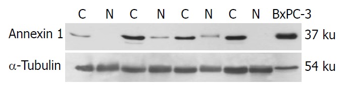
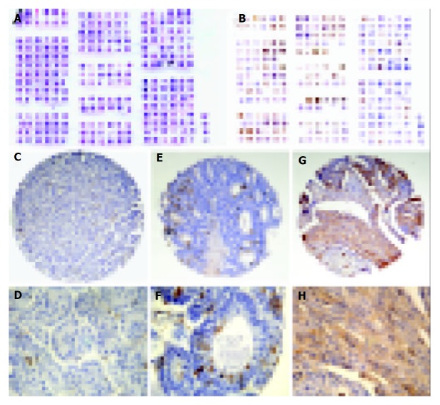
Methods: Annexin I expression was analyzed by Western blot and immunohistochemical staining in pancreatic adenocarcinoma and multi-tissue microarrays (MTAs).
Results: Western blot analysis showed that annexin I was overexpressed in 84.6% (11/13) pancreatic ductal adenocarcinomas. Immunohistochemistry analysis of pancreatic cancer in MTAs showed that annexin I protein was 71.4%(30/42) positive which was markedly increased compared with that in the tumor matched normal pancreas tissues 18.4%(7/38) (P<0.01). In the meantime, the high expression of annexin 1 was correlated with the poor differentiation of pancreatic adenocarcinoma.
Conclusion: Annexin 1 overexpression is a frequent biological marker and correlates with the differentiation of pancreatic cancer during tumorigenesis.
Figures
References
-
- Jemal A, Murray T, Samuels A, Ghafoor A, Ward E, Thun MJ. Cancer statistics, 2003. CA Cancer J Clin. 2003;53:5–26. - PubMed
-
- Rosty C, Christa L, Kuzdzal S, Baldwin WM, Zahurak ML, Carnot F, Chan DW, Canto M, Lillemoe KD, Cameron JL, et al. Identification of hepatocarcinoma-intestine-pancreas/pancreatitis-associated protein I as a biomarker for pancreatic ductal adenocarcinoma by protein biochip technology. Cancer Res. 2002;62:1868–1875. - PubMed
Publication types
MeSH terms
Substances
LinkOut - more resources
Full Text Sources
Other Literature Sources
Medical
Research Materials
