

Interactions of the allogeneic effector leukemic T cell line, TALL-104, with human malignant brain tumors
- PMID: 15134622
- PMCID: PMC1871983
- DOI: 10.1215/s1152851703000140
Interactions of the allogeneic effector leukemic T cell line, TALL-104, with human malignant brain tumors
Abstract
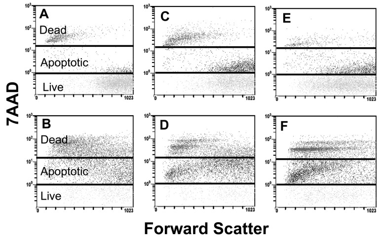
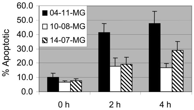
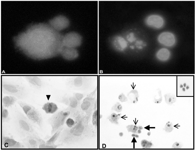
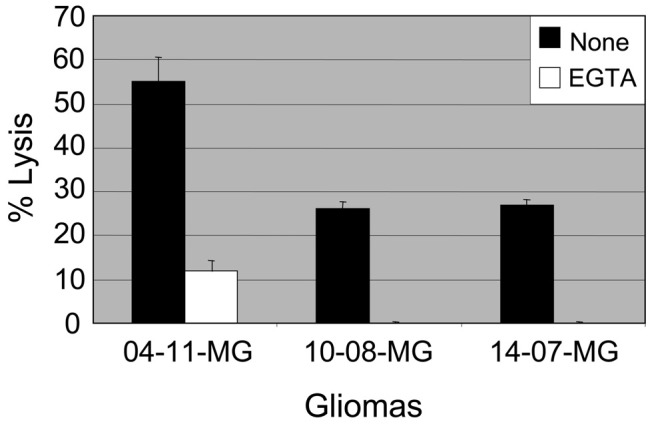
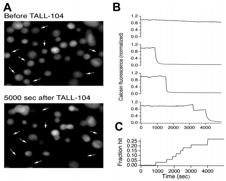
TALL-104 is a human leukemic T cell line that expresses markers characteristic of both cytotoxic T lymphocytes and natural killer cells. TALL-104 cells are potent tumor killers, and the use of lethally irradiated TALL-104 as cellular therapy for a variety of tumors has been explored. We investigated the interactions of TALL-104 cells with human brain tumor cells. TALL-104 cells mediated increased lysis of a panel of brain tumor cells at low effector-to-target ratios over time. We obtained evidence that TALL-104 cells injured glioma cells by both apoptotic and necrotic pathways. A 7-amino actinomycin D flow cytometry assay revealed that the percentages of both apoptotic and necrotic glioma cells increased after TALL-104 cell/glioma cell coincubations. Fluorescent microscopy studies and a quantitative morphologic assay confirmed that TALL-104 cell/glioma cell interactions resulted in tumor cell apoptosis. Cytokines are secreted when TALL-104 cells are coincubated with brain tumor cells; however, morphologic analysis assays revealed that the soluble factors contained within clarified supernates obtained from 4 h coincubates added back to brain tumor cell cultures did not trigger the glioma apoptosis. TALL-104 cells do not express Fas ligand, even upon coincubation with glioma targets, which suggests that the Fas/Fas ligand apoptotic pathway is not likely responsible for the cell injury observed. We obtained evidence that cell injury is calcium dependent and that lytic granule exocytosis is triggered by contact of TALL-104 cells with human glioma cells, suggesting that this pathway mediates glioma cell apoptosis and necrosis.
Figures
Similar articles
-
The human leukemic T-cell line, TALL-104, is cytotoxic to human malignant brain tumors and traffics through brain tissue: implications for local adoptive immunotherapy.Cancer Res. 2000 Oct 15;60(20):5731-9. Cancer Res. 2000. PMID: 11059767
-
Receptors and lytic mediators regulating anti-tumor activity by the leukemic killer T cell line TALL-104.J Leukoc Biol. 2005 Aug;78(2):359-71. doi: 10.1189/jlb.0604360. Epub 2005 Jun 3. J Leukoc Biol. 2005. PMID: 15937142
-
Antitumor activity of a human cytotoxic T-cell line (TALL-104) in brain tumor xenografts.Neuro Oncol. 2000 Apr;2(2):103-13. doi: 10.1093/neuonc/2.2.103. Neuro Oncol. 2000. PMID: 11303619 Free PMC article.
-
Human alloreactive CTL interactions with gliomas and with those having upregulated HLA expression from exogenous IFN-gamma or IFN-gamma gene modification.J Interferon Cytokine Res. 2003 Jul;23(7):379-93. doi: 10.1089/107999003322226032. J Interferon Cytokine Res. 2003. PMID: 14511464
-
Glioma Cell and Astrocyte Co-cultures As a Model to Study Tumor-Tissue Interactions: A Review of Methods.Cell Mol Neurobiol. 2018 Aug;38(6):1179-1195. doi: 10.1007/s10571-018-0588-3. Epub 2018 May 10. Cell Mol Neurobiol. 2018. PMID: 29744691 Free PMC article. Review.
Cited by
-
Immunoresistant human glioma cell clones selected with alloreactive cytotoxic T lymphocytes: downregulation of multiple proapoptotic factors.Gene Ther Mol Biol. 2008 Jun;12(1):101-110. Gene Ther Mol Biol. 2008. PMID: 19066635 Free PMC article.
-
Cellular and functional characterization of immunoresistant human glioma cell clones selected with alloreactive cytotoxic T lymphocytes reveals their up-regulated synthesis of biologically active TGF-beta.J Immunother. 2007 Apr;30(3):261-73. doi: 10.1097/01.cji.0000211339.81211.25. J Immunother. 2007. PMID: 17414317 Free PMC article.
-
Anti-Yo antibody-positive paraneoplastic cerebellar degeneration in a patient with possible cholangiocarcinoma: A case report and review of the literature.World J Clin Cases. 2021 Jun 16;9(17):4423-4432. doi: 10.12998/wjcc.v9.i17.4423. World J Clin Cases. 2021. PMID: 34141810 Free PMC article.
-
CD4 T-cell immune stimulation of HER2 + breast cancer cells alters response to trastuzumab in vitro.Cancer Cell Int. 2020 Nov 10;20(1):544. doi: 10.1186/s12935-020-01625-w. Cancer Cell Int. 2020. PMID: 33292267 Free PMC article.
-
Glioma cell integrin expression and their interactions with integrin antagonists: Research Article.Cancer Ther. 2005;3A:325-340. Cancer Ther. 2005. PMID: 16467916 Free PMC article.
References
-
- Berke G. The binding and lysis of target cells by cytotoxic lymphocytes: Molecular and cellular aspects. Annu Rev Immunol. 1994;12:735–773. - PubMed
-
- Bossi G, Griffiths GM. Degranulation plays an essential part in regulating cell surface expression of Fas ligand in T cells and natural killer cells. Nat Med. 1999;5:90–96. - PubMed
-
- Cappello P, Novelli F, Forni G, Giovarelli M. Death receptor ligands in tumors. J Immunother. 2002;25:1–15. - PubMed
-
- Cesano A, Santoli D. Two unique human leukemic T-cell lines endowed with a stable cytotoxic function and a different spectrum of target reactivity analysis and modulation of their lytic mechanisms. In Vitro Cell Dev Biol. 1992;28A:648–656. - PubMed
Publication types
MeSH terms
Substances
Grants and funding
LinkOut - more resources
Full Text Sources
Medical
Research Materials
Miscellaneous
