

Analysis of 1p, 19q, 9p, and 10q as prognostic markers for high-grade astrocytomas using fluorescence in situ hybridization on tissue microarrays from Radiation Therapy Oncology Group trials
- PMID: 15134623
- PMCID: PMC1871985
- DOI: 10.1215/s1152851703000231
Analysis of 1p, 19q, 9p, and 10q as prognostic markers for high-grade astrocytomas using fluorescence in situ hybridization on tissue microarrays from Radiation Therapy Oncology Group trials
Abstract
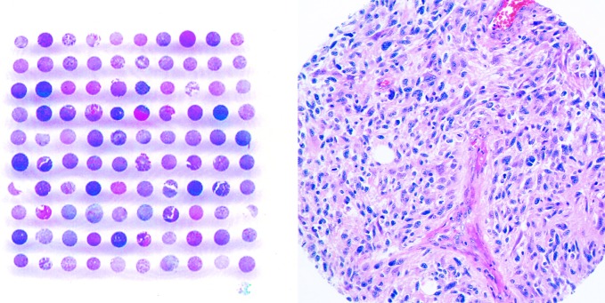
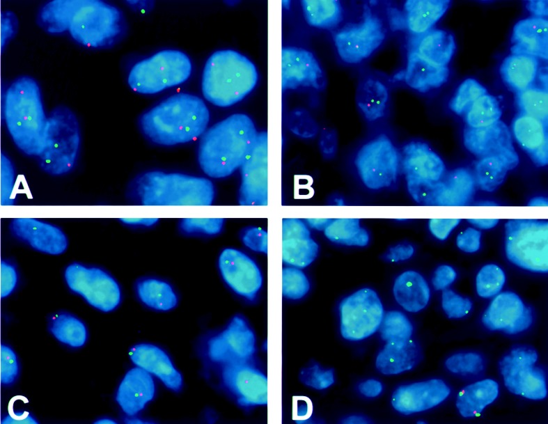
Survival periods vary considerably for patients with high-grade astrocytomas, and reliable prognostic markers are not currently available. We therefore investigated whether genetic losses from chromosomes 1p, 19q, 9p, or 10q were associated with survival in 89 high-grade astrocytomas using tissue microarrays (TMAs) derived from Radiation Therapy Oncology Group clinical trials. Cases included 15 anaplastic astrocytomas (AAs) and 74 glioblastomas (GBMs) selected on the basis of survival times significantly shorter or longer than the expected median. Genetic analysis was performed by TMA-fluorescence in situ hybridization (FISH) on array sections using 8 DNA probes, including those directed at 1p32, 19q13.4, 9p21 (p16/CDKN2A), and 10q (PTEN and DMBT1). Genetic status for each locus was correlated with patient survival group, and data were analyzed by using Fisher's exact test of association (adjusted P = 0.025). Losses of chromosome 1p, either alone or in combination with 19q, were encountered in only 2 cases, both AAs. This contrasts with oligodendrogliomas, in which combined 1p and 19q losses are frequent and predictive of prolonged survival. Solitary 19q loss was noted in 3/15 AAs and in 7/70 GBMs and was more frequent in the long-term survival group (P = 0.041, AA and GBM combined). Chromosome 9p loss was seen in 5/8 AAs and 39/57 GBMs, whereas chromosome 10q loss was detected in 4/15 AAs and 48/68 GBMs. The 9p and 10q deletions were slightly more frequent in short-term survivors, though none of the comparisons achieved statistical significance. Long-term and short-term survival groups of high-grade astrocytomas appear to have dissimilar frequencies of 19q, 9p, and 10q deletions. TMA-FISH is a rapid and efficient way of evaluating genetic alterations in such tumors.
Figures
Similar articles
-
Contribution of 1p, 19q, 9p and 10q Automated Analysis by FISH to the Diagnosis and Prognosis of Oligodendroglial Tumors According to WHO 2016 Guidelines.PLoS One. 2016 Dec 28;11(12):e0168728. doi: 10.1371/journal.pone.0168728. eCollection 2016. PLoS One. 2016. PMID: 28030632 Free PMC article.
-
Prognostic value of 1p, 19q, 9p, 10q, and EGFR-FISH analyses in recurrent oligodendrogliomas.J Neuropathol Exp Neurol. 2004 Apr;63(4):314-22. doi: 10.1093/jnen/63.4.314. J Neuropathol Exp Neurol. 2004. PMID: 15099021
-
Molecular analysis of anaplastic oligodendroglial tumors in a prospective randomized study: A report from EORTC study 26951.Neuro Oncol. 2009 Dec;11(6):737-46. doi: 10.1215/15228517-2009-011. Neuro Oncol. 2009. PMID: 19224764 Free PMC article. Clinical Trial.
-
Population-based studies on incidence, survival rates, and genetic alterations in astrocytic and oligodendroglial gliomas.J Neuropathol Exp Neurol. 2005 Jun;64(6):479-89. doi: 10.1093/jnen/64.6.479. J Neuropathol Exp Neurol. 2005. PMID: 15977639 Review.
-
Genetic markers in glioblastoma: prognostic significance and future therapeutic implications.Adv Anat Pathol. 2003 Jul;10(4):212-7. doi: 10.1097/00125480-200307000-00004. Adv Anat Pathol. 2003. PMID: 12826827 Review.
Cited by
-
Management of newly diagnosed glioblastoma: guidelines development, value and application.J Neurooncol. 2009 May;93(1):1-23. doi: 10.1007/s11060-009-9838-z. Epub 2009 May 9. J Neurooncol. 2009. PMID: 19430879 Review.
-
Correlation of histomorphologic prognostic markers and proliferative index with loss of heterozygosity 1p/19q and MGMT status in diffusely infiltrating gliomas.Med J Armed Forces India. 2013 Jul;69(3):228-36. doi: 10.1016/j.mjafi.2012.08.030. Epub 2012 Dec 1. Med J Armed Forces India. 2013. PMID: 24600115 Free PMC article.
-
Loss of Heterozygosity of 9p Is Associated with Poorer Survival in Patients with Gliomas.Mol Neurobiol. 2016 Nov;53(9):6407-6412. doi: 10.1007/s12035-015-9523-5. Epub 2015 Nov 19. Mol Neurobiol. 2016. PMID: 26582467
-
Glioblastoma multiforme with an abscess: case report and literature review.J Neurooncol. 2008 Jun;88(2):221-5. doi: 10.1007/s11060-008-9557-x. J Neurooncol. 2008. PMID: 18317688 Review.
-
Diagnosis of malignant glioma: role of neuropathology.J Neurooncol. 2008 Sep;89(3):287-311. doi: 10.1007/s11060-008-9618-1. Epub 2008 Aug 20. J Neurooncol. 2008. PMID: 18712282 No abstract available.
References
-
- Brat DJ, Castellano-Sanchez A, Kaur B, Van Meir EG. Genetic and biologic progression in astrocytomas and their relation to angiogenic dysregulation. Adv Anat Pathol. 2002;9:24–36. - PubMed
-
- Burton EC, Lamborn KR, Feuerstein BG, Prados M, Scott J, Forsyth P, Passe S, Jenkins RB, Aldape KD. Genetic aberrations defined by comparative genomic hybridization distinguish long-term from typical survivors of glioblastoma. Cancer Res. 2002;62:6205–6210. - PubMed
-
- CBTRUS. Central Brain Tumor Registry of the United States (2002) Statistical Report: Primary Brain Tumors in the United States, 1995 –1999.Chicago, Ill.: Central Brain Tumor Registry of the United States.
-
- Cairncross JG, Ueki K, Zlatescu MC, Lisle DK, Finkelstein DM, Hammond RR, Silver JS, Stark PC, Macdonald DR, Ino Y, Ramsay DA, Louis DN. Specific genetic predictors of chemotherapeutic response and survival in patients with anaplastic oligodendrogliomas. J Natl Cancer Inst. 1998;90:1473–1479. - PubMed
Publication types
MeSH terms
Substances
Grants and funding
LinkOut - more resources
Full Text Sources
Research Materials
Miscellaneous
