Review
. 2004 Apr;6(2):154-65.
doi: 10.1215/s115285170300067x.

Brain metastases in melanoma: roles of neurotrophins

Affiliations
Review

Brain metastases in melanoma: roles of neurotrophins

Yvonne Denkins et al. Neuro Oncol. 2004 Apr.

Abstract

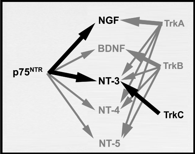
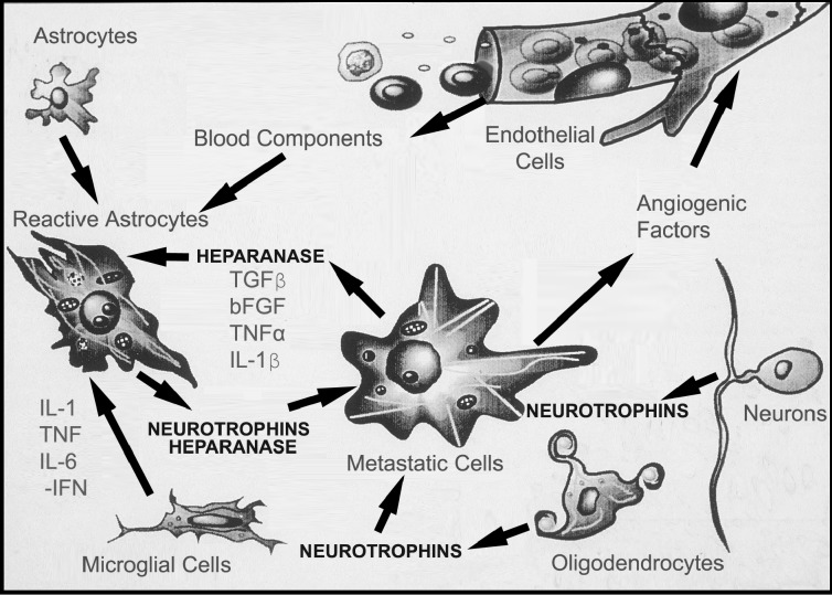
Brain metastasis, which occurs in 20% to 40% of all cancer patients, is an important cause of neoplastic morbidity and mortality. Successful invasion into the brain by tumor cells must include attachment to microvessel endothelial cells, penetration through the blood-brain barrier, and, of relevance, a response to brain survival and growth factors. Neurotrophins (NTs) are important in brain-invasive steps. Human melanoma cell lines express low-affinity NT receptor p75NTR in relation to their brain-metastatic propensity with their invasive properties being regulated by NGF, or nerve growth factor, the prototypic NT. They also express functional TrkC, the putative receptor for the invasion-promoting NT-3. In brain-metastatic melanoma cells, NTs promote invasion by enhancing the production of extracellular matrix (ECM)-degradative enzymes such as heparanase, an enzyme capable of locally destroying both ECM and the basement membrane of the blood-brain barrier. Heparanase is an endo-beta-d-glucuronidase that cleaves heparan sulfate (HS) chains of ECM HS proteoglycans, and it is a unique metastatic determinant because it is the dominant mammalian HS degradative enzyme. Brain-metastatic melanoma cells also produce autocrine/paracrine factors that influence their growth, invasion, and survival in the brain. Synthesis of these factors may serve to regulate NT production by brain cells adjacent to the neoplastic invasion front, such as astrocytes. Increased NT levels have been observed in tumor-adjacent tissues at the invasion front of human brain melanoma. Additionally, astrocytes may contribute to the brain-metastatic specificity of melanoma cells by producing NT-regulated heparanase. Trophic, autocrine, and paracrine growth factors may therefore determine whether metastatic cells can successfully invade, colonize, and grow in the CNS.


Figures

Fig. 1
Fig. 1
Embryologic relationship between melanocytes and the most common neuronal cell populations, both being neural-crest derived. Neuronal populations are neurotrophin-responsive and possess specific cell-surface neurotrophin receptors. Examples include neurons of peripheral nervous system sensory and sympathetic ganglia, Schwann cells, glial cells, and certain populations of CNS cholinergic neurons.
Fig. 2
Fig. 2
Schematic representation of the 2 different classes of neurotrophin receptors (p75NTR, TRK: TrkA, TrkB, TrkC). The p75NTR is a glycoprotein containing 4 cysteine-rich domains in its extracellular portion but no intracytoplasmic tyrosine kinase (TK) domain. However, p75NTR is capable of signaling independent of TRK presence, amplifying the TRK signal when TRK is present (Chao and Bothwell, 2002). TRK receptors contain a signal peptide sequence, cystein clusters, and leucine-rich and Ig-like regions in their extracellular domains and possess an intracytoplasmic TK domain. Percentages of similarity between the 3 main TRK receptors (TrkA, TrkB, TrkC) for their extracellular region/TK domain are respectively indicated.
Fig. 3
Fig. 3
Functional cross-reactivities among neurotrophins and neurotrophin receptors (p75NTR and TRK). P75NTR binds all NT members equally well. The primary ligand for each TRK receptor is indicated by heavy arrows. Black heavy arrows denote interactions between neurotrophin receptors and neurotrophins in brain-metastatic melanoma.
Fig. 4
Fig. 4
Reciprocal interactions between brain-invading melanoma cells and normal cells in the brain microenvironment. Tumor cells release cytokines that can affect host cells such as parenchymal cells, endothelial and glial cells, astrocytes, and brain tissue extracellular matrix (ECM). Reactive astrocytes can arise from stimulation by factors released by invading melanoma cells. In turn, brain cells can release factors that stimulate tumor cell motility and invasion. Astrocytes, oligodendrocytes, and neurons can release NT and ECM degradative enzymes, for example, heparanase produced by astrocytes (Marchetti et al., 2000) in response to brain-invading melanoma. Conversely, these cells secrete growth factors and cytokines which can synergistically regulate NT synthesis and activity in normal brain cells.

Similar articles

Cited by

References

    1. Albino AP, Davis BM, Nanus DM. Induction of growth factor RNA expression in human malignant melanoma: Markers of transformation. Cancer Res. 1991;51:4815–4820. - PubMed
    1. Aviezer D, Iozzo RV, Noonan DM, Yayon A. Suppression of autocrine and paracrine functions of basic fibroblast growth factor by stable expression of perlecan antisense cDNA. Mol Cell Biol. 1997;17:1938–1946. - PMC - PubMed
    1. Avruch J, Zhang XF, Kyriakis JM. Raf meets Ras: Completing the framework of a signal transduction pathway. Trends Biochem Sci. 1994;19:279–283. - PubMed
    1. Barbacid M. Nerve growth factor: A tale of two receptors. Oncogene. 1993;8:2033–2042. - PubMed
    1. Barrett GL, Bartlett PF. The p75 nerve growth factor receptor mediates survival or death depending on the stage of sensory neuron development. Proc Natl Acad Sci USA. 1994;91:6501–6505. - PMC - PubMed

Publication types

Substances