. 2004 May 7;565(1-3):111-6.
doi: 10.1016/j.febslet.2004.03.086.

Identification of a novel protein 3a from severe acute respiratory syndrome coronavirus

Affiliations

Identification of a novel protein 3a from severe acute respiratory syndrome coronavirus

Chia-Jung Yu et al. FEBS Lett. .

Abstract

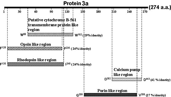
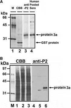
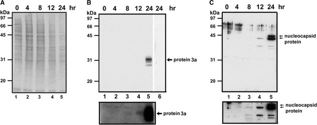
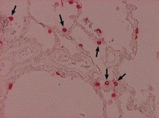
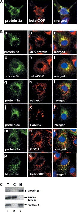
The open reading frame 3 of the severe acute respiratory syndrome coronavirus (SARS-CoV) genome encodes a predicted protein 3a, consisting of 274 amino acids, that lacks any significant similarities to any known protein. We generated specific antibodies against SARS protein 3a by using a synthetic peptide (P2) corresponding to amino acids 261-274 of the putative protein. Anti-P2 antibodies and the sera from SARS patients could specifically detect the recombinant SARS protein 3a expressed in Escherichia coli and in Vero E6 cells. Expression of SARS protein 3a was detected at 8-12 h after infection and reached a higher level after approximately 24 h in SARS-CoV-infected Vero E6 cells. Protein 3a was also detected in the alveolar lining pneumocytes and some intra-alveolar cells of a SARS-CoV-infected patient's lung specimen. Recombinant protein 3a expressed in Vero E6 cells and protein 3a in the SARS-CoV-infected cells was distributed over the cytoplasm in a fine punctate pattern with partly concentrated staining in the Golgi apparatus. Our study demonstrates that SARS-CoV indeed expresses a novel protein 3a, which is present only in SARS-CoV and not in other known CoVs.


Figures

Figure 1
Figure 1
Sequence analysis of putative SARS‐CoV protein 3a. Schematic representation of protein 3a aligned with the putative cytochrome B‐561 transmembrane protein from bacteria (R. solanacearum) (TrEMBL: http://Q8XVV8), opsin from hawkmoth (M. sexta) (TrEMBL: http://O02465), rodopsin from butterfly (P. glaucus) (TrEMBL: http://Q9UAM8), calcium pump from parasite (P. falciparum) (Swiss Prot: Q08853), and outer‐membrane porin from bacteria (S. oneidensis) (TrEMBL: http://Q8EAK6). The numbering system is based on the individual sequences.
Figure 2
Figure 2
Expression and immunodetection of recombinant SARS‐CoV protein 3a in E. coli or Vero E6 cells. (A) Recombinant SARS protein 3a expressed and partially purified from E. coli cells was separated by 12.5% SDS–PAGE and transferred to PVDF membrane. Purified protein was stained with Coomassie brilliant blue (lane 2) or probed with anti‐P2 antibody (lane 3) or pooled sera from SARS‐CoV‐infected patients (lane 4). Molecular weight markers were in lane 1. Recombinant protein 3a (∼33–34 kDa) containing additional 14 amino acids from pGEX 4T‐2 vector was indicated. GST protein degraded from GST‐protein 3a was also indicated. (B) A pcDNA3.1/myc‐His/protein 3a plasmid was transfected into vaccinia virus‐infected Vero E6 cells. After 48 h, non‐infected–transfected cell lysates (lanes 1 and 4), vaccinia virus‐infected Vero E6 cell lysates (lanes 2 and 5), and infected–transfected cell lysates (lanes 3 and 6) were prepared for Coomassie blue staining (lanes 1–3) and Western blot analysis with anti‐P2 antibody (lanes 4–6). This recombinant myc‐His tagged protein 3a (∼37 kDa) contains additional 38 amino acids from pcDNA3.1/myc‐His vector.
Figure 3
Figure 3
Detection of protein 3a in SARS‐CoV‐infected cells. (A) Vero E6 cells uninfected (lane 1) or infected with SARS‐CoV TW1 for 4, 8, 12, or 24 h (lanes 2–5) were harvested, treated with SDS‐sample buffer, and separated by 12.5% SDS–PAGE, transferred to PVDF membrane, and stained with Coomassie blue. (B) Protein samples in panel A were analyzed by Western blotting using an anti‐P2 antibody (lanes 1–5) or anti‐P2 antibody pre‐incubated with 5 μM of P2 peptide (lane 6). (C) Protein samples in panel A were analyzed by Western blotting using an anti‐nucleocapsid (anti‐N1) antibody. Lower panels in B and C show longer exposure time for lanes 1–5.
Figure 4
Figure 4
Immunohistochemical detection of protein 3a in lung section from an SARS‐CoV‐infected patient. The expression of protein 3a was detected using anti‐P2 antibody diluted 1:500 as described in Section 2. Arrowheads indicate protein 3a in the cytoplasm of some pneumocytes (100×).
Figure 5
Figure 5
Subcellular localization of protein 3a in SARS‐CoV‐infected cells. (A) Vaccinia virus‐infected Vero E6 cells were transfected with the pcDNA3.1/myc‐His/protein 3a plasmid and grown on glass coverslips. After 48 h, cells were fixed with formaldehyde and incubated with anti‐P2 or anti‐β‐COP antibody (Golgi marker). (B) Commercial slides with SARS‐CoV‐infected cells were examined by indirect immunofluorescence staining and confocal microscopy. The left panels show protein 3a or membrane (M) protein fluorescence in green, the middle panels show marker fluorescence including 58 K protein (Golgi marker), β‐COP (Golgi marker), calnexin (ER marker), LAMP‐2 (lysosome marker), and cytochrome c oxidase subunit 1 (COX 1) (mitochondria marker) in red, and the right panels show overlays of fluorescence images of protein 3a or M protein and the subcellular marker proteins where yellow indicates co‐localization of green (protein 3a or M protein) and red (marker proteins) signals. Hoechst 33258 nuclear DNA staining is shown in blue. The confocal fluorescence was visualized by Bio‐Rad Radiance 2100 confocal microscope (60×). (C) Subcellular distribution of protein 3a. Membrane (M), and cytosolic (C) fractions of recombinant protein 3a expressed in Vero E6 were prepared as described in Section 2. Equivalent amounts (from total homogenate, T) of each fraction were analyzed by electrophoresis and immunoblotting using specific antibodies against the P2 peptide of protein 3a, α‐tubulin (cytosolic marker), or calnexin (ER membrane marker).

Similar articles

Cited by

References

    1. Ksiazek T.G., Erdman D., Goldsmith C.S., Zaki S.R., Peret T., Emery S., Tong S., Urbani C., Comer J.A., Lim W., N. Engl. J. Med, 348, (2003), 1953– 1966. - PubMed
    1. Kuiken T., Fouchier R.A., Schutten M., Rimmelzwaan G.F., van Amerongen G., van Riel D., Laman J.D., de Jong T., van Doornum G., Lim W., Lancet, 362, (2003), 263– 270. - PMC - PubMed
    1. Holmes K.V., Enjuanes L., Science, 300, (2003), 1377– 1378. - PubMed
    1. Rota P.A., Oberste M.S., Monroe S.S., Nix W.A., Campagnoli R., Icenogle J.P., Penaranda S., Bankamp B., Maher K., Chen M.H., Science, 300, (2003), 1394– 1399. - PubMed
    1. Drosten C., Gunther S., Preiser W., van der Werf S., Brodt H.R., Becker S., Rabenau H., Panning M., Kolesnikova L., Fouchier R.A., N. Engl. J. Med, 348, (2003), 1967– 1976. - PubMed

Publication types

MeSH terms