. 2004 May 18;101(20):7641-5.
doi: 10.1073/pnas.0401654101. Epub 2004 May 10.

Cdx2 is essential for axial elongation in mouse development

Affiliations

Cdx2 is essential for axial elongation in mouse development

Kallayanee Chawengsaksophak et al. Proc Natl Acad Sci U S A. .

Abstract

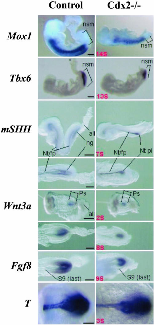
Inactivation of Cdx2 leads to preimplantation embryonic lethality. Rescue of the implantation defect by tetraploid fusion established that Cdx2 is necessary for trophoblastic development, vasculogenesis in the yolk sac mesoderm, allantoic growth, and chorioallantoic fusion. "Rescued" Cdx2 mutants die at late gastrulation stages because of failure of placental development. Cdx2 is also needed for the completion of the normal process of gastrulation and tail bud elongation. Presegmental paraxial mesoderm is severely restricted in amount and somites posterior to somite 5 are abnormal. The Cdx2 mutation, like mutations impairing Wnt and Fgf signaling, causes posterior truncations and disturbs axial patterning of the embryonic structures, indicated by changes in the Hox expression domains. The gene appears to be important in the integration of the pathways controlling embryonic axial elongation, and anterior-posterior patterning.


Figures

Fig. 1.
Fig. 1.
Section through the placentae of mutant (A) and wild-type (B) mice at a nominal age of 11.5 dpc created by injection of ES cells into tetraploid wild-type blastocysts bearing a LacZ transgene. The allantoic component of the wild-type placenta is clearly seen (arrow) but is absent from the mutant specimen. YS, yolk sac; D, maternal decidua. The section was stained for β-galactosidase and with hematoxylin and eosin. (Bar = 100 μmin A and 125 μmin B.) The diffuse blue staining on the fetal side of the mutant placenta is an artefact due to leaching of β-galactosidase stain from the trophoblastic nuclei into the surrounding cytoplasm. The tetraploid nuclei are dark due to β-galactosidase staining, whereas those of the allantoic mesoderm and of the decidua (D) of maternal host origin do not have LacZ activity.
Fig. 2.
Fig. 2.
Wild-type (A) and Cdx2-/- (B) mouse embryos at a nominal age of 10.5 dpc. The rostral region of the mutant is normal, though somewhat retarded in development when compared with the wild-type embryo. Caudally there is gross truncation distal to the forelimb bud (marked) in the mutant embryo. Arrows mark the tail tip in both mutant and wild-type. (Bar = 250 μmin A and 225 μmin B.) (C and D) An 8.5-dpc yolk sac from wild-type (C) and mutant (D) embryos immunologically stained with a PECAM-1 antibody. A coarse plexus of enlarged vessels is visible in the mutant (D), compared with the fine honeycomb appearance in the control (C). (Bar = 500 μm.) (E-J) Expression of Hox and Cdx genes in wild-type and mutant embryos. Whole-mount in situ hybridization of Hoxb1 (E) and Hoxd4 (F) transcripts at the five-somite stage in control and Cdx2-/- embryos. Note, in the Hoxd4 hybridized mutant embryo, the reduced amount of unsegmented mesoderm, the last somite being close to the anterior part of the streak. Expression of the more 5′ Hox gene, Hoxb8 (G and H) shows a posterior shift of the anterior boundary of the expression domain in the mesoderm (S13 instead of S11) and in the neural tube (level of S7 instead of S5) in the mutant. Expression levels of Cdx1 (I) appear unchanged in Cdx2 mutants at the early somite stage, whereas the expression level of Cdx4 (J) is much reduced compared to wild-type embryos. (Bar = 200 μm.) ov, optic vesicle; r, rhombomere; nsm, nonsegmented mesoderm; Ps, primitive streak; S, somite.
Fig. 3.
Fig. 3.
Expression of mesoderm, neurectoderm, and endoderm markers, and of Wnt and Fgf signals in Cdx2 mutant embryos and controls. Mox1 marks segmented paraxial mesoderm; note the important reduction in the amount of unsegmented mesoderm in the caudal aspect of the 14-somite Cdx2-/- embryo. The expression domain of Tbx6, marking nonsegmented mesoderm, is reduced in Cdx2-/- compared with controls. This pattern is complementary to that of Mox1. Shh is a marker of midline structures and of hindgut endoderm; a lateral view above, and a ventral view below both show delayed hindgut diverticulum formation in the mutant leading to an absence of Shh endodermal signal posteriorly in Cdx2-/- embryos. Wnt3a was assayed at a stage earlier (two somites, above), or later (eight somite embryos, below) than the first manifestation of an altered phenotype in Cdx2 mutant embryos. Expression of Fgf8 is shown in a nine-somite mutant and a control. Brachyury T was expressed in the posterior mesoderm of both mutant and control at the three- and two-somite stage, respectively. all, allantois; nsm, nonsegmented mesoderm; ps, primitive streak; Nt/fp, Notochord, floorplate; Nt pl, notochordal plate; hg, hindgut; S9, somite 9, the last formed somite. (Bar = 200 μm.)

References

    1. Moreno, E. & Morata, G. (1999) Nature 400, 873-877. - PubMed
    1. Duprey, P., Chowdhury, K., Dressler, G. R., Balling, R., Simon, D., Guenet, J. L. & Gruss, P. (1988) Genes Dev. 2, 1647-1654. - PubMed
    1. Gamer, L. W. & Wright, C. V. (1993) Mech. Dev. 43, 71-81. - PubMed
    1. James, R. & Kazenwadel, J. (1991) J. Biol. Chem. 266, 3246-3251. - PubMed
    1. Beck, F., Erler, T., Russell, A. & James, R. (1995) Dev. Dyn. 204, 219-227. - PubMed

Publication types

LinkOut - more resources