

doi: 10.1073/pnas.0400730101.
Epub 2004 May 10.
The effect of interspecific oocytes on demethylation of sperm DNA
Affiliations
- PMID: 15136736
- PMCID: PMC419658
- DOI: 10.1073/pnas.0400730101
Item in Clipboard
The effect of interspecific oocytes on demethylation of sperm DNA
Proc Natl Acad Sci U S A.
.
Abstract
In contrast to mice, in sheep no genome-wide demethylation of the paternal genome occurs within the first postfertilization cell cycle. This difference could be due either to an absence of a sheep demethylase activity that is present in mouse ooplasm or to an increased protection of methylated cytosine residues in sheep sperm. Here, we use interspecies intracytoplasmic sperm injection to demonstrate that sheep sperm DNA can be demethylated in mouse oocytes. Surprisingly, mouse sperm can also be demethylated to a limited extent in sheep oocytes. Our results suggest that the murine demethylation process is facilitated either by a sperm-derived factor or by male pronuclear chromatin composition.
Figures
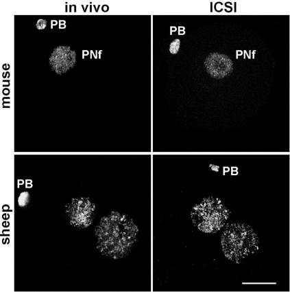
Immunodetection of 5mC in mouse or sheep embryos produced either in vivo or by ICSI. Independent of the protocol, we detected methylation in both pronuclei for the sheep and only in the female pronucleus (PNf) for the mouse. PB, polar body. (Bar, 20 μm.)
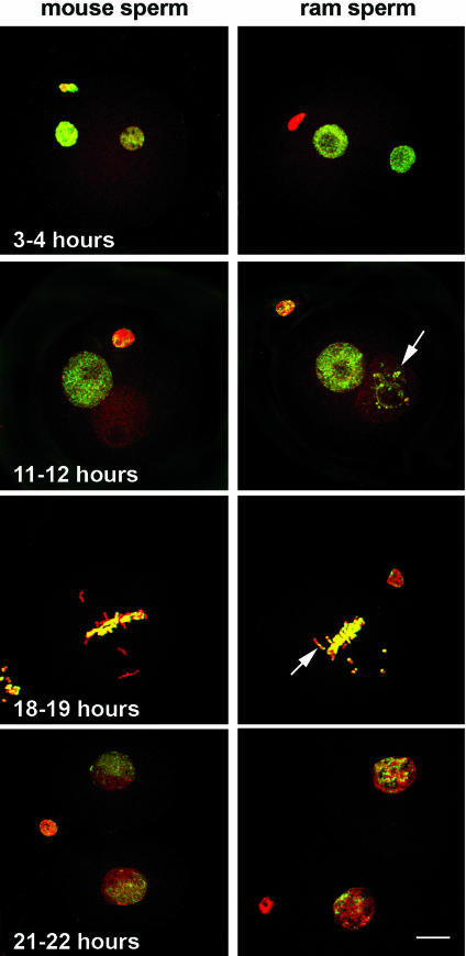
Confocal images of hybrid zygotes obtained by either mouse sperm ICSI or ram sperm ICSI into mouse oocytes. Zygotes were stained with propidium iodide for chromatin (red) and FITC-conjugated secondary antibody for 5mC (green). Fixation was performed at various times after fertilization: 3–4, 11–12, 18–19 h, at mitosis; 21–22 h at the two-cell stage. For both sperm types, genome-wide DNA methylation starts to decrease with sperm decondensation to finally become undetectable by the end of the first cell cycle, except for some condensed regions in the sheep-derived pronucleus (arrows). At the two-cell stage, maternal and paternal compartments are still separated as shown by the differential green staining. However, in sheep/mouse hybrids, the distinction is less striking. (Bar, 10 μm.)
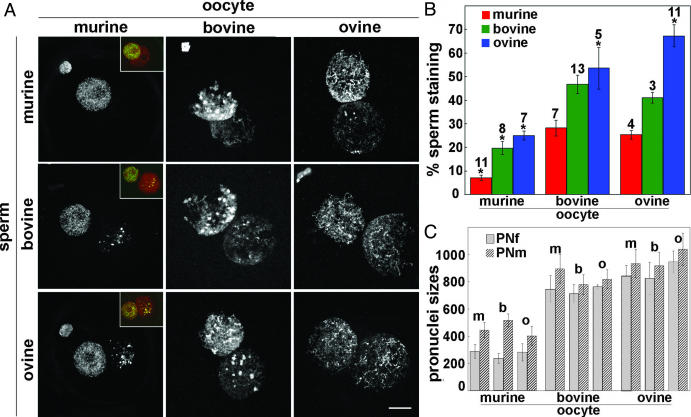
(A) Differential demethylating process in ICSI hybrid zygotes depends on oocyte source. Murine, bovine, or ovine sperm were microinjected into murine, bovine, and ovine oocytes. Hybrid zygotes were then processed for 5mC immunodetection at the end of the first cell cycle, after replication had taken place, i.e., 11–12, 20–22, and 18–20 h for mouse, cow, and sheep oocyte recipients, respectively. (Insets) Merged images of DNA (red) and methylation staining (green), with yellow showing overlapping signal. ICSI into mouse oocytes clearly demonstrates high demethylation capacity of the mouse cytoplasm regardlessofthe sperm origin. In contrast, sperm demethylation was lower in bovine oocytes and almost nonexistent in sheep oocytes. (Bar, 10 μm.) (B) Relative amounts of the methylation quantified in the male versus the female pronuclei (the number of hybrids analyzed is indicated above each column). Error bars represent the SE between hybrids within one group. Statistical analyses were performed with Student's t test. The asterisks denote values that are statistically different for one sperm origin between the different oocyte sources (P < 0.01). This demonstrates that, independently of the oocyte source, the mouse paternal pronucleus is significantly less methylated than bull- or ram-derived pronuclei (P < 0.01). In contrast, mouse and bull sperm have similar methylation levels in cow and sheep oocytes, whereas sheep sperm methylation is even higher after injection into sheep oocytes (P < 0.01). (C) Sizes of the female (PNf) and male (PNm) pronuclei (arbitrary units) quantified after injection of murine (m), bovine (b), or ovine (o) sperm into various oocytes. Error bars represent the SE between hybrids within one group.
References
-
- Jaenisch, R. & Bird, A. (2003) Nat. Genet. 33, 245-254. - PubMed
-
- Bestor, T. H. (2003) Ann. N.Y. Acad. Sci. 983, 22-27. - PubMed
-
- Robertson, K. D. (2002) Oncogene 21, 5361-5379. - PubMed
-
- Reik, W., Santos, F. & Dean, W. (2003) Theriogenology 59, 21-32. - PubMed
-
- Li, E. (2002) Nat. Rev. Genet. 3, 662-673. - PubMed
Publication types
MeSH terms
Substances
LinkOut - more resources
Full Text Sources
