Case Reports
. 2004 Jul;75(1):106-11.
doi: 10.1086/422103. Epub 2004 May 10.

Hypergonadotropic ovarian failure associated with an inherited mutation of human bone morphogenetic protein-15 (BMP15) gene

Affiliations
Case Reports

Hypergonadotropic ovarian failure associated with an inherited mutation of human bone morphogenetic protein-15 (BMP15) gene

Elisa Di Pasquale et al. Am J Hum Genet. 2004 Jul.

Abstract

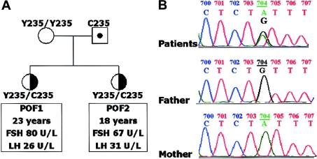
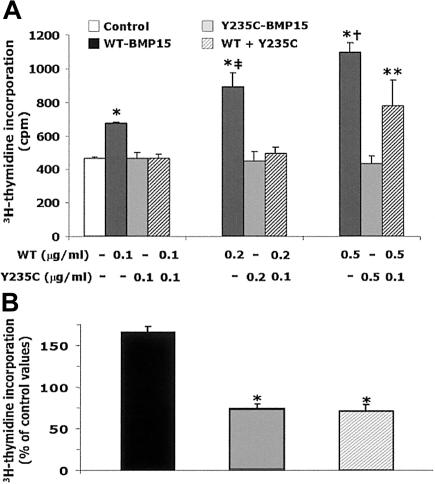
Hypergonadotropic ovarian failure is a common cause of female infertility. It is a heterogeneous disorder that, in the most severe forms, is a result of ovarian dysgenesis (OD). Most OD cases are associated with major X-chromosome abnormalities, but the pathogenesis of this disorder is still largely undefined in patients with a normal karyotype. Animal models showed the important role in female reproduction played by the product of a gene located at Xp11.2 in humans (BMP15). BMP15 is an oocyte-specific growth/differentiation factor that stimulates folliculogenesis and granulosa cell (GC) growth. We report two sisters with a normal karyotype who are affected with hypergonadotropic ovarian failure due to OD. The familial presentation suggested a genetic origin, and candidate genes were screened for mutations. A heterozygous nonconservative substitution in the pro region of BMP15 (Y235C) was identified in both sisters but not in 210 control alleles. This mutation was inherited from the father. Mutant BMP15 appears to be processed abnormally, is associated with reduced GC growth, and antagonizes the stimulatory activity of wild-type protein on GC proliferation. In conclusion, the first natural mutation in human BMP15 is associated with familial OD, indicating that the action of BMP15 is required for the progression of human folliculogenesis. This condition represents an exceptional example of X-linked human disease exclusively affecting heterozygous females who inherited the genetic alteration from the unaffected father. BMP15 defects are involved in the pathogenesis of hypergonadotropic ovarian failure in humans.


Figures

Figure  1
Figure 1
Pedigree and sequence analysis. A, Pedigree of the family. The elder sister (VB) and her younger sister (SB) were affected with idiopathic hypergonadotropic ovarian failure and presented with primary amenorrhea. After a 3-mo estrogen withdrawal, estradiol was low (<0.1 nmol/L) and gonadotropin values rose into the postmenopausal range, as shown (premenopausal female values: FSH = 1.0–8.0 U/L; LH = 0.5–9.0 U/L). The mother had regular menses and was postmenopausal at the time of the study (physiological menopause at 49 years), and the father had normal fertility and gonadal function (FSH = 5.3 U/L; LH = 2.5 U/L; testosterone = 15.6 nmol/L). B, BMP15 sequence analyses in VB, SB, and the parents. In the proband (VB), automated sequencing revealed a heterozygous A→G transition at base pair 704 of the BMP15 gene located at Xp11.2. The same heterozygous transition was seen in the younger sister, whereas the father was a hemizygous carrier of the same 704A→G transition and the mother was normal. The grandmother, two uncles, and two aunts of the paternal family had normal fertility (five, one, one, four, and three births, respectively). One of these aunts had a normal BMP15 sequence; therefore, the mutation should have arisen de novo in the hemizygous father. This transition generates a missense substitution (Y235C) located in the propeptide region of the BMP15 protein.
Figure  2
Figure 2
Alignment of the amino acid sequence of part of the BMP15 propeptide region from several mammalian species with its human homologue GDF9. The tyrosine affected by the mutation and the surrounding residues are highly conserved.
Figure  3
Figure 3
Western immunoblotting was performed under reducing and nonreducing conditions (with or without β-mercaptoetanol [β-ME]) by use of anti-His-HRP antibody (Roche) or anti-Myc antibody (Invitrogen), as appropriate. Immunofluorescent signals were detected with the use of the ECL system (Amersham). Western blots performed with anti-Myc antibody are shown. A, Analysis performed in the presence of β-ME. Bands corresponding to mature (duplet at ∼21–23 kDa) and precursor (at ∼50–55 kDa) BMP15-Myc-His chimeras were seen in the WT and mutant (MUT) lanes but were not seen in the control media from nontransfected (NT) cells. B, Analysis performed under nonreducing conditions allowing the visualization of dimeric products. Bands corresponding to mature (∼45 kDa) and precursor (>110 kDa) dimers were seen in both WT and MUT lanes. Additional bands corresponding to precursor monomers (∼50–55 kDa) or to stable precursor-mature dimers (∼80 kDa) were detected in the mutant Y235C-BMP15 but not in WT lanes, even when double the amount of protein was tested (WT 2x). The band at 65–70 kDa in both panels corresponds to a nonspecific cross-reacting protein present in all media.
Figure  4
Figure 4
Granulosa cell-growth assay evaluated by direct cell count (see methods in appendix A [online only]). Results (mean ± SD) of a representative assay using different doses of BMP15-Myc-His fusion proteins. Tagged proteins were purified on nickel columns, and doses of purified proteins (0.1 or 0.2 μg/ml) were tested in triplicate wells. An asterisk (*) indicates P<.05 versus control and mutant preparations (two-tailed, unpaired Student’s t test).
Figure  5
Figure 5
Granulosa cell-growth assay evaluated by 3H-thymidine incorporation (see methods in appendix A [online only]). A, Results (mean ± SD) of a representative assay using different doses of BMP15-Myc-His fusion proteins. Tagged proteins were obtained after affinity chromatography on nickel columns. Mutant BMP15 and WT-BMP15 were tested either separately or in combination (1:1 or 1:5 mixtures) in triplicate wells. An asterisk (*) indicates P<.01 versus control and mutant alone; a double dagger (‡) indicates P<.02 versus WT 0.1 μg/ml; a dagger (†) indicates P<.03 versus WT 0.2 μg/ml; and two asterisks (**) indicate P<.03 versus control and mutant alone (two-tailed, unpaired Student’s t test). B, Effects of recombinant human BMP15 tagged proteins (0.2 μg/ml) on GC growth, tested in eight experiments with the use of different GC preparations from eight women undergoing ovarian hyperstimulation. Owing to variable growth rates among these different GC preparations, results (mean ± SE; n=8) obtained with WT, mutant, or 1:1 mixtures are expressed as the percentage of 3H-thymidine incorporation in control wells. In WT-BMP15 wells, growth rate was 165% ± 6% (mean ± SE) of control wells (P<.001). In contrast, growth rates were reduced significantly, by >2-fold, when mutant BMP15 was tested separately or in the presence of equal amounts (1:1) of WT (74% ± 4% or 69% ± 6% of controls, respectively). An asterisk (*) indicates P<.001 versus WT-BMP15 (two-tailed, unpaired Student’s t test).

References

Electronic-Database Information

    1. GenBank, http://www.ncbi.nlm.nih.gov/Genbank/ (for human BMP15 genomic sequence [accession number AF082349] and human GDF9 mRNA sequence [accession number NM_005260])
    1. Online Mendelian Inheritance in Man (OMIM), http://www.ncbi.nlm.nih.gov/Omim/

References

    1. Aaltonen J, Laitinen MP, Vuojolainen K, Jaatinen R, Horelli-Kuitunen N, Seppa L, Louhio H, Tuuri T, Sjoberg J, Butzow R, Hovata O, Dale L, Ritvos O (1999) Human growth differentiation factor 9 (GDF-9) and its novel homolog GDF-9B are expressed in oocytes during early folliculogenesis. J Clin Endocrinol Metab 84:2744–275010.1210/jc.84.8.2744 - DOI - PubMed
    1. Aittomaki K, Lucena JLD, Pakarinen P, Sistonen P, Tapanainen J, Gromoll J, Kaskikari R, Sankila EM, Lehvaslaiho H, Engel AR, Nieschlag E, Huhtaniemi I, de la Chapelle A (1995) Mutation in the follicle-stimulating hormone receptor gene causes hereditary hypergonadotropic ovarian failure. Cell 82:959–968 - PubMed
    1. Brunner AM, Marquardt H, Malacko AR, Lioubin MN, Purchio AF (1989) Site directed mutagenesis of cysteine residues in the pro region of the transforming growth factor β1 precursor. J Biol Chem 264:13660–13664 - PubMed
    1. Chang H, Brown CW, Matzuk MM (2002) Genetic analysis of the mammalian transforming growth factor-β superfamily. Endocr Rev 23:787–82310.1210/er.2002-0003 - DOI - PubMed
    1. Dube JL, Wang P, Elvin J, Lyons KM, Celeste AJ, Matzuk MM (1998) The bone morphogenetic protein 15 gene is X-linked and expressed in oocytes. Mol Endocrinol 12:1809–181710.1210/me.12.12.1809 - DOI - PubMed

Publication types

MeSH terms

Substances