. 2004 May;78(10):5311-23.
doi: 10.1128/jvi.78.10.5311-5323.2004.

Nef binds p6* in GagPol during replication of human immunodeficiency virus type 1

Affiliations

Nef binds p6* in GagPol during replication of human immunodeficiency virus type 1

Luciana J Costa et al. J Virol. 2004 May.

Abstract

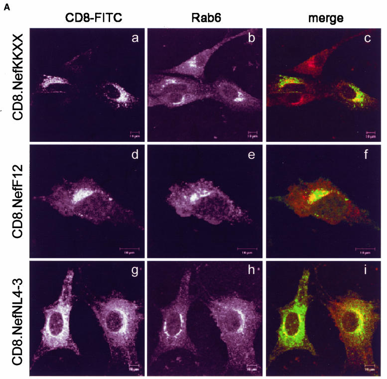
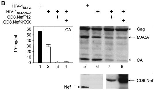
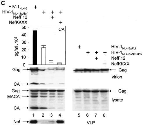
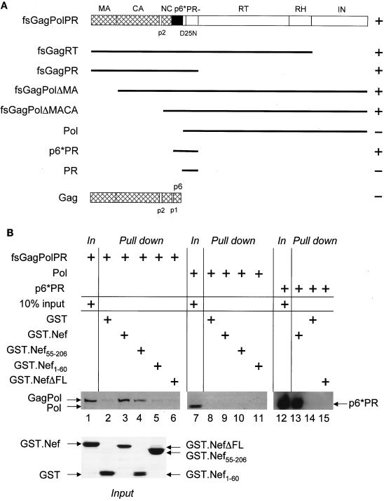
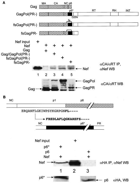
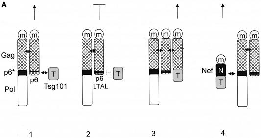
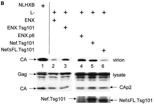
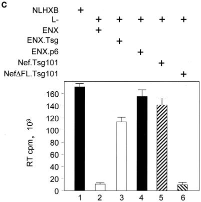
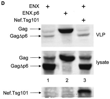
The atypical Nef protein (NefF12) from human immunodeficiency virus type 1 strain F12 (HIV-1(F12)) interferes with virion production and infectivity via a mysterious mechanism. The correlation of these effects with the unusual perinuclear subcellular localization of NefF12 suggested that the wild-type Nef protein could bind to assembly intermediates in late stages of viral replication. To test this hypothesis, Nef from HIV-1(NL4-3) was fused to an endoplasmic reticulum (ER) retention signal (NefKKXX). This mutant NefKKXX protein recapitulated fully the effects of NefF12 on on Gag processing and virion production, either alone or as a CD8 fusion protein. Importantly, the mutant NefKKXX protein also localized to the intermediate compartment, between the ER and the trans-Golgi network. Furthermore, Nef bound the GagPol polyprotein in vitro and in vivo. This binding mapped to the C-terminal flexible loop in Nef and the transframe p6* protein in GagPol. The significance of this interaction was demonstrated by a genetic assay in which the release of a mutant HIV-1 provirus lacking the PTAP motif in the late domain that no longer binds Tsg101 was rescued by a Nef.Tsg101 chimera. Importantly, this rescue as well as incorporation of Nef into HIV-1 virions correlated with the ability of Nef to interact with GagPol. Our data demonstrate that the retention of Nef in the intermediate compartment interferes with viral replication and suggest a new role for Nef in the production of HIV-1.


Figures

FIG. 1.
FIG. 1.
(A) The mutant CD8.NefKKXX chimera localizes to the intermediate compartment between the ER and TGN as determined by confocal microscopy. Shown is the immunostaining of NIH 3T3 cells, which expressed the indicated proteins. The subcellular distribution of expressed proteins and of the TGN was visualized with fluorescein isothiocyanate-conjugated anti-CD8 (green staining but left black and white for better resolution) and anti-Rab6 (red staining but left black and white for better resolution) antibodies, respectively. The right panels show the merge of the two pictures on the left. Cells expressed the hybrid mutant CD8.NefKKXX (a to c), the hybrid CD8.NefF12 (d to f), and the hybrid CD8.NefNL4-3 (g to i) proteins. (B) The hybrid mutant CD8.NefKKXX protein interferes with virion production and Gag processing in 293T cells. The mutant CD8.NefKKXX chimera inhibits the production of HIV-1. Left panel, virion production from 293T cells, which expressed HIV-1NL4-3 (bar 1) or HIV-1NL4-3ΔNef (bar 2) or coexpressed HIV-1NL4-3ΔNef and CD8.NefF12 (bar 3) or mutant CD8.NefKKXX (bar 4) chimeras, was measured by CA capture ELISA. Means and standard deviations from three independent experiments are shown. Right panels, Western blotting of these cell lysates was performed with anti-CA (top panel) and anti-Nef (bottom panel) antibodies. (C). Mutant NefKKXX protein inhibits Gag processing and virion production. 293T cells expressed HIV-1NL4-3 (bar 1) or mutant HIV-1NL4-3ΔNef (bar 2) viruses or coexpressed the mutant HIV-1NL4-3ΔNef virus and NefF12 (bar 3) or mutant NefKKXX (bar 4) proteins. Viral supernatants and cell lysates (lysate) were monitored for CA as described for panel B (left panels). Additionally, anti-CA Western blotting was performed on viral supernatants (virion). As controls, effects of NefF12 and NefKKXX on VLPs were analyzed (lanes 5 to 8). 293T cells expressed mutant HIV-1NL4-3ΔPol (lane 5) or HIV-1NL4-3ΔNef ΔPol (lane 6) viruses or coexpressed mutant HIV-1NL4-3ΔNef ΔPol virus and NefF12 (lane 7) or mutant NefKKXX (lane 8) proteins. Arrows indicate the Gag (55 kDa) and MACA (capsid, matrix) (41 kDa) precursors, the CA (24 kDa) and Nef (27 kDa) proteins, and the 57-kDa CD8.Nef chimera.
FIG. 1.
FIG. 1.
(A) The mutant CD8.NefKKXX chimera localizes to the intermediate compartment between the ER and TGN as determined by confocal microscopy. Shown is the immunostaining of NIH 3T3 cells, which expressed the indicated proteins. The subcellular distribution of expressed proteins and of the TGN was visualized with fluorescein isothiocyanate-conjugated anti-CD8 (green staining but left black and white for better resolution) and anti-Rab6 (red staining but left black and white for better resolution) antibodies, respectively. The right panels show the merge of the two pictures on the left. Cells expressed the hybrid mutant CD8.NefKKXX (a to c), the hybrid CD8.NefF12 (d to f), and the hybrid CD8.NefNL4-3 (g to i) proteins. (B) The hybrid mutant CD8.NefKKXX protein interferes with virion production and Gag processing in 293T cells. The mutant CD8.NefKKXX chimera inhibits the production of HIV-1. Left panel, virion production from 293T cells, which expressed HIV-1NL4-3 (bar 1) or HIV-1NL4-3ΔNef (bar 2) or coexpressed HIV-1NL4-3ΔNef and CD8.NefF12 (bar 3) or mutant CD8.NefKKXX (bar 4) chimeras, was measured by CA capture ELISA. Means and standard deviations from three independent experiments are shown. Right panels, Western blotting of these cell lysates was performed with anti-CA (top panel) and anti-Nef (bottom panel) antibodies. (C). Mutant NefKKXX protein inhibits Gag processing and virion production. 293T cells expressed HIV-1NL4-3 (bar 1) or mutant HIV-1NL4-3ΔNef (bar 2) viruses or coexpressed the mutant HIV-1NL4-3ΔNef virus and NefF12 (bar 3) or mutant NefKKXX (bar 4) proteins. Viral supernatants and cell lysates (lysate) were monitored for CA as described for panel B (left panels). Additionally, anti-CA Western blotting was performed on viral supernatants (virion). As controls, effects of NefF12 and NefKKXX on VLPs were analyzed (lanes 5 to 8). 293T cells expressed mutant HIV-1NL4-3ΔPol (lane 5) or HIV-1NL4-3ΔNef ΔPol (lane 6) viruses or coexpressed mutant HIV-1NL4-3ΔNef ΔPol virus and NefF12 (lane 7) or mutant NefKKXX (lane 8) proteins. Arrows indicate the Gag (55 kDa) and MACA (capsid, matrix) (41 kDa) precursors, the CA (24 kDa) and Nef (27 kDa) proteins, and the 57-kDa CD8.Nef chimera.
FIG. 2.
FIG. 2.
Nef binds GagPol and p6*PR polypeptides in GST pull-down assays. (A) Shown are the frameshifted, protease-negative GagPol (fsGagPolPR−) and Gag polyproteins and their truncated derivatives. MA, CA, NC, p6, PR, RT, RT, RNase H (RH), the spacer peptides p2 and p1, and p6* are shown. Note that p6 and p6* (frameshifted p6), which share no similarity in their sequences, are present only in Gag and GagPol, respectively. Black lines represent the truncated GagPol and Gag precursors. GagPol and its truncated derivatives contained an inactive protease (PR−). The plus and minus signs next to each polyprotein reflect its binding to the GST.Nef chimera (data not shown). (B) GST and GST.Nef fusion proteins were expressed in E. coli and purified from GST beads (bottom panel, Coomassie blue staining). They were incubated with 35S-labeled Gag, GagPol, or their truncated derivatives, which were transcribed and translated in vitro by using the rabbit reticulocyte lysate. Bound proteins were resolved by SDS-10% PAGE followed by autoradiography. Shown are three examples of GST pull-down assays. Arrows indicate the 160-kDa GagPol, the 115-kDa Pol, and the 17-kDa p6*PR polyproteins. GST was used as the negative control (lanes 2, 8, and 14).
FIG. 3.
FIG. 3.
Nef binds GagPol and p6* in cells. (A) Genomic regions of Gag and GagPol from HIV-1NL4-3 are represented as in Fig. 2. Gag which contained a deletion in the pol open reading frame was used for the expression of only Gag. Gag/GagPol directed the expression of Gag and GagPol. GagPol bears mutations in the ribosomal frameshifting site (TTTTTT→CTCCTC) and expressed only GagPol (indicated by brackets between NC and p6*). GagPR contained a stop codon at the end of the pr open reading frame and encodes only GagPR. PR in all these polyproteins was inactive due to the mutation of aspartate to alanine at position 25 in its active site. For coimmunoprecipitations, these proviruses were expressed in 293T cells. Cells were lysed in RIPA buffer, followed by incubation with anti-CA (αCA) polyclonal (for Gag, Gag/GagPol, and GagPro) or anti-RT monoclonal (for Gag/GagPol and GagPol) antibodies. After the immunoprecipitation, Western blotting (WB) was performed with the anti-Nef antibody (middle panel). A control Western blotting for protein inputs was performed with anti-CA and anti-RT antibodies and represents 50% of the input proteins (bottom panel). Immunoprecipitation of Gag was used as the negative control for the binding between Nef and GagPol (lane 2). (B). Shown are p6 and p6*. p1 is the coding region where the frameshifting sites are located. Note that the p6* open reading frame starts in p1 (the arrow indicates the frameshifting position) and is flanked by NC at the N terminus and by PR at the C terminus. The first amino acid residues of p6 and p6* are represented by boldface letters. HA epitope-tagged p6 and p6* proteins were expressed alone or coexpressed with wild-type NefNL4-3 protein in 293T cells. Cells were lysed and immunoprecipitated with the anti-HA polyclonal antibody. Immune complexes were processed as described above, and Western blotting was performed with the anti-Nef antibody (middle panel). A control Western blot for protein input was performed with the anti-HA antibody (bottom panel). Immunoprecipitation of p6 was used as the negative control for the binding between Nef and p6* (lane 3). Arrows indicate the 27-kDa Nef protein, the 6-kDa p6 and p6* proteins, the 55-kDa Gag protein, and the 160-kDa GagPol and 72-kDa GagPR precursors.
FIG. 4.
FIG. 4.
Mutant NefΔFLKKXX protein no longer interferes with Gag processing or virion production, and mutant NefΔFL protein is not incorporated into viral particles. (A) Virion production from 293T cells, which expressed HIV-1NL4-3 (bar 1) or HIV-1NL4-3ΔNef (bar 2) or coexpressed HIV-1NL4-3ΔNef and the mutant NefΔFLKKXX (bar 3) or the wild-type Nef (bar 4) proteins, was measured by CA capture ELISA. Means and standard deviations from three independent experiments are shown. Western blotting of these cell lysates was performed with anti-CA and anti-Nef antibodies. Arrows indicate the Gag (55 kDa) and MACA (41 kDa) precursors and the CA (24 kDa) and the Nef (27 kDa) proteins. (B) Nef lacking the flexible loop (NefΔFL) is not incorporated into progeny virions. From culture supernatants, virions were separated by centrifugation through a sucrose cushion and examined for the presence of Gag and Nef by Western blotting. Arrows point to different Nef proteins in cell lysates (top panel) and virions (middle panel) as well as structural proteins in virions (bottom panel).
FIG. 4.
FIG. 4.
Mutant NefΔFLKKXX protein no longer interferes with Gag processing or virion production, and mutant NefΔFL protein is not incorporated into viral particles. (A) Virion production from 293T cells, which expressed HIV-1NL4-3 (bar 1) or HIV-1NL4-3ΔNef (bar 2) or coexpressed HIV-1NL4-3ΔNef and the mutant NefΔFLKKXX (bar 3) or the wild-type Nef (bar 4) proteins, was measured by CA capture ELISA. Means and standard deviations from three independent experiments are shown. Western blotting of these cell lysates was performed with anti-CA and anti-Nef antibodies. Arrows indicate the Gag (55 kDa) and MACA (41 kDa) precursors and the CA (24 kDa) and the Nef (27 kDa) proteins. (B) Nef lacking the flexible loop (NefΔFL) is not incorporated into progeny virions. From culture supernatants, virions were separated by centrifugation through a sucrose cushion and examined for the presence of Gag and Nef by Western blotting. Arrows point to different Nef proteins in cell lysates (top panel) and virions (middle panel) as well as structural proteins in virions (bottom panel).
FIG. 5.
FIG. 5.
The Nef.Tsg101 chimera but not the mutant NefΔFL.Tsg101 chimera rescues mutant HIV-1 in the PTAP motif of the late domain. (A) Schematic representation of the trans-complementation assay for virus release. By recruiting Tsg101 to the PTAP domain of viral protein p6, cells expressing the HIV-1NLHXB provirus are able to release new virions to the supernatant (panel 1). To create the L− proviral clone, the two prolines in the PTAP motif in p6 of HIV-1NLHXB were mutated to leucines (PTAP→LTAL). Cells expressing the L− provirus no longer release virions to the supernatant (panel 2). To rescue this release, this L− mutant provirus was coexpressed with compensatory late domains (added back to a HIV-1NLHXB mutant, named ENX, with p6, pol, vif, vpr, and nef deleted) in 293T cells (panel 3). In another scenario, the L− provirus was coexpressed with a plasmid expressing the Nef.Tsg101 chimera (panel 4). (B) Cellular supernatants were harvested, and virions were precipitated by ultracentrifugation. Western blotting of virion preparations and cell lysates were performed with the anti-CA antibody. Lysates were also analyzed by Western blotting with the anti-Nef antibody for the presence of the Nef.Tsg101 chimera. Arrows indicate the Gag (55 kDa) and MACA (41 kDa) precursors, the CA (24 kDa) protein, and the Nef.Tsg101 (∼83 kDa) and the NefΔFL.Tsg101 (∼80 kDa) chimeras. (C) An aliquot of each virion preparation was used to determine amounts of virus present in supernatants of transfected cells (RT). The wild-type HIV-1NLHXB (bar 1) and the L− proviruses plus ENX.p6 (bar 4) were used as positive controls for virion production. The L− provirus plus the plasmid ENX were used as negative control for the trans-complementation assay (bar 2). The mutant NefΔFL.Tsg101 fusion protein (bar 6) was used as the negative control for the binding between HIVNef and GagPol (bar 5). (D) The Nef.Tsg101 chimera does not rescue the release of VLPs from ENX. VLPs from supernatants of cells expressing ENX, expressing ENX.p6 as a positive control, or coexpressing ENX with the Nef.Tsg101 chimera were collected and purified. Viral and cell-associated proteins were analyzed by Western blotting with the anti-CA antibody. Arrows indicate the Gag (55 kDa) and mutant GagΔp6 (49 kDa) proteins. The presence of the Nef.Tsg101 chimera was also confirmed.
FIG. 5.
FIG. 5.
The Nef.Tsg101 chimera but not the mutant NefΔFL.Tsg101 chimera rescues mutant HIV-1 in the PTAP motif of the late domain. (A) Schematic representation of the trans-complementation assay for virus release. By recruiting Tsg101 to the PTAP domain of viral protein p6, cells expressing the HIV-1NLHXB provirus are able to release new virions to the supernatant (panel 1). To create the L− proviral clone, the two prolines in the PTAP motif in p6 of HIV-1NLHXB were mutated to leucines (PTAP→LTAL). Cells expressing the L− provirus no longer release virions to the supernatant (panel 2). To rescue this release, this L− mutant provirus was coexpressed with compensatory late domains (added back to a HIV-1NLHXB mutant, named ENX, with p6, pol, vif, vpr, and nef deleted) in 293T cells (panel 3). In another scenario, the L− provirus was coexpressed with a plasmid expressing the Nef.Tsg101 chimera (panel 4). (B) Cellular supernatants were harvested, and virions were precipitated by ultracentrifugation. Western blotting of virion preparations and cell lysates were performed with the anti-CA antibody. Lysates were also analyzed by Western blotting with the anti-Nef antibody for the presence of the Nef.Tsg101 chimera. Arrows indicate the Gag (55 kDa) and MACA (41 kDa) precursors, the CA (24 kDa) protein, and the Nef.Tsg101 (∼83 kDa) and the NefΔFL.Tsg101 (∼80 kDa) chimeras. (C) An aliquot of each virion preparation was used to determine amounts of virus present in supernatants of transfected cells (RT). The wild-type HIV-1NLHXB (bar 1) and the L− proviruses plus ENX.p6 (bar 4) were used as positive controls for virion production. The L− provirus plus the plasmid ENX were used as negative control for the trans-complementation assay (bar 2). The mutant NefΔFL.Tsg101 fusion protein (bar 6) was used as the negative control for the binding between HIVNef and GagPol (bar 5). (D) The Nef.Tsg101 chimera does not rescue the release of VLPs from ENX. VLPs from supernatants of cells expressing ENX, expressing ENX.p6 as a positive control, or coexpressing ENX with the Nef.Tsg101 chimera were collected and purified. Viral and cell-associated proteins were analyzed by Western blotting with the anti-CA antibody. Arrows indicate the Gag (55 kDa) and mutant GagΔp6 (49 kDa) proteins. The presence of the Nef.Tsg101 chimera was also confirmed.

References

    1. Adachi, A., H. E. Gendelman, S. Koenig, T. Folks, R. Willey, A. Rabson, and M. A. Martin. 1986. Production of acquired immunodeficiency syndrome-associated retrovirus in human and nonhuman cells transfected with an infectious molecular clone. J. Virol. 59:284-291. - PMC - PubMed
    1. Alexander, L., Z. Du, A. Y. Howe, S. Czajak, and R. C. Desrosiers. 1999. Induction of AIDS in rhesus monkeys by a recombinant simian immunodeficiency virus expressing Nef of human immunodeficiency virus type 1. J. Virol. 73:5814-5825. - PMC - PubMed
    1. Bresnahan, P. A., W. Yonemoto, S. Ferrell, D. Williams-Herman, R. Geleziunas, and W. C. Greene. 1998. A dileucine motif in HIV-1 Nef acts as an internalization signal for CD4 downregulation and binds the AP-1 clathrin adaptor. Curr. Biol. 8:1235-1238. - PubMed
    1. Bukovsky, A., and H. Gottlinger. 1996. Lack of integrase can markedly affect human immunodeficiency virus type 1 particle production in the presence of an active viral protease. J. Virol. 70:6820-6825. - PMC - PubMed
    1. Cassan, M., V. Berteaux, P. O. Angrand, and J. P. Rousset. 1990. Expression vectors for quantitating in vivo translational ambiguity: their potential use to analyse frameshifting at the HIV gag-pol junction. Res. Virol. 141:597-610. - PMC - PubMed

Publication types

MeSH terms

Substances