

Dynamic imaging of pancreatic diseases by contrast enhanced coded phase inversion harmonic ultrasonography
- PMID: 15138213
- PMCID: PMC1774066
- DOI: 10.1136/gut.2003.029934
Dynamic imaging of pancreatic diseases by contrast enhanced coded phase inversion harmonic ultrasonography
Abstract
Background: Coded phase inversion harmonic ultrasonography, a newly available sonographic technique, enables visualisation of slow flow in minute vessels in a real time fashion with the use of a sonographic contrast agent containing monosaccharide. Our purpose was to employ this novel technique to observe microvessels in pancreatic tumours.
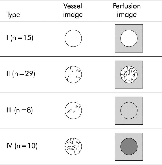
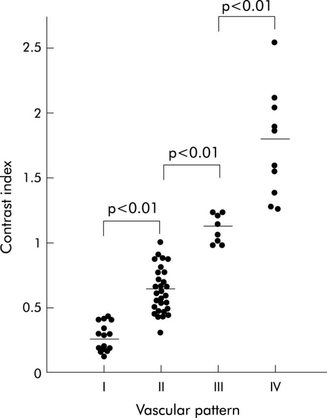
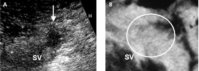
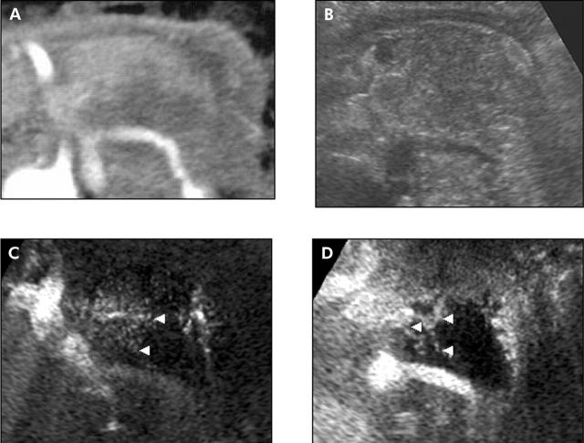
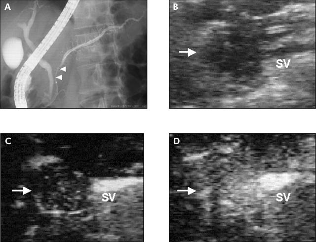
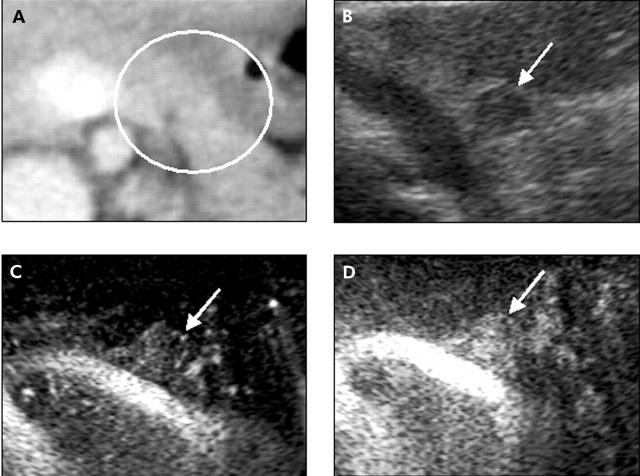
Subjects and methods: Sixty five patients with suspicious pancreatic tumours received contrast enhanced coded phase inversion harmonic ultrasonography, contrast enhanced computed tomography, and endosonography. Final diagnoses based on histological findings were pancreatic ductal carcinomas in 49 patients, inflammatory pseudotumours with chronic pancreatitis in seven, and endocrine tumours in nine. For contrast enhanced coded harmonic ultrasonography, Levovist, a contrast agent, was injected intravenously as a bolus. When the first microbubble signal appeared in the pancreas, images of the ideal scanning plane were displayed in a real time continuous fashion (vessel images). Subsequently, interval delay scanning (perfusion images) was taken to demonstrate parenchymal flow. Tumour vascularity was evaluated by using the two types of imaging. Sensitivities for depicting pancreatic tumours were compared between three examinations.
Results: Contrast enhanced ultrasonography demonstrated tumour vessels in 67% of pancreatic ductal carcinomas, although most were relatively hypovascular compared with the surrounding pancreatic tissue. The vascular patterns of tumours obtained by contrast enhanced ultrasonography were closely correlated with those obtained by contrast enhanced computed tomography. Values for sensitivity in depicting pancreatic tumours of 2 cm or less in size were 68% for contrast enhanced computed tomography, 95% for endosonography, and 95% for contrast enhanced ultrasonography.
Conclusion: Contrast enhanced coded phase inversion harmonic ultrasonography successfully visualised fine vessels in pancreatic tumours and may play a pivotal role in the depiction and differential diagnosis of pancreatic tumours.
Figures
References
-
- Ueno N, Tomiyama T, Tano S, et al. Contrast enhanced color Doppler ultrasonography in diagnosis of pancreatic tumor: two case reports. J Ultrasound Med 1996;15:527–30. - PubMed
-
- Bhutani MS, Hoffman BJ, Velse A, et al. Contrast-enhanced endoscopic ultrasonography with galactose microparticles:SHU508A (Levovist). Endoscopy 1997;29:635–9. - PubMed
-
- Ricke J, Hänninen LE, Amthauer H, et al. Assessment of the vascularisation of endocrine tumors by stimulated acoustic emission of SHU 508A ultrasound contrast agent and color or power Doppler sonography. Invest Radiol 2000;4:253–9. - PubMed
-
- Becker D, Strobel D, Bernatik T, et al. Echo-enhanced color- and power-Doppler EUS for the discrimination between focal pancreatitis and pancreatic carcinoma. Gastrointest Endosc 2001;53:784–9. - PubMed
-
- Ding H, Kudo M, Onda H, et al. Hepatocellular carcinoma: depiction of tumor parenchymal flow with intermittent harmonic power Doppler US during the early arterial phase in dual-display mode. Radiology 2001;220:349–56. - PubMed
Publication types
MeSH terms
Substances
LinkOut - more resources
Full Text Sources
Medical