Review
. 2004 Jun;53(6):899-910.
doi: 10.1136/gut.2003.025478.

Stem cell in gastrointestinal structure and neoplastic development

Affiliations
Review

Stem cell in gastrointestinal structure and neoplastic development

M Brittan et al. Gut. 2004 Jun.
No abstract available


Figures

Figure 1
Figure 1
Clonal origin of normal human intestinal crypts and small intestinal villi. (A) Monoclonal origin of human colonic crypts: normal colonic mucosa in an XO/XY mosaic individual stained by in situ hybridisation for a Y chromosome specific probe showing a XO crypt (central) surrounded by two XY crypts (courtesy of M Novelli). (B) Villi, receiving cells from more than one crypt of different clonal derivation, show a polyclonal pattern in this X0/XY patient. Apart from the occasional Y chromosome positive inflammatory cell (red chromosome label) the majority of cells on the right of this villus are XO (green chromosome paint) whereas on the left side the cells are XY (red and green chromosome paint) (courtesy of R Poulsom).
Figure 2
Figure 2
Demonstration of patch size is vital in studies to determine clonal origin of intestinal crypts. Glucose-6-phosphate dehydrogenase staining pattern in cross section of crypts within normal colonic mucosa showing large patches of crypts with irregular patch borders (from Novelli and colleagues with permission).
Figure 3
Figure 3
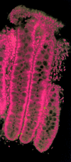
Musashi-1 expression within human colon crypts—a putative stem cell marker. Confocal imaging of human colon crypts (red: propidium iodide; green: Musashi-1). Several Musashi-1 positive cells were found near the base of the crypts (from Nishimura and colleagues with permission).
Figure 4
Figure 4
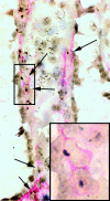
Bone marrow derived intestinal subepithelial myofibroblasts (ISEMF) are present in cellular columns in the lamina propria of the mouse colon, reaching from the base of the crypt to the intestinal lumen. Female mouse colon six weeks following a bone marrow transplant from a male mouse donor. Bone marrow derived ISEMFs are present as Y chromosome positive cells, immunoreactive for α-smooth muscle actin (SMA). The Y chromosome is seen as a brown/black punctate density, and red cytoplasmic staining indicates immunoreactivity for α-SMA (courtesy of M Brittan).
Figure 5
Figure 5
The Wnt signalling pathway. (A) In the absence of Wnt signalling, Dishevelled is inactive (Dshi) and Drosophila Zeste-white 3 or its mammalian homologue glycogen synthase kinase 3 (Zw3/GSK3) is active. β-Catenin (black dumbbell), via association with the APC-Zw3/GSK3 complex, undergoes phosphorylation and. degradation by the ubiquitin-proteasome pathway. Meanwhile, T cell factor (Tcf) is bound to its DNA binding site in the nucleus where it represses expression of genes such as Siamois in Xenopus. (B) In the presence of a Wnt signal, Dishevelled is activated (Dsha) leading to inactivation of Zw3/GSK3 by an unknown mechanism. β-Catenin fails to be phosphorylated and is no longer targeted into the ubiquitin-proteosome pathway, instead it accumulates in the cytoplasm and enters the nucleus by an unknown pathway where it interacts with Tcf to alleviate repression of the downstream genes and provide a transcriptional activation domain (from Willert and Nusse with permission).
Figure 6
Figure 6
The Math1 signalling pathway. (A) Low power section of adult mouse small intestine. Precursor cells (brown) are stained for cyclin PCNA (proliferating cell nuclear antigen); enterocytes (red) express intestinal alkaline phosphatase; goblet cells (blue) secrete mucins. The inset shows a high power image of small intestinal enterocytes and goblet cells. (B) Math1, a component of the Notch signalling pathway, influences intestinal epithelial cell fate decisions. In crypt progenitor stem cells that express high levels of Notch, the Hes1 transcription factor is switched on and expression of Math1 and of other “prosecretory” genes is blocked. The result is that the precursor cells become enterocytes. In cells expressing low amounts of Notch, levels of Delta are high, production of Hes1 is blocked, and Math1 expression is induced. Production of the Math1 helix-loop-helix transcription factor allows precursor cells to make a choice: whether to become goblet cells, Paneth cells, or enteroendocrine cells (Vi, villus; Cr, crypt) (A and B from van Den Brink and colleagues with permission). (C) Math1 is essential for secretory cells. Whether Math1 expressing cells descend directly from stem cells or an intermediate progenitor remains unknown. Sec, secretin; L, glucagon/peptide YY; CCK, cholecystokinin; SP, substance P; 5HT, serotonin; Som, somatostatin; GIP, gastric inhibitory peptide; Gas, gastrin (from Yang and colleagues with permission).
Figure 7
Figure 7
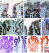
(A) Contrasting theories for the morphogenesis of adenomas and the part played by stem cells. (A) Haematoxylin and eosin stained sections of a small tubular adenoma. Dysplastic epithelium is superficial within the crypts, with histologically normal underlying epithelium. (B) Abrupt transition between dysplastic and normal appearing epithelial cells at the mid point of this crypt. Proliferative activity assessed with the Ki-67 antibody distributed throughout the dysplastic epithelium at the top of the crypts. (C) Nuclear β-catenin is highly expressed and distributed throughout the dysplastic epithelium at the top of the crypts but not in the crypt bases. (D) β-Catenin in the nuclei of adenomatous crypts from a tiny tubular adenoma. (E) Nuclear β-catenin extends to the bottom of crypts in early adenomas, including the very bases of the crypts. (F) β-catenin staining in nuclei of budding crypts (A–F, from Shih and colleagues with permission). (G) Junction between early adenomatous crypts, showing a sharp junction on the surface with accumulation of nuclear β-catenin, giving way sharply to membranous staining in the normal surface cells. (H) High power serial sections, demonstrating the sharp junction between nuclear staining in the adenomatous cells and membranous staining in normal surface epithelial cells. (I) Surface continuity between crypts showing nuclear β-catenin staining. (J) Crypts from a larger adenoma stained for β-catenin showing invasion of adjacent crypt territories in a top down fashion (G–J, from Preston and colleagues with permission).
Figure 8
Figure 8
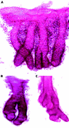
Microdissected crypts from normal colonic mucosae and adenomas. (A) Symmetrical fission of normal colonic crypts. (B) Isolated adenomatous crypt showing frequent crypt fission with atypical and asymmetrical branching. (C) Another crypt from an adenoma with a bizarre shape, asymmetrical branching, and multiple budding (A–C, from Preston and colleagues with permission).
Figure 9
Figure 9
Adenomas from an XO/XY mosaic individual with familial adenomatous polyposis, stained by in situ hybridisation for a Y chromosome specific probe (from Preston and colleagues with permission). (A) Clonal monocryptal adenoma. (B) Polyclonal adenoma, with a mixture of XO and XY crypts. Note the sharp margin between the territories of XO and XY adenomatous crypts at the surface, with no evidence of invasion.

Similar articles

Cited by

References

    1. Brittan M. Bone marrow-derived stem cells contribute to multiple cell lineages in experimental colitis. J Pathol 2003;201(suppl):1A. - PubMed
    1. Brittan M, Hunt T, Jeffery R, et al. Bone marrow derivation of pericryptal myofibroblasts in the mouse and human small intestine and colon. Gut 2002;50:752–7. - PMC - PubMed
    1. Krause DS, Theise ND, Collector MI, et al. Multi-organ, multi-lineage engraftment by a single bone marrow-derived stem cell. Cell 2001;105:369–77. - PubMed
    1. Korbling M, Katz RL, Khanna A, et al. Hepatocytes and epithelial cells of donor origin in recipients of peripheral-blood stem cells. N Engl J Med 2002;346:738–46. - PubMed
    1. Jiang Y, Jahagirdar BN, Reinhardt RL, et al. Pluripotency of mesenchymal stem cells derived from adult marrow. Nature 2002;418:41–9. - PubMed