. 2004 Jul 16;279(29):30287-97.
doi: 10.1074/jbc.M402960200. Epub 2004 May 11.

TALE homeodomain proteins regulate gonadotropin-releasing hormone gene expression independently and via interactions with Oct-1

Affiliations

TALE homeodomain proteins regulate gonadotropin-releasing hormone gene expression independently and via interactions with Oct-1

Naama Rave-Harel et al. J Biol Chem. .

Abstract

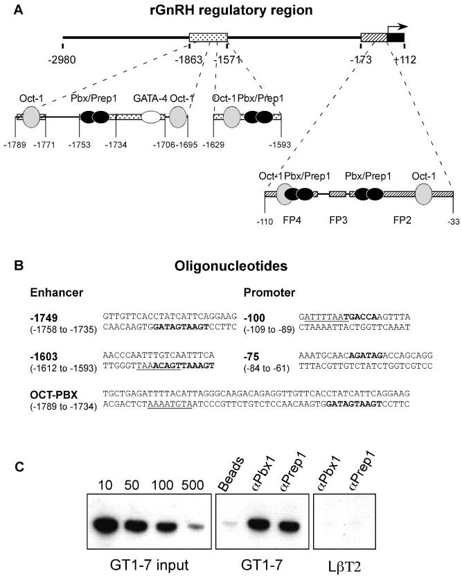
Gonadotropin-releasing hormone (GnRH) is the central regulator of reproductive function. Expression of the GnRH gene is confined to a rare population of neurons scattered throughout the hypothalamus. Restricted expression of the rat GnRH gene is driven by a multicomponent enhancer and an evolutionarily conserved promoter. Oct-1, a ubiquitous POU homeodomain transcription factor, was identified as an essential factor regulating GnRH transcription in the GT1-7 hypothalamic neuronal cell line. In this study, we conducted a two-hybrid interaction screen in yeast using a GT1-7 cDNA library to search for specific Oct-1 cofactors. Using this approach, we isolated Pbx1b, a TALE homeodomain transcription factor that specifically associates with Oct-1. We show that heterodimers containing Pbx/Prep1 or Pbx/Meis1 TALE homeodomain proteins bind to four functional elements within the GnRH regulatory region, each in close proximity to an Oct-1-binding site. Cotransfection experiments indicate that TALE proteins are essential for GnRH promoter activity in the GT1-7 cells. Moreover, Pbx1 and Oct-1, as well as Prep1 and Oct-1, form functional complexes that enhance GnRH gene expression. Finally, Pbx1 is expressed in GnRH neurons in embryonic as well as mature mice, suggesting that the associations between TALE homeodomain proteins and Oct-1 regulate neuron-specific expression of the GnRH gene in vivo.


Figures

Fig. 1
Fig. 1. TALE proteins present in GT1-7 cells specifically associate with Oct-1
A, Pbx proteins are expressed in GT1-7 cells. Western blots of nuclear extracts were probed with antibodies specific to Pbx1, Pbx2, and Pbx3. Each lane contained 12 μg of total protein from mouse hypothalamus (HYP) or the cell line indicated. Numbers on the left are molecular masses in kilodaltons. The arrows mark the positions of the identified proteins. The two Pbx1 forms, Pbx1a (arrow a) and Pbx1b (arrow b), are indicated. B, Pbx cofactors Prep1 and Meis1 are expressed in GT1-7 cells. Western blot analysis was performed using antibodies specific to Prep1 and Meis1. C, mapping the interaction between Prep1 and Pbx1b with Oct-1. In vitro translated 35S-labeled proteins were used for binding assays with GST or GST fusion proteins adsorbed to glutathione-Sepharose beads. One-tenth of each of the in vitro translated proteins used for binding was run in the input lanes to visualize the protein products. Numbers on the left are molecular masses in kilodaltons. D, schematic diagram of the protein constructs used in our GST pull-down assay. Both the Prep1 and Pbx proteins contain N-terminal conserved domains that serve for heterodimerization with each other (gray boxes) and a TALE (three-amino acid loop extension) homeodomain (HD; black boxes). The Prep1 N-terminal non-conserved domain is depicted by the hatched boxes. The E2A-Pbx1b fusion protein contains the transactivation domain of E2A (dotted box) fused to the homeodomain and C-terminal part of Pbx1b. The DNA-binding domain of Oct-1 consists of the POU-specific domain and the POU homeodomain (PS and PH, respectively; black boxes).
Fig. 2
Fig. 2. Heterodimers of TALE homeodomain proteins bind four motifs within the GnRH enhancer and promoter
EMSA was performed on nuclear proteins isolated from the GT1-7 cell line. The DNA probes consisted of radiolabeled oligonucleotides containing the −1749 and −1603 sites of the enhancer, the −75 and −100 elements of the promoter, and a Pbx/Hox consensus site, PBX, which served as a control for Pbx-binding activity. A, TALE homeodomain proteins bind to the −1749 probe of the GnRH enhancer. The PBX probe is shown in lanes 1–6, and the −1749 probe is shown in lanes 7–20. A 100-fold excess of the unlabeled PBX oligonucleotide, the PBX-mut oligonucleotide, or the wild-type −1749 oligonucleotide used for competition and various antibodies used for supershift assays were included in the reaction mixtures as indicated. The arrow mark the positions of supershifted complexes. B, comparison of the DNA-binding abilities of in vitro translated Pbx/Meis1 and Pbx/Prep1 using the OCT-PBX probe from the GnRH enhancer. EMSA was performed with GT1-7 nuclear extracts (GT1 NE; lanes 1) or with in vitro translated proteins as indicated. A control reaction containing the unprogrammed reticulocyte lysate (R.L.) is shown in lane 2. C, TALE homeodomain protein complexes bind to the −75, −100, and −1603 sites within the GnRH regulatory region. Lanes 1 and 2, the −1603 probe; lanes 3 and 4, the −100 probe; lanes 5 and 6, the −75 probe. A 100-fold excess of the unlabeled −1749 oligonucleotide was included in the reaction mixtures as indicated above the gel.
Fig. 3
Fig. 3. TALE proteins interact with the GnRH promoter in vivo
A and B, the rat GnRH upstream regulatory region contains binding sites for the TALE and Oct-1 transcription factors. A, the upper diagram shows the positions of the elements in the rat sequence that confer neuron specificity, the enhancer (dotted box) and the proximal promoter (hatched box); the lower diagrams depict the central and proximal portions of the enhancer and the proximal portion of the promoter. The boxes enclose the regions footprinted (FP) by GT1-7 nuclear extract (5, 35). The ovals illustrate the transcription factors: Oct-1 (gray), GATA-4 (white), and Pbx/Prep1 (black). B, the sequences of the rat GnRH enhancer and promoter elements that were used as oligonucleotide probes in EMSA are shown. The octamer motifs are underlined, and the Pbx-binding elements are in boldface. C, Pbx1 and Prep1 interact with the GnRH promoter in vivo. A chromatin immunoprecipitation experiment was performed in GT1-7 cells using anti-Pbx1 and anti-Prep1 antibodies. The primers used for PCR amplification of the mouse GnRH promoter are located at −34 and −267 relative to the transcription start site and correspond to the conserved promoter elements depicted in the rat sequence. Immunoprecipitation of chromatin with either of the antibodies followed by PCR amplification gave the appropriate ∼230-bp product. Immunoprecipitation with no antibody (Beads) was used as a negative control. Dilutions of 1:10 to 1:500 of the total input chromatin showed that PCR amplification of the immunoprecipitated chromatin was performed in the linear range. Immunoprecipitation of chromatin from LβT2 cells was used as an inactive GnRH promoter control. PCR amplification of the LβT2 chromatin total input was shown previously (36).
Fig. 4
Fig. 4. TALE proteins in combination with Oct-1 regulate GnRH enhancer activity
A and B, transient transfections were conducted in GT1-7 cells with the GnRH enhancer (GnRHe-RSVp) and the GnRH enhancer containing a mutation of the −1749 Pbx/Prep1-binding element (GnRHe–1749mut-RSVp) driving luciferase expression. A, the dominant-negative form of Prep1 interferes with GnRH enhancer activity. The cells were cotransfected with the indicated expression vectors: pcDNA1.1 (empty vector, negative control; white bars) and human dnPrep1 (black bars). The luciferase activity of the empty expression vector (pcDNA1.1) normalized to the activity of the cotransfected β-galactosidase internal control was set at 1 for each experiment. The normalized values for the expression plasmids are reported relative to the value of pcDNA1.1. Error bars represent S.E. *, p < 0.05 (Tukey-Kramer Honestly Significant Difference). The data shown are from five independent experiments, each performed in triplicate. B, TALE proteins in combination with Oct-1 activate transcription of the GnRH enhancer. Transient transfections were performed with expression vectors, all in the pcDNA1.1 backbone, as indicated. *, p < 0.05 for the wild-type versus mutant reporter (t test); #, p < 0.1 for the empty expression vector (Tukey-Kramer HSD). The data shown are from four independent experiments, each performed in triplicate. C, TALE proteins and Oct-1 associate with DNA concurrently. The OCT-PBX DNA probe of the GnRH enhancer (Fig. 3B) was incubated with GT1-7 nuclear extract (GT1-7 NE) and antibodies as indicated. The arrows mark the positions of the identified complexes. The slower mobility complexes are labeled A and B.
Fig. 5
Fig. 5. Pbx1 and Oct-1 co-localize with GnRH neurons
A and C, Pbx1 co-localizes with GnRH neurons in 13.5 dpc mouse embryos. In situ hybridization/immunohistochemical analysis of mouse embryos was carried out using an antisense probe specific for Pbx1 (blue). Subsequently, anti-GnRH antibody (Ab) was incubated with the tissue section and visualized with DAB (brown). A and C are ×10 and ×40 magnifications of the same section, respectively. B and D, Oct-1 co-localizes with GnRH neurons in embryonic day 13.5 mouse embryos. In situ hybridization and immunohistochemical analysis were performed with an antisense probe specific for Oct-1 (blue; shown in B) and then anti-GnRH antibody (brown; shown in D) as described under “Experimental Procedures.” E, the Pbx1 protein co-localizes with GnRH neurons in 13.5 dpc mouse embryos. Transgenic animals carrying GnRHe-GnRHp driving the β-galactosidase (β-gal) reporter gene targeted LacZ expression to GnRH neurons (blue) (6). An antibody specific to Pbx1 was incubated with the tissue and visualized with DAB (brown precipitates). F and G, Pbx1 co-localizes with GnRH neurons in adult mice. Co-immunohistochemistry was conducted on a wild-type adult female mouse brain. Anti-Pbx1 antibody was incubated with the tissue section and visualized with nickel (black). Subsequently, anti-GnRH antibody was incubated with the tissue and visualized with DAB (brown). Co-localization of GnRH and Pbx1 was detected in hypothalamic neurons of the medial preoptic area and the organum vasculosum of the lamina terminalis. F and G are is ×10 and ×40 magnifications of the same section, respectively. Arrows mark the positions of some co-localized neurons. Sense probes for Oct-1 and Pbx1 showed no specific hybridization. OA, olfactory area, CP, cribriform plate; AF, anterior forebrain.

Similar articles

Cited by

References

    1. Wolberger C. Curr Opin Genet Dev. 1998;8:552–559. - PubMed
    1. Vale W, Rivier C, Brown M. Annu Rev Physiol. 1977;39:473–527. - PubMed
    1. Mellon PL, Windle JJ, Goldsmith P, Pedula C, Roberts J, Weiner RI. Neuron. 1990;5:1–10. - PubMed
    1. Eraly SA, Mellon PL. Mol Endocrinol. 1995;9:848–859. - PubMed
    1. Whyte DB, Lawson MA, Belsham DD, Eraly SA, Bond CT, Adelman JP, Mellon PL. Mol Endocrinol. 1995;9:467–477. - PubMed

Publication types

MeSH terms

LinkOut - more resources