. 2004 May 10;165(3):421-32.
doi: 10.1083/jcb.200311045.

Regulation of cell migration and survival by focal adhesion targeting of Lasp-1

Affiliations

Regulation of cell migration and survival by focal adhesion targeting of Lasp-1

Yi Hsing Lin et al. J Cell Biol. .

Abstract

Large-scale proteomic and functional analysis of isolated pseudopodia revealed the Lim, actin, and SH3 domain protein (Lasp-1) as a novel protein necessary for cell migration, but not adhesion to, the extracellular matrix (ECM). Lasp-1 is a ubiquitously expressed actin-binding protein with a unique domain configuration containing SH3 and LIM domains, and is overexpressed in 8-12% of human breast cancers. We find that stimulation of nonmotile and quiescent cells with growth factors or ECM proteins facilitates Lasp-1 relocalization from the cell periphery to the leading edge of the pseudopodium, where it associates with nascent focal complexes and areas of actin polymerization. Interestingly, although Lasp-1 dynamics in migratory cells occur independently of c-Abl kinase activity and tyrosine phosphorylation, c-Abl activation by apoptotic agents specifically promotes phosphorylation of Lasp-1 at tyrosine 171, which is associated with the loss of Lasp-1 localization to focal adhesions and induction of cell death. Thus, Lasp-1 is a dynamic focal adhesion protein necessary for cell migration and survival in response to growth factors and ECM proteins.


Figures

Figure 1.
Figure 1.
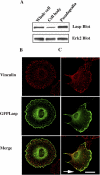
Subcellular localization of Lasp-1 in spreading and migrating cells. (A) Pseudopodia and cell body fractions were purified as described in Materials and methods. 10 μg of proteins were analyzed for relative expression of Lasp-1 by Western blotting using anti-Lasp-1 and anti-Erk2 antibodies, which served as a loading control. (B) Deconvolution images of NIH 3T3 cells transfected with GFP Lasp. Cells were allowed to attach and spread on fibronectin-coated coverslips for 120 min or (C) to migrate into a wound for 18 h. Cells were fixed and stained with anti-vinculin antibodies to visualize focal adhesions. Merged images show colocalization (yellow) of focal adhesions (red) and GFP Lasp (green). Note that GFP Lasp strongly localized to focal adhesions proximal to the cell edge at the leading front of the pseudopodium. Arrow indicates the direction of cell migration. Bar, 28 μm.
Figure 2.
Figure 2.
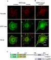
Lasp-1 is a dynamic protein that transits from the cell periphery to focal adhesions upon stimulation with growth factors. (A) Deconvolution images of NIH 3T3 cells transfected with GFP Lasp and stained for vinculin-positive focal adhesions as described in Materials and methods. Cells were serum starved overnight (−serum) and stimulated with 10% FBS for 15 min. Merged images represent colocalization (yellow) of focal adhesions (red) and GFP Lasp (green). (B) GFP Lasp with its COOH-terminal region truncated (GFP LaspΔC) was examined for localization to focal adhesions upon serum stimulation as described in Materials and methods. Arrows indicate membrane ruffles. GFP LaspΔC cells without serum showed a similar pattern of expression as of cells with serum (not depicted). (C) Schematic representation of the LaspΔC and SH3 domain mutants (LaspSH3) used in this experiment and the experiments shown in Fig. 4 C. Bar, 28 μm.
Figure 3.
Figure 3.
Lasp-1 localizes to cortical F-actin and actin cables in quiescent cells and dorsal membrane ruffles in serum stimulated cells. Deconvolution images of NIH 3T3 cells transfected with GFP Lasp and stained for F-actin with rhodamine-phalloidin. Cells were serum starved overnight (NT) and then stimulated with PDGF-BB for 30 min. Merged images represent colocalization (yellow) of F-actin (red) and GFP Lasp (green). Bar, 28 μm.
Figure 4.
Figure 4.
Lasp-1 is necessary for cell migration, but not adhesion to the ECM. (A) Cos-7 cells were depleted of endogenous Lasp-1 protein using siRNA specific for Lasp-1 or control siRNA to GL2 luciferase. Cells were allowed to either migrate using Boyden chambers coated on the bottom with 10 μg/ml fibronectin or (B) to attach to culture dishes coated with the same ECM as described in Materials and methods. The number of migratory or adherent cells per microscopic field was counted. Each bar represents the mean ± SEM of cells in triplicate migration/adhesion chambers of three independent experiments. An aliquot of cell lysates prepared from cells treated as described above were Western blotted for expression of Lasp-1 or actin, which served as a loading control. (C) COS-7 cells were transfected with the empty vector (Mock), wild-type Lasp-1 (Wt), LaspΔC (ΔC), or the SH3 domain tagged with HA (SH3) along with a β-galactosidase reporter construct to mark transfected cells. Cells were allowed to chemotax with or without LPA in the lower chamber for 3 h. Migratory cells per microscopic field on the underside of the membrane were stained and counted. Each bar represents the mean ± SEM of blue cells in triplicate migration chambers of three independent experiments. An aliquot of cell lysates prepared from cells treated as described above were Western blotted for the expression of the appropriate protein or tag as indicated. (D) HEK 293 cells stably expressing the empty vector (mock) or the vector encoding full-length Lasp-1 were tested for chemotaxis with or without 10 μg/ml insulin in the lower chamber as indicated in C. Lasp-1 protein was Western blotted to confirm the protein expression. HEK 293 cells show low levels of endogenous Lasp-1 protein, which is not detected in the short exposure time to the film. (E) Deconvolution images of COS-7 cells transfected with either Lasp-1 or LaspΔC fused with the membrane-targeting sequence (myr) along with a GFP reporter construct. Cells were then fixed and examined for membrane blebs 16 h after transfection, and the number of transfected cells with prominent blebs were counted per microscopic field relative to the total number of GFP-positive cells.
Figure 5.
Figure 5.
Abl kinase associates with the COOH terminus of Lasp-1 and tyrosine phosphorylates Y171. (A) COS-7 cells were transfected with HA-Abl together with Lasp-1 or LaspΔC. HA-Abl was immunoprecipitated and Western blotted for associated Lasp-1 protein with anti-Lasp-1 antibodies. The blot was then stripped and reprobed for phosphotyrosine (PY). Western blots of whole-cell lysates are included to confirm expression of the appropriate protein (bottom). (B) GST-tagged Lasp or GST LaspΔC was precipitated with glutathione beads and Western blotted for associated Abl protein with anti-HA antibodies from COS-7 cells expressing these constructs. Blots were stripped and reprobed for PY as indicated above, and whole-cell lysates were included to confirm protein expression. (C) GST Lasp was precipitated and Western blotted for associated Abl or Abl without its polyproline domain (AblΔPro) using anti-Abl antibodies. The blot was then stripped and blotted for PY to detect Lasp-1 phosphorylation. The expression levels of wild-type Abl and AblΔpro in whole-cell lysates are shown below. (D) Bcr-Abl–transformed NIH 3T3 cells (+) and mock control NIH 3T3 cells (−) transfected with GST Lasp were starved and treated with 2 μM STI 571 or buffer only for 16 h. GST Lasp was precipitated and Western blotted using anti-phosphotyrosine antibodies and then was stripped and reprobed with GST-specific antibodies. (E) Purified activated Abl was incubated with bacterial purified GST Lasp in the presence of ATP and kinase reaction buffer or purified GST alone as a control. Western blotting with phosphotyrosine, GST, or Lasp-1 was performed as indicated. (F) Cos-7 cells with or without HA-Abl were transfected along with Lasp-1 or Lasp with the Abl consensus tyrosine phosphorylation site Y171 substituted with phenylalanine (LaspY171F). Lasp-1 from these cells was Western blotted using anti-phosphotyrosine or Lasp-1-specific antibodies.
Figure 6.
Figure 6.
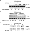
Apoptotic agents induce tyrosine phosphorylation of Lasp-1, and this requires Abl and Arg kinase activity. (A) Cos-7 cells transfected with GST Lasp were treated with 1 mM H2O2 for the indicated times in the presence or absence of 5 μM STI 571. GST Lasp was precipitated and Western blotted using anti-phosphotyrosine or GST antibodies. (B) Embryonic fibroblast cells isolated from abl−/−arg−/− animals or these cells stably reconstituted with Abl were transfected with GST Lasp and were then serum starved and treated with 1 mM H2O2 for the indicated times. GST Lasp expression and tyrosine phosphorylation were determine as described above. (C) Cos-7 cells transfected with GST Lasp were incubated with 25 μM cisplatin for the indicated times in the presence or absence of 2 μM STI 571. Lasp-1 expression and tyrosine phosphorylation were determined as described above.
Figure 7.
Figure 7.
Tyrosine phosphorylation Y171 of Lasp-1 prevents its translocation from the cell periphery to focal adhesions in response to growth factor stimulation. Deconvolution images of NIH 3T3 cells expressing GFP Lasp or GFP LaspY171F were treated for 30 min with 1 mM H2O2 and for 15 min with FBS. Cells were fixed and stained with anti-vinculin antibodies to visualize focal adhesions as described above. In some cases, GFP Lasp transfected cells were preincubated with 5 μM STI 571 before H2O2 and FBS treatment to block Abl kinase activity as described in Materials and methods. Merged images represent colocalization (yellow) of focal adhesions (red) and GFP Lasp (green). Bars: 28 μm (right and left panels) and 20 μm (middle panel).
Figure 8.
Figure 8.
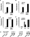
Depletion of Lasp-1 protein increases cell apoptosis induced by H 2 O 2 and cisplatin, but not TSA. COS-7 cells were depleted of endogenous Lasp-1 protein using siRNA specific for Lasp-1 or control siRNA to GL2 luciferase as described in the Materials and methods. Cells were plated onto fibronectin-coated glass coverslips, and were treated with either (A) 25 μM cisplatin, (B) 1 mM H2O2, or (C) 300 ng/ml TSA for 24 h, or were treated with the indicated vehicle. (D) Cells were treated with or without (NT) H2O2 or TSA as described above in the presence or absence of 5 μM STI 571. Apoptotic cells were determined after 24 h by staining with propidium iodide and counting the number of cells per microscopic field with condensed nuclei as described previously (Kain et al., 2003). The bars represent the mean ± SEM of three independent experiments. (E) COS-7 cells were treated with 300 ng/ml TSA or with DMSO for 24 h, and were then lysed in detergent and Western blotted for the indicated proteins. Note that TSA does not induce a mobility shift of c-CrkII and Lasp-1 protein, indicating that these proteins are not phosphorylated and Abl kinase is not activated under these apoptotic conditions. Asterisk shows the cleaved form of PARP, indicating that TSA induced apoptosis in these cells. The top band is a nonspecific protein that served as a loading control.
Figure 9.
Figure 9.
Cells expressing LaspY171F show reduced apoptosis in response to cisplatin and H 2 O 2 , but not TSA. COS-7 cells were transfected with the empty vector (Mock), wild-type Lasp-1, or Lasp-1 with tyrosine 171 mutated to phenylalanine (LaspY171F) along with GFP, which served as a reporter for transfected cells. Cells were then treated with either (A) cisplatin, (B) H2O2, or (C) TSA, and the number of GFP-positive apoptotic cells was determined as described in Fig. 8. Bars represent the mean ± SEM of three independent experiments.

Similar articles

Cited by

References

    1. Agami, R., G. Blandino, M. Oren, and Y. Shaul. 1999. Interaction of c-Abl and p73α and their collaboration to induce apoptosis. Nature. 399:809–813. - PubMed
    1. Aizawa, H., Y. Fukui, and I. Yahara. 1997. Live dynamics of Dictyostelium cofilin suggests a role in remodeling actin latticework into bundles. J. Cell Sci. 110:2333–2344. - PubMed
    1. Allen, W.E., D. Zicha, A.J. Ridley, and G.E. Jones. 1998. A role for Cdc42 in macrophage chemotaxis. J. Cell Biol. 141:1147–1157. - PMC - PubMed
    1. Bailly, M., L. Yan, G.M. Whitesides, J.S. Condeelis, and J.E. Segall. 1998. Regulation of protrusion shape and adhesion to the substratum during chemotactic responses of mammalian carcinoma cells. Exp. Cell Res. 241:285–299. - PubMed
    1. Bailly, M., I. Ichetovkin, W. Grant, N. Zebda, L.M. Machesky, J.E. Segall, and J. Condeelis. 2001. The F-actin side binding activity of the Arp2/3 complex is essential for actin nucleation and lamellipod extension. Curr. Biol. 11:620–625. - PubMed

Publication types

MeSH terms