. 2004 May;2(5):E110.
doi: 10.1371/journal.pbio.0020110. Epub 2004 May 11.

Mre11 assembles linear DNA fragments into DNA damage signaling complexes

Affiliations

Mre11 assembles linear DNA fragments into DNA damage signaling complexes

Vincenzo Costanzo et al. PLoS Biol. 2004 May.

Erratum in

  • PLoS Biol. 2004 Jun;2(6):876

Abstract

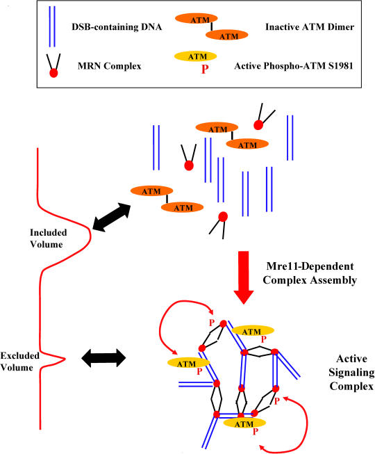
Mre11/Rad50/Nbs1 complex (MRN) is essential to suppress the generation of double-strand breaks (DSBs) during DNA replication. MRN also plays a role in the response to DSBs created by DNA damage. Hypomorphic mutations in Mre11 (which causes an ataxia-telangiectasia-like disease [ATLD]) and mutations in the ataxia-telangiectasia-mutated (ATM) gene lead to defects in handling damaged DNA and to similar clinical and cellular phenotypes. Using Xenopus egg extracts, we have designed a simple assay to define the biochemistry of Mre11. MRN is required for efficient activation of the DNA damage response induced by DSBs. We isolated a high molecular weight DNA damage signaling complex that includes MRN, damaged DNA molecules, and activated ATM. Complex formation is partially dependent upon Zn(2+) and requires an intact Mre11 C-terminal domain that is deleted in some ATLD patients. The ATLD truncation can still perform the role of Mre11 during replication. Our work demonstrates the role of Mre11 in assembling DNA damage signaling centers that are reminiscent of irradiation-induced foci. It also provides a molecular explanation for the similarities between ataxia-telangiectasia (A-T) and ATLD.


Conflict of interest statement

The authors have declared that no conflicts of interest exist.

Figures

Figure 1
Figure 1. Functional MRN Is Required for the Response to DSBs, and Mre11–ATLD Separates Essential and Nonessential Mre11 Functions
(A) The activity of protein kinases responsive to DSBs in Xenopus laevis egg extracts was monitored by incorporation of 32P from γ-32P-ATP into H2AX-derived peptides in the presence (plus DSB) or absence (minus DSB) of fragmented DNA. Labels: Wild-Type, H2AX substrate peptide containing serine 134 and serine 139; S134A, H2AX substrate peptide with a substitution of serine 134 to alanine; S139A, H2AX substrate peptide with a substitution of serine 139 to alanine; S134A/S139A, H2AX substrate peptide with a substitution of both serines to alanine. (B) Extract incubated with linear DNA at 50 ng/μl (equivalent to 4.5 × 10¹⁰ breaks/μl) was assayed with H2AX peptide in the presence of buffer (Control), ATM-neutralizing antibodies (ATM Ab), ATR-neutralizing antibodies (ATR Ab), ATM- and ATR-neutralizing antibodies (ATM/ATR Abs), ATM- and ATR-neutralizing antibodies in Ku70-depleted extracts (ATM/ATR Abs; Ku depletion), 5 mM caffeine (Caffeine). (C) DSB-responsive kinase activity was measured in the presence of 0, 5, 10, 25, and 50 ng/μl of linear DNA in control extract (filled diamonds), mock-depleted extract (open diamonds), Mre11-depleted extract (open squares), Mre11-depleted extract supplemented with 500 nM of recombinant MRN (filled squares), or Mre11-depleted extract supplemented with 500 nM MRN-ATLD1/2 (filled triangles). (D) DSB accumulation during DNA replication was monitored by TUNEL assay. Postreplicative nuclei were isolated from a control extract (stripes), Mre11-depleted extract (dots), Mre11-depleted extract supplemented with MRN (diamonds), Mre11-depleted extract supplemented with MRN-ATLD1/2 (gray) or mock-depleted extract (white).
Figure 2
Figure 2. Requirements for the Assembly of DNA–Protein Complexes
Elution profiles of α-32P-dATP-labeled 1 kb linear DNA from BioGel A15m chromatography columns. After loading, fractions 1–31 were collected and radioactivity was counted in a scintillation counter. (A–E) Complete elution profile. (A) Linear DNA alone. (B) Linear DNA incubated 2 h in extract at 22°C. (C) α-32P-dATP-labeled circular plasmid incubated for 2 h in extract at 22°C. (D) Linear DNA incubated with extract treated with 1 mg/ml proteinase K immediately prior to loading. (E) Linear DNA incubated in Mre11-depleted extract. (F and G) Excluded volume (fractions 6–14). (F) Linear DNA incubated in the following extracts: Mre11-depleted extract (open triangles), Mre11-depleted extract supplemented with 500 nM of MRN (filled triangles), Mre11-depleted extract supplemented with 500 nM of MRN-ATLD1/2 (open squares), or control extract supplemented with MRN (filled squares). (G) Linear DNA incubated in the following extracts: control extract (filled circles), extract treated with 5 mM caffeine (open circles), extract treated with TPEN at 100 μM (open diamonds).
Figure 3
Figure 3. Mre11 Tethers DSB-Containing DNA
(A) Control and treated extracts were incubated with α-32P-dATP-labeled DNA fragments and loaded onto BioGel A15 columns. Fractions 10 and 25 were collected and incubated with polyclonal antibodies against Mre11 or protein A beads alone. Beads were collected and washed, and radioactivity was counted in a scintillation counter. Shown are control extract (stripes), Mre11-depleted extract (dots), or Mre11-depleted extract supplemented with Mre11 that had been immunoprecipitated from the extract (diamonds), and extract incubated with beads alone (black). (B) Biotinylated DNA fragments were mixed with α-32P-dATP-labeled DNA fragments and incubated with various extracts. The extracts were then loaded onto BioGel A15 columns. Fractions 10 and 25 were collected and incubated with streptavidin-magnetic beads. Beads were collected and washed, and radioactivity was counted in a scintillation counter. Shown are control extract (stripes), Mre11-depleted extract (dots), Mre11-depleted extract supplemented with 500 nM MRN (diamonds), and streptavidin beads (black).
Figure 4
Figure 4. DNA–Protein Complexes Are Signaling Centers Containing Active Mre11 and ATM
(A) Western blot analysis of eluted fractions. Fraction numbers are indicated at bottom. Fractions were collected following chromatography of extracts incubated with fragmented (plus DSBs) or without fragmented DNA (minus DSBs). Samples from fractions were processed for SDS-PAGE and blotted with polyclonal antibodies against Mre11, ATM, and phosphorylated ATM. (B) Activity of ATM and ATR kinases in fractions 10 and 25. Extracts were incubated with DNA fragments and applied to BioGel A15m columns. Fraction 10 and fraction 25 from control extract were assayed for H2AX activity in presence of buffer (light gray), ATM-neutralizing antibodies (checks), ATR-neutralizing antibodies (dark gray), 300 μM vanillin (stripes), or 5 mM caffeine (black). (C) Activity of ATM and ATR kinases in fraction 10 and total extract. Control extracts or extracts supplemented with 500 nM recombinant MRN were incubated with DSBs and loaded onto BioGel A15m columns. Total control extract and fraction 10 were assayed for H2AX activity in the presence of buffer (light gray), ATM-neutralizing antibodies (checks), ATR-neutralizing antibodies (dark gray), or 5 mM caffeine (black).
Figure 5
Figure 5. Schematic Representation of the Mre11-Dependent Assembly of DNA Damage Signaling Complexes
MRN promotes the assembly of DNA–protein structures containing linear DNA fragments enriched with active ATM molecules. These active signaling complexes resemble IRIF in that they are the morphological and functional unit of the DNA damage response.

References

    1. Abraham RT. Cell cycle checkpoint signaling through the ATM and ATR kinases. Genes Dev. 2001;15:2177–2196. - PubMed
    1. Andegeko Y, Moyal L, Mittelman L, Tsarfaty I, Shiloh Y, et al. Nuclear retention of ATM at sites of DNA double strand breaks. J Biol Chem. 2001;276:38224–38230. - PubMed
    1. Aten JA, Stap J, Krawczyk PM, van Oven CH, Hoebe RA, et al. Dynamics of DNA double-strand breaks revealed by clustering of damaged chromosome domains. Science. 2004;303:92–95. - PubMed
    1. Bakkenist CJ, Kastan MB. DNA damage activates ATM through intermolecular autophosphorylation and dimer dissociation. Nature. 2003;421:499–506. - PubMed
    1. Burma S, Chen BP, Murphy M, Kurimasa A, Chen DJ. ATM phosphorylates histone H2AX in response to DNA double-strand breaks. J Biol Chem. 2001;276:42462–42467. - PubMed

Publication types

MeSH terms