. 2004 May;2(5):E129.
doi: 10.1371/journal.pbio.0020129. Epub 2004 May 11.

Early myocardial function affects endocardial cushion development in zebrafish

Affiliations

Early myocardial function affects endocardial cushion development in zebrafish

Thomas Bartman et al. PLoS Biol. 2004 May.

Abstract

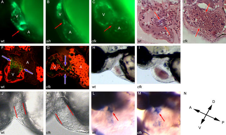
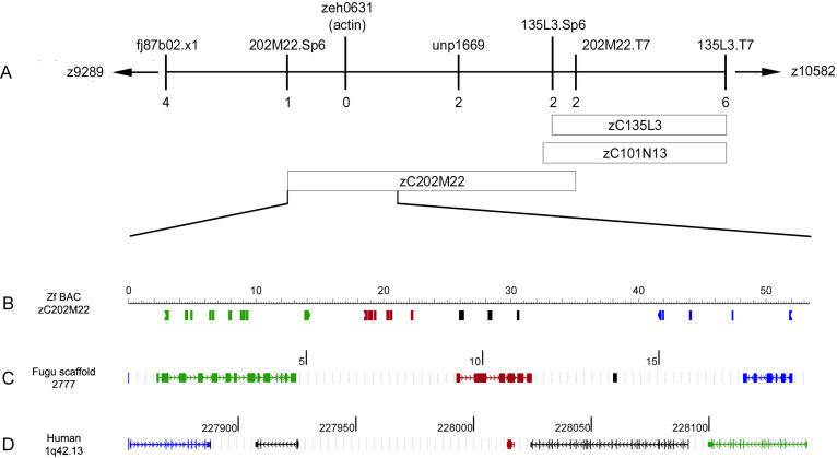
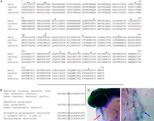
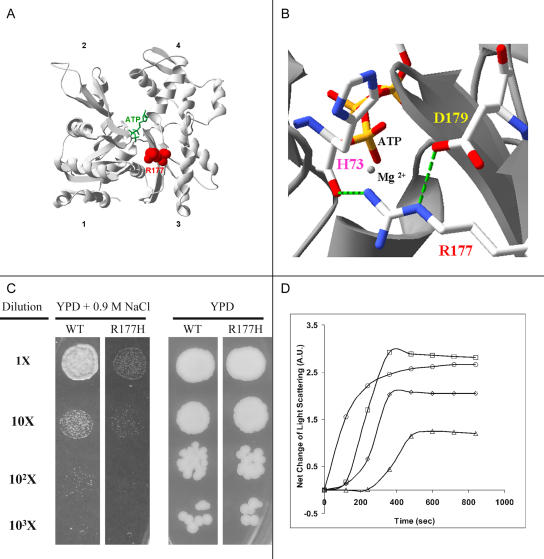
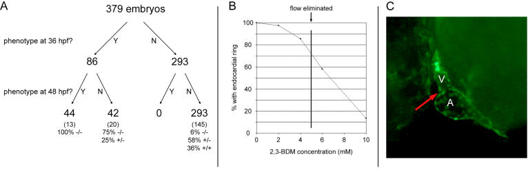
Function of the heart begins long before its formation is complete. Analyses in mouse and zebrafish have shown that myocardial function is not required for early steps of organogenesis, such as formation of the heart tube or chamber specification. However, whether myocardial function is required for later steps of cardiac development, such as endocardial cushion (EC) formation, has not been established. Recent technical advances and approaches have provided novel inroads toward the study of organogenesis, allowing us to examine the effects of both genetic and pharmacological perturbations of myocardial function on EC formation in zebrafish. To address whether myocardial function is required for EC formation, we examined silent heart (sih(-/-)) embryos, which lack a heartbeat due to mutation of cardiac troponin T (tnnt2), and observed that atrioventricular (AV) ECs do not form. Likewise, we determined that cushion formation is blocked in cardiofunk (cfk(-/-)) embryos, which exhibit cardiac dilation and no early blood flow. In order to further analyze the heart defects in cfk(-/-) embryos, we positionally cloned cfk and show that it encodes a novel sarcomeric actin expressed in the embryonic myocardium. The Cfk(s11) variant exhibits a change in a universally conserved residue (R177H). We show that in yeast this mutation negatively affects actin polymerization. Because the lack of cushion formation in sih- and cfk-mutant embryos could be due to reduced myocardial function and/or lack of blood flow, we approached this question pharmacologically and provide evidence that reduction in myocardial function is primarily responsible for the defect in cushion development. Our data demonstrate that early myocardial function is required for later steps of organogenesis and suggest that myocardial function, not endothelial shear stress, is the major epigenetic factor controlling late heart development. Based on these observations, we postulate that defects in cardiac morphogenesis may be secondary to mutations affecting early myocardial function, and that, in humans, mutations affecting embryonic myocardial function may be responsible for structural congenital heart disease.


Figures

Figure 1
Figure 1. Embryos with Defective Myocardial Function Do Not Form AV ECs
(A–C) Fluorescence micrographs of embryos carrying a tie2::GFP transgene, visualized at 48 hpf. In (A), the endocardial ring is visible as a collection of GFP-positive cells at the AV boundary in wild-type (wt) embryos (red arrow). In (B), sih−/− embryos fail to form an AV ring at 48 hpf. In (C), cfk−/− embryos fail to form an AV ring at 48 hpf. (D and E) Cushion development remains defective in cfk−/− embryos. In (D), a 5 μm hematoxylin and eosin-stained plastic section shows the initial stages of cushion development at the AV boundary (red arrows) in a 72 hpf wild-type embryo, with the ECs being two to three cell layers thick at this stage. In (E), a cfk −/− embryo at 72 hpf shows dilation of both chambers of a blood-filled heart with no evidence of cushion formation at the AV boundary (red arrows). (F and G) cfk−/− embryos fail to form ECs at late stages. Embryos were visualized at identical magnification after counter-staining with rhodamine phalloidin. Red blood cells (RBCs) are seen in the atria of the hearts. In (F), confocal microscopy of a 96 hpf wild-type heart from a tie2::GFP line shows triangular ECs at the AV boundary (blue arrows). In (G), cfk−/− embryos at 96 hpf lack cushion formation and clustering of GFP-positive cells at the AV boundary (blue arrows). (H) At 72 hpf, wild-type embryos have narrow hearts with forward blood flow through the embryo. (I) At 72 hpf, cfk−/− embryos have dilated hearts filled with blood that regurgitates freely from the ventricle to the atrium. (J and K) The initial phenotype in cfk−/− embryos is cardiac dilation at 36 hpf. In (J), wild-type embryos have a narrow ventricle and generate pulsatile flow at 36 hpf. In (K), cfk−/− embryos have an increased end-diastolic diameter (on average 1.18× wild-type, p < 0.01) and do not generate blood flow at 36 hpf. (L and M) Increased bmp-4 expression at the AV boundary (red arrow) is observed in wild-type (L) and cfk−/− (M) embryos at 42 hpf in anticipation of endocardial ring formation. (N) Orientation of the embryos shown in (L) and (M).
Figure 2
Figure 2. Positional Cloning of cfk
(A) The locus on zebrafish LG13 containing cfk is shown, with the number of recombinants indicated below each marker. The one recombinant at the Sp6 end of BAC zC202M22 and the two recombinants at the T7 end of the same BAC define the critical region. (B) The first 52 kb of zebrafish BAC zC202M22 is shown, including the Sp6 end. One of the two recombinants from the T7 end of the BAC is still present at a marker at 52 kb, narrowing the critical region to this span. GenScan and BLAST analyses identified four coding sequences in this span, including abc-b10 (green), actin (red), a novel EST fv49b10 (black), and rab4a (blue). (C) Each of the three known genes identified on BAC zC202M22 has a homologue on scaffold 2777 of Fugu rubripes, in the same orientation (green, abc-b10; red, actin; blue, rab4a). (D) The same three genes lie in proximity to each other and in the same order on human Chromosome 1q42.13 (green, abc-b10; red, actin; blue, rab4a). Units in black are genes, predicted genes, or ESTs, which are unique in this region to that particular organism.
Figure 3
Figure 3. Sequence and Expression Analysis of cfk
(A) cfk encodes a sarcomeric actin highly homologous to zebrafish α-cardiac and α-skeletal actins. Cfk differs from zebrafish α-cardiac actin at six residues and from zebrafish α-skeletal actin at four residues, but from zebrafish β-actin at 28 residues. Residues in red are those that differ from Cfk. Dots above the sequence indicate residues that universally distinguish sarcomeric from cytoplasmic actins. The arrow at R177 indicates the location of histidine in Cfks11. (B) The arginine at position 177 is universally conserved in all actin proteins examined. (C and D) cfk is expressed in the myocardium during development. In (C), whole-mount in situ hybridization on a cmlc2::GFP embryo at 36 hpf shows that cfk is expressed throughout the AP extent of the heart tube. Blue staining indicates areas of cfk expression; green is the region of cmlc2 expression. The red arrow indicates the heart tube. In (D), a plastic section of stained embryo shows cfk expression in the myocardium of the heart (blue arrow), but not in the endocardial cells (red arrow). From the onset of its expression in the heart region (around the 16-somite stage), cfk does not appear to be expressed in endothelial and endocardial cells. Weak cfk expression is also seen in the somites (data not shown).
Figure 4
Figure 4. An Arginine to Histidine Change at Position 177 of Cfk Alters the Location of Positive Charges in the Nucleotide Binding Cleft of Actin
(A) The position of R177 (red) in the structure of the yeast-actin monomer. The bound ATP is shown in green. The structure shown is based on a crystal structure of actin (PDB:1YAG). (B) Magnification of the cleft region showing the hydrogen bonding (green dashed lines) involving R177, which will be disrupted in H177. (C) The R177H mutation restricts yeast growth under hyperosmolar stress. Wild-type (wt) and mutant cells were grown to a density of 3 × 10⁶ per milliliter, and aliquots were plated either on YPD or YPD plus 0.9 M NaCl at different dilutions of the culture. The cells were then incubated at 30°C for 72 h. The normosmolar and hyperosmolar experiments were done at different times. (D) Purified R177H actin has a higher critical concentration of polymerization and a delayed nucleation phase. Wild-type or mutant actin was purified from yeast cultures and polymerization mea-sured by light diffraction. Symbols and abbreviations: circles, 5 μM wild-type actin; triangles, 5 μM R177H actin; diamonds, 7.5 μM R177H actin; squares, 10 μM R177H actin; ATP, adenine triphosphate; wt, wild-type.
Figure 5
Figure 5. Lack of EC Formation Is Secondary to Defective Myocardial Function
(A) Embryos were observed for defective myocardial function and lack of blood flow at 36 hpf and lack of endocardial ring formation at 48 hpf. Approximately half of the embryos with defective myocardial function at 36 hpf did not develop endocardial rings. All embryos with a lack of endocardial rings previously demonstrated a myocardial function phenotype at 36 hpf. Further analyses including the genotyping of a subset of embryos showed that a small percentage of cfk−/− embryos were unaffected at 36 hpf and that they subsequently developed ECs. (B) Embryos treated from 24 to 48 hpf with 2,3-BDM to decrease myocardial force failed to develop endocardial rings in a dose-dependent manner. Loss of ring formation was not linked to loss of blood flow—14% of embryos treated at 4 mM 2,3-BDM did not form rings despite the presence of blood flow, and 58% of embryos treated at 6 mM did form rings despite the absence of blood flow. (C) Example of an embryo treated with 10 mM 2,3-BDM that failed to develop an endocardial ring at the AV boundary (red arrow).

References

    1. Alexander J, Stainier DY, Yelon D. Screening mosaic F1 females for mutations affecting zebrafish heart induction and patterning. Dev Genet. 1998;22:288–299. - PubMed
    1. Biben C, Weber R, Kesteven S, Stanley E, McDonald L, et al. Cardiac septal and valvular dysmorphogenesis in mice heterozygous for mutations in the homeobox gene Nkx2-5. Circ Res. 2000;87:888–895. - PubMed
    1. Bruneau BG, Nemer G, Schmitt JP, Charron F, Robitaille L, et al. A murine model of Holt–Oram syndrome defines roles of the T-box transcription factor Tbx5 in cardiogenesis and disease. Cell. 2001;106:709–721. - PubMed
    1. Burge C, Karlin S. Prediction of complete gene structures in human genomic DNA. J Mol Biol. 1997;268:78–94. - PubMed
    1. Camenisch TD, Spicer AP, Brehm-Gibson T, Biesterfeldt J, Augustine ML, et al. Disruption of hyaluronan synthase-2 abrogates normal cardiac morphogenesis and hyaluronan-mediated transformation of epithelium to mesenchyme. J Clin Invest. 2000;106:349–360. - PMC - PubMed

Publication types

MeSH terms