. 2004 May;2(5):E130.
doi: 10.1371/journal.pbio.0020130. Epub 2004 May 11.

Pax7 is necessary and sufficient for the myogenic specification of CD45+:Sca1+ stem cells from injured muscle

Affiliations

Pax7 is necessary and sufficient for the myogenic specification of CD45+:Sca1+ stem cells from injured muscle

Patrick Seale et al. PLoS Biol. 2004 May.

Abstract

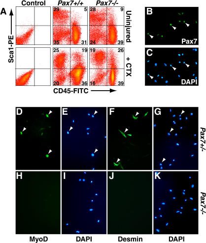
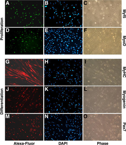
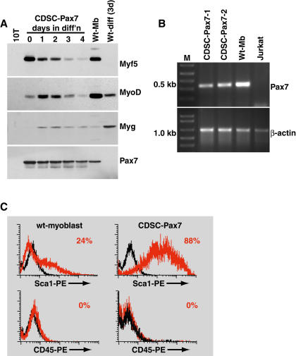
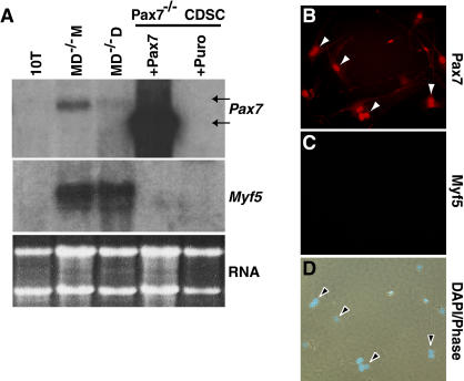
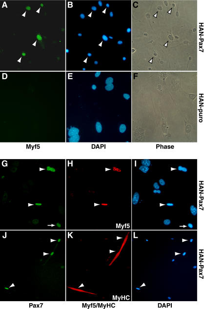
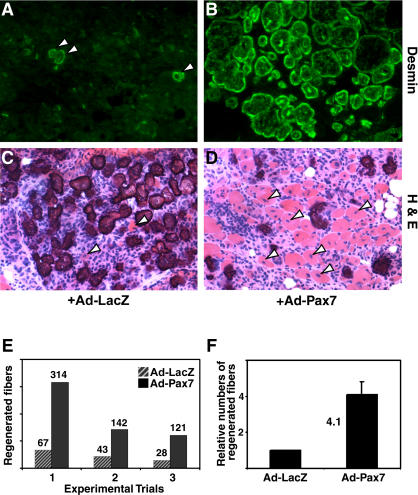
CD45(+):Sca1(+) adult stem cells isolated from uninjured muscle do not display any myogenic potential, whereas those isolated from regenerating muscle give rise to myoblasts expressing the paired-box transcription factor Pax7 and the bHLH factors Myf5 and MyoD. By contrast, CD45(+):Sca1(+) isolated from injured Pax7( -/-) muscle were incapable of forming myoblasts. Infection of CD45(+):Sca1(+) cells from uninjured muscle with retrovirus expressing Pax7 efficiently activated the myogenic program. The resulting myoblasts expressed Myf5 and MyoD and differentiated into myotubes that expressed myogenin and myosin heavy chain. Infection of CD45(-):Sca1(-) cells from Pax7( -/-) muscle similarly gave rise to myoblasts. Notably, infection of Pax7-deficient muscle with adenoviral Pax7 resulted in the de novo formation of regenerated myofibers. Taken together, these results indicate that Pax7 is necessary and sufficient to induce the myogenic specification of CD45(+) stem cells resident in adult skeletal muscle. Moreover, these experiments suggest that viral transduction of Pax7 is a potential therapeutic approach for the treatment of neuromuscular degenerative diseases.


Conflict of interest statement

The authors have declared that no conflicts of interest exist.

Figures

Figure 1
Figure 1. Pax7 Is Required for the Myogenic Specification of CD45+:Sca1+ Cells
(A) Flow cytometric analysis of cell suspensions derived from uninjured and regenerating wild-type and Pax7  −/− muscle (4 d after ctx injection) showed an increased proportion of CD45+ cells in Pax7  −/− samples. (B and C) Pax7 protein was expressed in approximately 6%–10% of CD45+:Sca1+ cells purified from regenerating Pax7 +/− muscle. (D–K) MyoD (D and E) and Desmin (F and G) were induced in CD45+:Sca1+ cells from regenerating Pax7 +/− but were not expressed in CD45+:Sca1+ cells from regenerating Pax7  −/− muscle (H–K).
Figure 2
Figure 2. Pax7 Induces Myogenic Commitment in CD45+:Sca1+ Cells
(A) Western blot analysis with anti-Pax7 antibody confirmed high levels of ectopic Pax7 in C3H10T1/2 cells infected with retrovirus-Pax7 (HAN-Pax7) but not with control virus expressing a puromycin-resistance marker (HAN-puro). (B and C) HAN-Pax7 did not induce expression of myogenin in C3H10T1/2 cells. (D and E) By contrast, MyoD virus (HAN-MyoD) efficiently converted C3H10T1/2 cells to myogenin-expressing myocytes (green) . (F–I) HAN-Pax7 (F and G) but not HAN-puro (H and I) activated expression of MyoD (red) in CD45+:Sca1+ cells from uninjured muscle. (J–M) HAN-Pax7 (J) but not HAN-puro (K) also induced Myf5nLacZ expression in CD45+:Sca1+ cells. Furthermore, HAN-Pax7-infected CD45+:Sca1+ cultures differentiated into MyHC-expressing myocytes (green) under differentiation conditions (L), whereas HAN-puro-infected cells did not undergo myogenic differentiation (M). DAPI staining (blue) was used to visualize all nuclei.
Figure 3
Figure 3. CDSC-Pax7 Cells Become Myogenic Progenitors
Myf5 (A–C) and MyoD (D–F) protein (green) are expressed in proliferating CDSC-Pax7 cells. Exposure of CD45+:Sca1+ cultures to low mitogen medium induced the formation of multinucleated myotubes and expression of myogenic differentiation markers including MyHC (red) (G–I) and myogenin (red) (J–L). Sustained expression of Pax7 (red) (M–O) in these cultures did not interfere with their differentiation. DAPI staining (blue) was used to visualize all nuclei.
Figure 4
Figure 4. CDSC-Pax7 Cells Express High Levels of Myf5 and Sca1
(A) Western blot analysis of CDSC-Pax7 cells in proliferation conditions (day 0) and during differentiation (days 1–4) revealed high levels of Myf5 expression and low levels of MyoD expression. By contrast, satellite-cell-derived myoblasts (Wt-Mb) displayed the opposite profile of Myf5 and MyoD expression. Myogenin (Myg) was upregulated during the differentiation of CDSC-Pax7 and satellite-cell-derived myoblasts (Wt-diff). Note the sustained expression of Pax7 during the differentiation of CDSC-Pax7 cells. C3H10T1/2 (10T) lysate was used as a negative control. (B) RT-PCR analysis indicated that CDSC-Pax7 cells (two different lines) upregulated the endogenous Pax7 mRNA. Satellite-cell-derived myoblasts (Wt-Mb) and Jurkat cells were used as positive and negative controls, respectively. (C) Flow cytometry indicated that CDSC-Pax7 cells lost expression of CD45 but retained high levels of Sca1. About 24% of satellite-cell-derived myoblasts (wt-myoblasts) expressed low levels of Sca1. (Black graph depicts staining with IgG-PE control antibody; red graph shows target staining using Sca1-PE or CD45-PE.)
Figure 5
Figure 5. CDSC-Pax7 Cells Efficiently Contribute to the Repair of Dystrophic Muscle
(A) Wild-type muscle expressed dystrophin at the plasmalemma of all myofibers. (B) Dystrophin protein was not detected in muscle sections from dystrophin-deficient mdx:nude mice (mdx:nu). (C–F) CDSC-Pax7 cells differentiated in vivo after transplantation, readily forming large numbers of dystrophin-expressing myofibers (green) in mdx:nude muscle (C and D). Serial cross sections showing the viral expression of Pax7 protein in central nuclei of regenerated fibers (red staining in [E]) confirmed the donor origin of dystrophin-positive myofibers (red staining in [F]).
Figure 6
Figure 6. Pax7 Does Not Induce Myogenesis in CD45+:Sca1+ Cells from Pax7  −/− Muscle
(A) Northern analysis shows that MyoD −/− satellite-cell-derived myoblasts (MD −/− M) and differentiating cells (MD −/− D) express endogenous Pax7 (upper arrow, Pax7 blot) and Myf5 transcripts. Pax7−/− CD45+:Sca1+ cells (CDSC) transduced with HAN-Pax7 (+Pax7) or HAN-puro (+puro) did not initiate expression of Myf5 mRNA. The retroviral transcript producing Pax7 (lower arrow) is smaller than the endogenous Pax7 mRNA (e.g., lower arrow). (B–D) Ectopic expression of Pax7 (red) (B) in Pax7  −/− CDSC cells did not induce Myf5 protein expression (C). DAPI staining (blue) was used to visualize nuclei (D).
Figure 7
Figure 7. Pax7 Promotes Myogenesis in CD45:Sca1 Cells from Pax7  −/− Muscle
(A–C) Ectopic expression of Pax7 (HAN-Pax7) induced Myf5 expression (green) and myogenic commitment in CD45:Sca1 cells from Pax7  −/− muscle. (D–F) By contrast, Myf5-expressing cells were completely absent from HAN-puro-infected cultures after selection. (G–L) CD45:Sca1 cells from Pax7  −/− muscle expressed Myf5 (red) (H) and MyHC (red) (K) only in cells that also coexpressed high levels of Pax7 protein (G and J). Arrowheads indicate cells coexpressing Pax7 and Myf5/MyHC. Arrow in (G) and (I) depicts a Pax7+, Myf5 cell.
Figure 8
Figure 8. Adenovirus-Pax7 Significantly Improves Regeneration In Vivo
(A and B) Infection of ctx-damaged Pax7  −/− muscles with Ad-Pax7 resulted in markedly improved muscle integrity and a significantly increased number of Desmin immunoreactive (green) regenerated fibers (B) relative to muscles treated with Ad-LacZ (A). (C and D) Hematoxylin and Eosin staining similarly showed an increased number of centrally nucleated fibers in Ad-Pax7-treated Pax7  −/− muscles. (E) In three separate experimental trials, the number of regenerated fibers was markedly increased in Ad-Pax7-treated muscles relative to Ad-LacZ; however, the response was biologically variable between groups. On average, Ad-Pax7 infection resulted in a 4.1 ± 0.72–fold increase in regenerated Pax7  −/− myofibers (F).

Similar articles

Cited by

References

    1. Asakura A, Seale P, Girgis-Gabardo A, Rudnicki MA. Myogenic specification of side population cells in skeletal muscle. J Cell Biol. 2002;159:123–134. - PMC - PubMed
    1. Bendall AJ, Ding J, Hu G, Shen MM, Abate-Shen C. Msx1 antagonizes the myogenic activity of Pax3 in migrating limb muscle precursors. Development. 1999;126:4965–4976. - PubMed
    1. Bischoff R. New York: McGraw-Hill; 1994. The satellite cell and muscle regeneration. In: Myogenesis, AG Engel, C Franszini-Armstrong, editors; pp. 97–118.
    1. Blaveri K, Heslop L, Yu DS, Rosenblatt JD, Gross JG, et al. Patterns of repair of dystrophic mouse muscle: Studies on isolated fibers. Dev Dyn. 1999;216:244–256. - PubMed
    1. Borycki AG, Li J, Jin F, Emerson CP, Epstein JA. Pax3 functions in cell survival and in pax7 regulation. Development. 1999;126:1665–1674. - PubMed

Publication types

MeSH terms

Associated data