. 2004 May;2(5):E139.
doi: 10.1371/journal.pbio.0020139. Epub 2004 May 11.

Many ribosomal protein genes are cancer genes in zebrafish

Affiliations

Many ribosomal protein genes are cancer genes in zebrafish

Adam Amsterdam et al. PLoS Biol. 2004 May.

Abstract

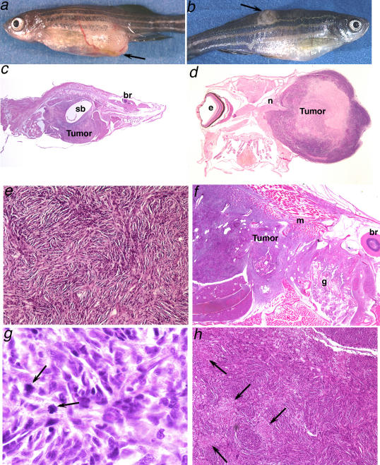
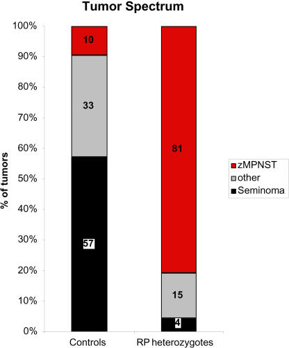
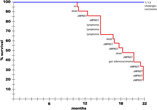
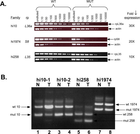
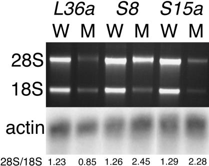
We have generated several hundred lines of zebrafish (Danio rerio), each heterozygous for a recessive embryonic lethal mutation. Since many tumor suppressor genes are recessive lethals, we screened our colony for lines that display early mortality and/or gross evidence of tumors. We identified 12 lines with elevated cancer incidence. Fish from these lines develop malignant peripheral nerve sheath tumors, and in some cases also other tumor types, with moderate to very high frequencies. Surprisingly, 11 of the 12 lines were each heterozygous for a mutation in a different ribosomal protein (RP) gene, while one line was heterozygous for a mutation in a zebrafish paralog of the human and mouse tumor suppressor gene, neurofibromatosis type 2. Our findings suggest that many RP genes may act as haploinsufficient tumor suppressors in fish. Many RP genes might also be cancer genes in humans, where their role in tumorigenesis could easily have escaped detection up to now.


Conflict of interest statement

The authors have declared that no conflicts of interest exist.

Figures

Figure 1
Figure 1. Spindle Cell Tumors Resembling MPNSTs in Zebrafish Heterozygous for Mutations in RP Genes
(A and B) Fish with apparent masses, as indicated by the arrows, or other evident pathology were selected for histological analysis: (A) a hi2582 fish, (B) a hi1034B fish. (C–H) Histopathology of representative tumors stained with hematoxylin and eosin reveals patterns consistent with the diagnosis of MPNST in hi10 fish (C and D), hi1974 fish (E–G), and hi1807 fish (H). (C) Tumors typically filled the entire abdomen (sb, swim bladder; br, brain) (80×). (D) A large tumor with central necrosis is seen emanating from the optic nerve (n) (e, eye) (20×). (E) Tumors consist of spindle cells that stack into short fascicles, typically organizing into whorls (400×). (F) Tumor is aggressively invading muscle (m) and gill (g) (br, brain) (100×). (G) Mitotic figures (arrows) are evident (1000×). (H) Areas of focal necrosis (arrows) are frequently seen (200×).
Figure 2
Figure 2. The Tumor Spectrum in Fish Heterozygous for Mutations in RP Genes Shows an Increased Proportion of zMPNSTs
Fish with apparent masses were selected and processed for histological analysis. Numbers are shown as percent of the total number of diagnosed tumors from either population. The control group includes 42 tumors from 41 fish, including both wild-type and non-RP family transgenics. The RP group includes 68 tumors from 65 RP heterozygotes from 18 different lines representing mutations in 16 different genes. The “other” tumor category includes pancreatic islet adenomas, ultimobranchial gland tumors, neuroblastomas, retinoblastomas, lymphomas, ganglioneuromas, ductal carcinomas, gastrointestinal adenocarcinomas, hepatocellular carcinomas, leukemias, meningiomas, and histiocytic sarcomas.
Figure 3
Figure 3. Rate of Tumor Appearance in hi10 Heterozygotes
A cohort of 28 hi10 fish and 13 of their noncarrier siblings were observed over 22 mo for the appearance of ill health or externally visible tumors. Symptomatic individuals were sacrificed, fixed, and sectioned for histological analysis. The graph represents the percentage of fish remaining over time, with the diagnosis of each removed fish. Three fish labeled “dead” died before fixation and had too much tissue damage to establish a diagnosis. Also, seven of the carrier fish (though none of the noncarriers) were lost to unknown causes over the course of the experiment; while they most likely died, to be conservative these were removed from the total number of fish charted. At 22 mo, the remaining externally healthy fish (4/21 carriers, 13/13 noncarriers) were also histologically examined, and the status of these fish is indicated.
Figure 4
Figure 4. RP Genes Appear to Be Haploinsufficient Tumor Suppressors
(A) RP mutations decrease the amount of RP gene expression. RNA was prepared from 3-d-old homozygous mutant embryos and their wild-type siblings, and serial dilutions of first strand cDNA were used as templates for PCR. The decrease in expression in the mutants can be determined by the difference in the dilution between wild type and mutant where the PCR product amount diminishes. The actin control shows that the total amount of mRNA was the same between samples. (B) LOH is not observed in RP mutant tumors. DNA was prepared from tumors (T) and normal tissue (N) from the same fish, and PCR was conducted with three primers that show the presence or absence of both the insert-bearing (mutant) and wild-type chromosomes. In each case, the upper band is the wild-type chromosome and the lower band is the insert-bearing one. hi10 fish #1 normal (lane 1), tumor (lane 2); hi10 fish #2 normal (lane 3), tumor (lane 4); hi258 fish normal (lane 5), tumor (lane 6); hi1974 fish normal (lane 7), tumor (lane 8).
Figure 5
Figure 5. Ribosomal RNA Levels Are Reduced in RP Mutants
RNA was prepared from 3-d-old homozygous mutant embryos or their wild-type siblings from lines hi10 (L36a), hi1974 (S8), and hi2649 (S15a), and RNA content was visualized by electrophoresis and ethidium bromide staining. The ratio of 28S/18S as determined by densitometry is shown below each lane. Note that L36a mutants show a preferential loss of the 28S band by 1.5-fold, while S8 and S15a mutants show a preferential loss of the 18S band by 1.9- and 1.8-fold, respectively. These RNAs were also northern blotted and probed for beta actin as an mRNA content control.

References

    1. Amatruda JF, Zon LI. Zebrafish as a cancer model system. Cancer Cell. 2002;1:229–231. - PubMed
    1. Amsterdam A, Burgess SB, Golling G, Chen W, Sun Z, et al. A large-scale insertional mutagenesis screen in zebrafish. Genes Dev. 1999;13:2713–2724. - PMC - PubMed
    1. Beck-Engeser GB, Monach PA, Mumberg D, Yang F, Wanderling S, et al. Point mutation in essential genes with loss or mutation of the second allele: Relevance to the retention of tumor-specific antigens. J Exp Med. 2001;194:285–300. - PMC - PubMed
    1. Beckwith LG, Moore JL, Tsao-Wu GS, Harshbarger JC, Cheng KC. Ethylnitrosourea induces neoplasia in zebrafish (Danio rerio) . Lab Invest. 2000;80:379–385. - PubMed
    1. Ben-Ishai R, Scharf R, Sharon R, Kapten I. A human cellular sequence implicated in trk oncogene activation is DNA damage inducible. Proc Natl Acad Sci. 1990;87:6039–6043. - PMC - PubMed

Publication types