. 2004 May;25(5):880-91.

MR imaging presentation of intracranial disease associated with Langerhans cell histiocytosis

Affiliations

MR imaging presentation of intracranial disease associated with Langerhans cell histiocytosis

Daniela Prayer et al. AJNR Am J Neuroradiol. 2004 May.

Abstract

Background and purpose: Intracranial manifestations of Langerhans cell histiocytosis (LCH) are underestimated in frequency and diversity. We categorized the spectrum of MR imaging changes in LCH.

Methods: We retrospectively reviewed 474 MR images in 163 patients with LCH and 55 control subjects. Lesions were characterized by anatomic region and signal intensity. Brain atrophy was assessed.

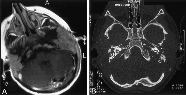
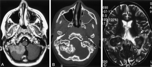
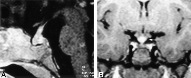
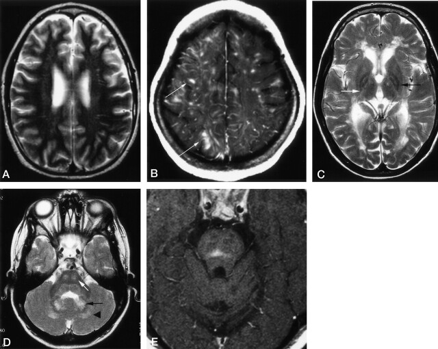
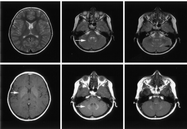
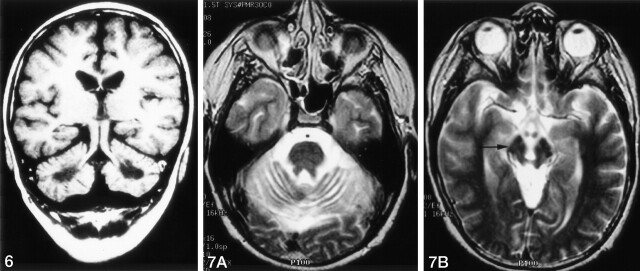
Results: We noted osseous lesions in the craniofacial or skull bones in 56% of patients, meningeal lesions in 29%, and choroid-plexus involvement in 6%. In the hypothalamic-pituitary region, infundibular thickening occurred in 50%; pronounced hypothalamic mass lesions in 10%; and infundibular atrophy in 29%. The pineal gland had a cystic appearance in 28%, and pineal-gland enlargement (>10 mm) was noted in 14%. Nonspecific paranasal-sinus or mastoid opacifications were seen in 55% of patients versus 20% of controls, and accentuated Virchow-Robin spaces occurred in 70% of patients versus 27% of controls (P <.001). Intra-axial, white-matter parenchymal changes resulted in a leukoencephalopathy-like pattern in 36%. Enhancing lesions in a vascular distribution were noted in 5%. Gray-matter changes suggestive of neurodegeneration were identified in the cerebellar dentate nucleus in 40% and in the supratentorial basal ganglia in 26%. All patients with neurodegenerative lesions had lesions in the extra-axial spaces. Cerebral atrophy was found in 8%.

Conclusion: In LCH, cranial and intracranial changes at MR imaging include 1) lesions of the craniofacial bone and skull base with or without soft-tissue extension; 2) intracranial, extra-axial changes (hypothalamic-pituitary region, meninges, circumventricular organs); 3) intracranial, intra-axial changes (white matter and gray matter); and 4) cerebral atrophy.


Figures

F<sc>ig</sc> 1.
Fig 1.
Mastoid involvement at the diagnosis of LCH in a 3-year-old patient. A, Axial contrast-enhanced T1WI shows enhancing lesions in both mastoids. B, Axial bone-window CT scan shows bilateral osseous destruction of the mastoids.
F<sc>ig</sc> 2.
Fig 2.
Images in two patients with LCH. A and B, Lesions in a 13-year-old male patient at the diagnosis of LCH. Axial contrast-enhanced T1WI in A shows an extra-axial, enhancing, space-occupying lesion originating from the meninges. Axial bone-window CT scan in B shows calcification of the extra-axial lesion on the right side. Note the opacification of the left maxillary sinus. C, Choriod plexus lesion in a 6-year-old girl with a 4.5-year history of LCH. Axial T2WIs show bilateral, hypointense masses in the choroid plexus and hyperintense changes in the parieto-occipital white matter.
F<sc>ig</sc> 3.
Fig 3.
Images in two patients with LCH. A, Thickened, enhancing pituitary stalk in a 2-year-old girl with a 1-year history of LCH and new-onset diabetes insipidus B, Coronal contrast-enhanced T1WI in a 6-year-old girl obtained 2 years after the onset of diabetes insipidus. Image shows a thickened pituitary stalk at the cranial portion, in the region of the median eminence (arrow).
F<sc>ig</sc> 4.
Fig 4.
Axial images in patients with LCH. A, T2WI in a 16-year-old male patient with LCH diagnosed 1 year before this study. CSF-intense VRSs are visible in the deep white matter of both hemispheres. The patients had additional hyperintense changes in the dentate nucleus (not shown) and midbrain atrophy (Fig 7). B, Contrast-enhanced T1WI in a 15-year-old female patient with a 10-year history of LCH and progressive cerebellar symptoms since 18 months of age. Enhancing lesions show a vascular pattern; some even have a space-occupying effect (arrows). Additional T1WIs (not shown) depicted hyperintense changes in the dentate nucleus, cerebellar white matter, and basal ganglia. C, T2WI in an 8-year-old patient with a 6-year history of LCH and severe neurologic disabilities. Hyperintense changes in the posterior limb of the internal capsule (white arrow) and periventricular region have a leukodystrophy-like pattern. Image also shows hypointensity of the pallidum with a hyperintense center (black arrow). D, T2WI in the same patient as in C shows hyperintense changes in the central pons (white arrow), dentate nucleus (black arrow), and surrounding white matter (arrowhead). E, Contrast-enhanced T1WI in a 28-year-old man with a 1-year history of LCH and moderate dysarthria and ataxia. Image shows an enhancing lesion in the center of the pons.
F<sc>ig</sc> 5.
Fig 5.
Axial images in a 9-year-old asymptomatic boy with a 7-year history of LCH. Top row, T2WI show the hyperintense appearance of the dentate nucleus (white arrow) and its surrounding white matter (black arrow). Note the normal appearance of the lentiform nucleus. Bottom row, T1WI with magnetization transfer contrast show the hyperintense appearance of the dentate nucleus and the lentiform nucleus (white arrows).
F<sc>ig</sc> 6.
Fig 6.
Coronal T1WI in a 12-year-old boy with a 10-year history of LCH, severe neurologic symptoms, and intellectual impairment. Image shows CSF-intense holes in the regions of the dentate nuclei. Fig 7. Axial T2WIs in the same patient as in Figure 4A. A, Cerebellar atrophy with thinned cerebellar peduncles. B, Midbrain atrophy with wide interpeduncular cistern and distant mammillary bodies, with hypointensity of the pars compacta of the substantia nigra (arrow)

References

    1. A multicentre retrospective survey of Langerhans’ cell histiocytosis: 348 cases observed between 1983 and 1993: The French Langerhans’ Cell Histiocytosis Study Group. Arch Dis Child 1996;75:17–24 - PMC - PubMed
    1. Arceci RJ. The histiocytoses: the fall of the Tower of Babel. Eur J Cancer 1999;35:747–767 - PubMed
    1. Egeler RM, D’Angio GJ. Langerhans cell histiocytosis. J Pediatr 1995;127:1–11 - PubMed
    1. Grois N, Flucher-Wolfram B, Heitger A, Mostbeck GH, Hofmann J, Gadner H. Diabetes insipidus in Langerhans cell histiocytosis: results from the DAL-HX 83 study. Med Pediatr Oncol 1995;24:248–256 - PubMed
    1. Nanduri VR, Bareille P, Pritchard J, Stanhope R. Growth and endocrine disorders in multisystem Langerhans’ cell histiocytosis. Clin Endocrinol (Oxf) 2000;53:509–515 - PubMed