. 2004 Jun;63(6):636-43.
doi: 10.1136/ard.2003.007229.

Galectin-3 surface expression on human adult chondrocytes: a potential substrate for collagenase-3

Affiliations

Galectin-3 surface expression on human adult chondrocytes: a potential substrate for collagenase-3

M Guévremont et al. Ann Rheum Dis. 2004 Jun.

Abstract

Background: Galectin-3 is a lectin detected in mature and early hypertrophic chondrocytes; osteoarthritic (OA) chondrocytes can re-express hypertrophic markers.

Objective: To investigate the synthesis and subcellular localisation of galectin-3 in adult chondrocytes as well as the possibility of cleavage of galectin-3 by collagenase-1 and -3.

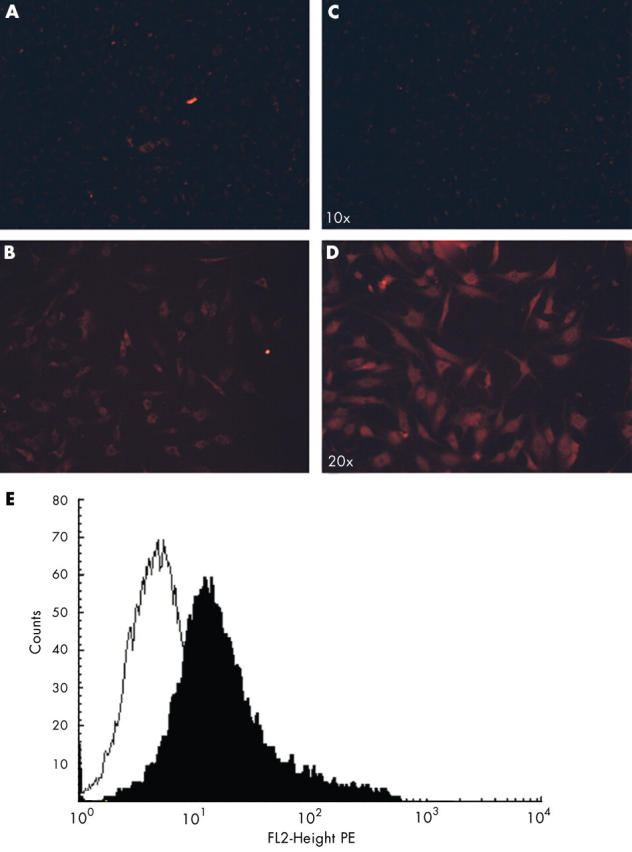
Methods: Galectin-3 was assessed by immunohistochemistry and real time polymerase chain reaction (PCR) in normal and OA cartilage. Its localisation was investigated by subcellular fractionation, immunocytology, and flow cytometry. Proteolysis of galectin-3 by collagenase-1 and -3 was determined by in vitro assay.

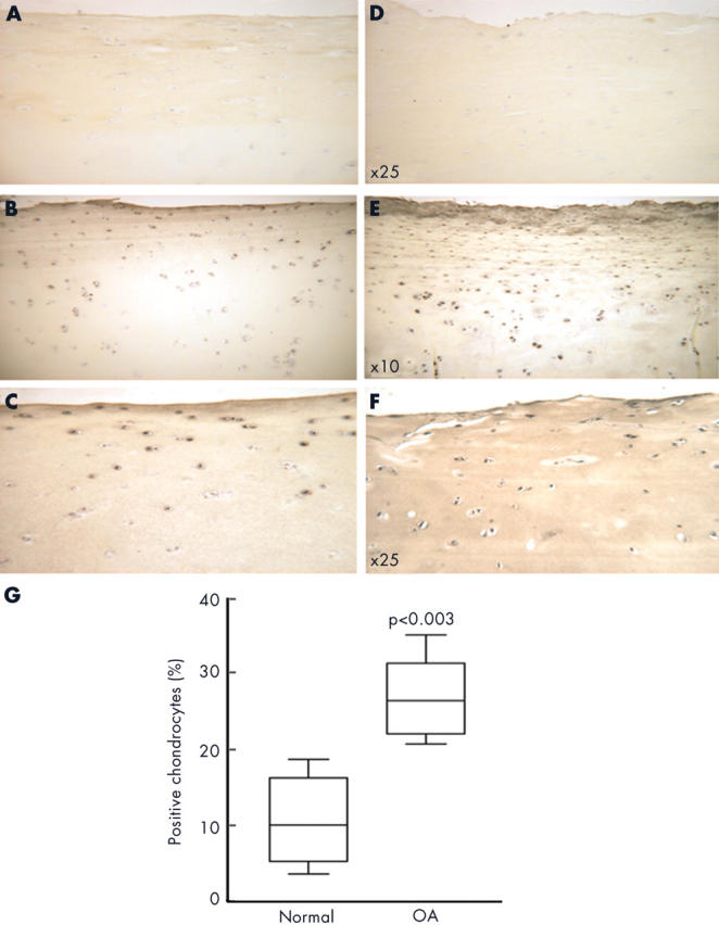
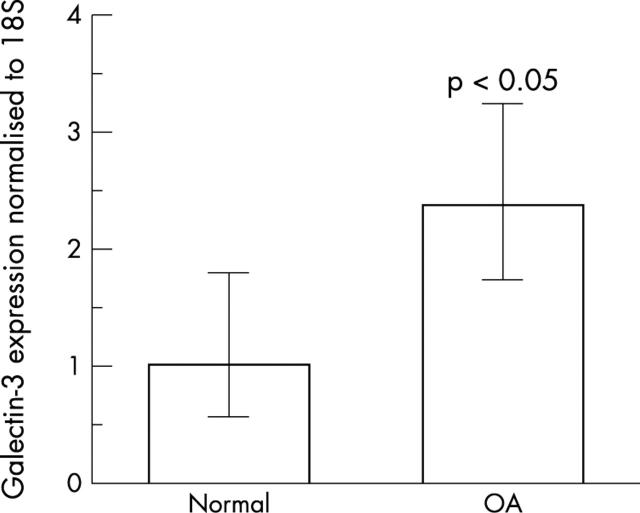
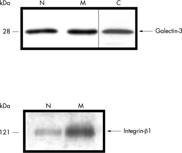
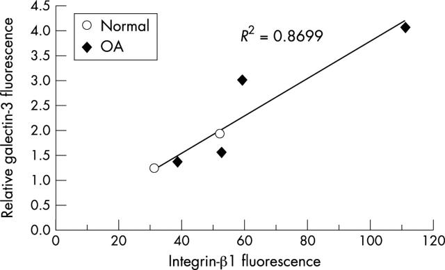
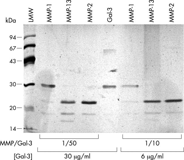
Results: Galectin-3 expression was increased 2.4-fold as measured by reverse transcriptase (RT)-PCR (p<0.05, n = 5) and threefold by immunohistochemistry (p<0.003 n = 6) in OA cartilage compared with normal cartilage. In adult chondrocytes, galectin-3 was found in the cytosol and membrane enriched fractions. Both immunocytology and flow cytometry confirmed the presence of galectin-3 at the surface of chondrocytes. A strong correlation was found between integrin-beta1 and galectin-3 expression at the surface of chondrocytes. Moreover, collagenase-3 cleaved galectin-3 with a higher activity than collagenase-1. The proteolysed sites generated were identical to those produced by gelatinases A and B.

Conclusion: Galectin-3 may play a part in OA, having two roles, one intracellular and not yet identified, and another at the cell surface, possibly related to the interaction of chondrocytes and the cartilage matrix.


Figures

Figure 1
Figure 1
Representative section of cartilage, showing immunostaining for galectin-3 from normal (A–C) and OA cartilage (D–F). (A and D) Non-immune serum; (B, C, E, and F) polyclonal anti-galectin-3. Original magnification x25; (G) galectin-3 levels over the entire cartilage thickness. Data are presented as box plots, where the boxes represent the first and third quartiles, the lines within the boxes represent the median, and the lines outside the boxes represent the spread of the galectin-3 cell scores outside the first and third quartiles. The p value, obtained with the Mann-Whitney test, indicates the difference compared with the normal group.
Figure 2
Figure 2
Expression of galectin-3 mRNA in human normal and OA cartilage was studied using real time RT-PCR as described in "Materials and methods". The PCR analysis was performed by normalising the PCR products of the galectin-3 to the 18S PCR products. Bars show the mean (SD) intensity of five normal and OA specimens. The p value, obtained with Student's t test, indicates the difference compared with the normal group.
Figure 3
Figure 3
Subcellular OA chondrocyte fractionation was performed as described in "Materials and methods". Proteins were quantified for each fraction, 10 µg were blotted, and immunodetection was performed with either a polyclonal anti-galectin-3 or a monoclonal anti-integrin-ß1 antibody. N, nuclear fraction; M, membrane enriched fraction; C, cytosol.
Figure 4
Figure 4
Immunofluorescence and flow cytometry detection of galectin-3 in OA chondrocytes. Cells were incubated with a specific anti-galectin-3 polyclonal antibody (B, D) or non-immune-serum (A, C) and detected with a fluorescent conjugated secondary antibody. (A, B) non-permeabilised cells; (C, D) permeabilised cells; (E) cell surface expression of galectin-3 by primary OA articular chondrocytes. Non-permeabilised cells were immunostained with a polyclonal anti-galectin-3 (filled histogram) or with non-immune serum (open histogram) followed by phycoerythrin (PE) conjugated secondary antibody. The assessment that chondrocytes were not permeabilised was performed by using the 7-AAD dye, which penetrates into the cells, only when the membrane is disrupted.
Figure 5
Figure 5
Correlation of galectin-3 presence with integrin-ß1 at the chondrocyte surface. Cell surface fluorescence was measured with either galectin-3 serum or integrin-ß1 antibody on non-permeabilised cells, as explained in "Materials and methods". As non-immune serum produced background level discrepancies, galectin-3 fluorescence was normalised to the non-immune signal. Relative galectin-3 fluorescence = galectin-3 fluorescence divided by non-immune fluorescence.
Figure 6
Figure 6
Proteolytic activity of collagenase-3 (MMP-13) on galectin-3. rh-galectin-3 was incubated in the presence of activated MMPs as described in "Materials and methods". As galectin-3 at high concentration could multimerise,26 two galectin-3 concentrations were used: 30 µg/ml (multimerisation) and 6 µg/ml (monomerisation). The reaction mixture was stopped with a solution of 6xLaemmli buffer, loaded onto an SDS-PAGE system, and the galectin-3 was stained with silver nitrate. MMP-1, collagenase-1; MMP-13, collagenase-3; MMP-2, gelatinase A; Gal-3, galectin-3.
Figure 7
Figure 7
Schematic representation of galectin-3 sites generated by collagenase-3.

References

    1. Cell Mol Biol (Noisy-le-grand). 1999 Dec;45(8):1191-202 - PubMed
    1. J Rheumatol. 1999 Sep;26(9):2002-14 - PubMed
    1. Osteoarthritis Cartilage. 2000 Mar;8(2):96-105 - PubMed
    1. Osteoarthritis Cartilage. 2000 Jul;8(4):294-302 - PubMed
    1. Matrix Biol. 2000 Jul;19(3):245-55 - PubMed

Publication types

MeSH terms