. 2004 Jun;14(6):1025-35.
doi: 10.1101/gr.1993504. Epub 2004 May 12.

Systems level insights into the stress response to UV radiation in the halophilic archaeon Halobacterium NRC-1

Affiliations

Systems level insights into the stress response to UV radiation in the halophilic archaeon Halobacterium NRC-1

Nitin S Baliga et al. Genome Res. 2004 Jun.

Abstract

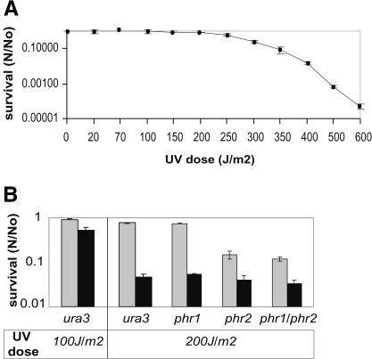
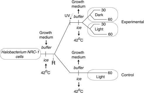
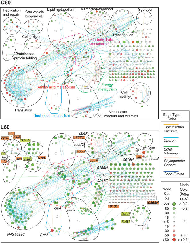
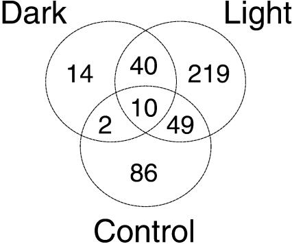
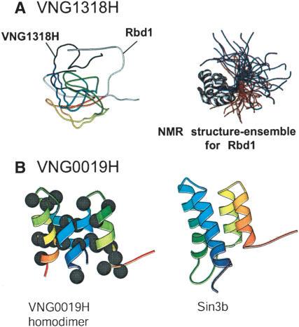
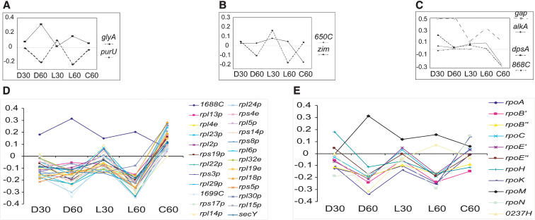
We report a remarkably high UV-radiation resistance in the extremely halophilic archaeon Halobacterium NRC-1 withstanding up to 110 J/m2 with no loss of viability. Gene knockout analysis in two putative photolyase-like genes (phr1 and phr2) implicated only phr2 in photoreactivation. The UV-response was further characterized by analyzing simultaneously, along with gene function and protein interactions inferred through comparative genomics approaches, mRNA changes for all 2400 genes during light and dark repair. In addition to photoreactivation, three other putative repair mechanisms were identified including d(CTAG) methylation-directed mismatch repair, four oxidative damage repair enzymes, and two proteases for eliminating damaged proteins. Moreover, a UV-induced down-regulation of many important metabolic functions was observed during light repair and seems to be a phenomenon shared by all three domains of life. The systems analysis has facilitated the assignment of putative functions to 26 of 33 key proteins in the UV response through sequence-based methods and/or similarities of their predicted three-dimensional structures to known structures in the PDB. Finally, the systems analysis has raised, through the integration of experimentally determined and computationally inferred data, many experimentally testable hypotheses that describe the metabolic and regulatory networks of Halobacterium NRC-1.


Figures

Figure 1
Figure 1
Survival of Halobacterium NRC-1 wild-type and putative photolyase mutants following UV-C irradiation. (A) Wild-type Halobacterium NRC-1 survival (N/No; x-axis) in light following increasing doses of UV irradiation (J/m2; y-axis). (B) Survival of single (phr1 or phr2) and double (phr1/phr2) knockout mutants on recovery in light (gray bars) and dark (black bars) post-UV irradiation at 200 J/m2. Relative difference in effects of UV-C irradiation at 100 and 200 J/m2 on viability of the wild-type organism is shown. (N) Number of viable cells in challenged sample; (No) number of viable cells in control; error bars represent standard error for at least three independent experiments.
Figure 2
Figure 2
Experiment design for global analysis of UV-response in Halobacterium NRC-1. Halobacterial cells were harvested from the growth medium, resuspended at a low density in a clear isotonic buffer, and placed on ice. Total RNA prepared from an aliquot of the cells at this point was used as the reference (R) in microarray analyses. Subsequently, one-half of the cell suspension was kept aside on ice for use as control. The remainder was irradiated on ice with UV-C light and post-irradiation the cells were diluted twofold in CM and allowed to recover at 42°C under light or dark conditions. Total RNA was prepared at 30 and 60 min from both the light and dark repair samples. The control was processed in a manner identical to the 60-min light repair sample, except that it was not irradiated.
Figure 4
Figure 4
Systems-level visualization of the Halobacterium NRC-1 transcriptome in C60 and L60 with layout organized by general function. The mRNA changes are visualized as a network of genes (nodes) and their interactions (edges). A total of 420 mRNAs that changed during repair or in control are shown with shades of red for increased and shades of green for decreased levels; node size correlates to statistical significance (λ) of change (see inset key). The five types of edges connecting the genes are color coded and described in the text (see inset key for color codes). Representative genes with mRNA changes during repair masked by mRNA changes resulting from the experimental procedure alone are labeled and highlighted with red for a masked up-regulated change during repair and green for a masked down-regulated change during repair. For a complete list of 147 potential masked changes, see Supplemental Table 3. The asterisk indicates the ntp gene, a transposase, which was originally misannotated as a neutral proteinase (see text for details).
Figure 3
Figure 3
Venn diagram of mRNA changes observed during repair and in the control (see text for details).
Figure 5
Figure 5
Functional annotation of proteins through ab inito structure prediction by Rosetta. (A) Alignment of the Rosetta-predicted structure for VNG1318H and Rbd1 (left). The NMR structure for Rbd1 is shown on the right. (B) Rosetta-predicted structure for the VNG0019H homodimer (left) and its closest match, the NMR structure for Sin3b. The black dots indicate hydrophobic residues located primarily at the predicted protein–protein interface.
Figure 6
Figure 6
Transcript levels for select genes involved with DNA repair, transcription, and translation. (A) The glyA and purU genes are involved with 5,10-CH=THF biosynthesis pathway. (B) The two genes, zim (d(CTAG) methylase) and VNG0650C (RecJ exonuclease), are involved with a putative d(CTAG) methylation-directed mismatch repair genes. (C) Four genes involved with photo-oxidative DNA damage repair. (D) The large ribosomal operon (cluster 1). (E) RNA polymerase synthesis. The y-axis indicates log10 ratios of mRNA changes with respect to the reference RNA, and the x-axis indicates 30 and 60 min during dark and light repair, as well as the control. The lines connecting the dots are for the benefit of the reader to correlate mRNA changes for a given gene in the five experiments, and do not indicate a temporal ordinate except from D30 to D60 and L30 to L60.
Figure 7
Figure 7
Cobalamin biosynthesis pathway in Halobacterium NRC-1. (A) Cofactor and vitamin biosynthesis biomodules are linked to five genes of unknown function visualized in Cytoscape; (see Fig. 4 for key to network display). Genes implicated in cobalamin biosynthesis are shown in a larger font size. (B) mRNA changes for the cobalamin biosynthesis genes (the x- and y-axes are defined in Fig. 7). (C) Cobyrininc acid biosynthesis pathway. The proteins catalyzing the various steps are shown along with the standard correlation between mRNA changes in adjacent biochemical steps. The question mark (?) indicates enzymes yet to be identified. (D) Hierarchical clustering of mRNA changes using standard correlation. Red denotes up-regulation and green denotes down-regulation. The dendogram on the left represents closeness between two genes on the basis of the similarity in their expression level changes in the five perturbations.

Similar articles

Cited by

References

    1. Amillet, J.M., Buisson, N., and Labbe-Bois, R. 1995. Positive and negative elements involved in the differential regulation by heme and oxygen of the HEM13 gene (coproporphyrinogen oxidase) in Saccharomyces cerevisiae. Curr. Genet. 28: 503–511. - PubMed
    1. Baliga, N.S., Pan, M., Goo, Y.A., Yi, E.C., Goodlett, D.R., Dimitrov, K., Shannon, P., Aebersold, R., Ng, W.V., and Hood, L. 2002. Coordinate regulation of energy transduction modules in Halobacterium sp. analyzed by a global systems approach. Proc. Natl. Acad. Sci. 99: 14913–14918. - PMC - PubMed
    1. Bateman, A., Birney, E., Durbin, R., Eddy, S.R., Howe, K.L., and Sonnhammer, E.L. 2000. The Pfam protein families database. Nucleic Acids Res. 28: 263–266. - PMC - PubMed
    1. Birrell, G.W., Brown, J.A., Wu, H.I., Giaever, G., Chu, A.M., Davis, R.W., and Brown, J.M. 2002. Transcriptional response of Saccharomyces cerevisiae to DNA-damaging agents does not identify the genes that protect against these agents. Proc. Natl. Acad. Sci. 99: 8778–8783. - PMC - PubMed
    1. Bonneau, R., Tsai, J., Ruczinski, I., Chivian, D., Rohl, C., Strauss, C.E., and Baker, D. 2001. Rosetta in CASP4: Progress in ab initio protein structure prediction. Proteins Suppl: 119–126. - PubMed

WEB SITE REFERENCES

    1. http://bioinfo.pl/meta/; A Meta server provides access to various fold recognition and local structure prediction methods. This server also provides translation of the standard formats like PDB, CASP, or PIR, and is coupled to several consensus servers such as pcons.
    1. http://www.sbc.su.se/∼arne/pcons/; A consensus fold recognition predictor.

Publication types

MeSH terms