

Evidence that climbing fibers control an intrinsic spike generator in cerebellar Purkinje cells
- PMID: 15140921
- PMCID: PMC6729399
- DOI: 10.1523/JNEUROSCI.4530-03.2004
Evidence that climbing fibers control an intrinsic spike generator in cerebellar Purkinje cells
Erratum in
- J Neurosci. 2004 Jun ;24(23):following 5456
Abstract
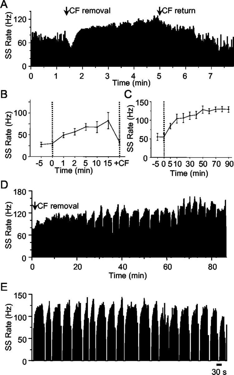
It is well established that the climbing fiber (CF) input to a cerebellar Purkinje cell (PC) can exert a controlling influence on the background simple spike (SS) activity of the cell, in that repetitive stimulation of CFs causes a decrease in SS activity, and removal or inactivation of CFs is followed by a rise in activity. In the present study, the effects of inactivation of CFs in the short term and longer term (hours) were investigated in anesthetized rats to determine how the CFs control the PC SS activity. Inactivation of the CF input to a PC was accomplished by either reversibly inactivating with lignocaine or by microlesioning the inferior olive. Consistent with previous findings, CF removal caused a transformation of the PC firing pattern, with SSs discharging more regularly and rising to an exceptionally high level. In cases in which CF activity resumed, SS rate declined to control levels within a few seconds. However, with sustained CF inactivation (30 min to 5 hr), SS activity continues to rise progressively and develops an oscillating firing pattern, consisting of alternating bursts of high-frequency discharge at up to 100-150 Hz followed by 10-20 sec periods of electrical quiescence. No accompanying changes in the threshold for evoking SSs via the parallel fibers were seen to accompany the increases in tonic SS activity. We conclude that the increase in SS activity that follows CF inactivation could be caused by the removal of an inhibitory action that CFs exert on the intrinsic pacemaker present in PCs under normal conditions.
Figures


Similar articles
-
Feedback control of Purkinje cell activity by the cerebello-olivary pathway.Eur J Neurosci. 2004 Dec;20(11):2999-3005. doi: 10.1111/j.1460-9568.2004.03789.x. Eur J Neurosci. 2004. PMID: 15579154
-
Ontogenesis of olivocerebellar relationships. II. Spontaneous activity of inferior olivary neurons and climbing fibermediated activity of cerebellar Purkinje cells in developing rats.J Neurosci. 1981 Jul;1(7):703-9. doi: 10.1523/JNEUROSCI.01-07-00703.1981. J Neurosci. 1981. PMID: 7346579 Free PMC article.
-
Climbing fibers mediate vestibular modulation of both "complex" and "simple spikes" in Purkinje cells.Cerebellum. 2015 Oct;14(5):597-612. doi: 10.1007/s12311-015-0725-1. Cerebellum. 2015. PMID: 26424151 Review.
-
Cerebellar climbing fibers modulate simple spikes in Purkinje cells.J Neurosci. 2003 Aug 27;23(21):7904-16. doi: 10.1523/JNEUROSCI.23-21-07904.2003. J Neurosci. 2003. PMID: 12944521 Free PMC article.
-
Complex Spike Wars: a New Hope.Cerebellum. 2018 Dec;17(6):735-746. doi: 10.1007/s12311-018-0960-3. Cerebellum. 2018. PMID: 29982917 Free PMC article. Review.
Cited by
-
Effects of climbing fiber driven inhibition on Purkinje neuron spiking.J Neurosci. 2012 Dec 12;32(50):17988-97. doi: 10.1523/JNEUROSCI.3916-12.2012. J Neurosci. 2012. PMID: 23238715 Free PMC article.
-
Complex Dynamics in Simplified Neuronal Models: Reproducing Golgi Cell Electroresponsiveness.Front Neuroinform. 2018 Dec 3;12:88. doi: 10.3389/fninf.2018.00088. eCollection 2018. Front Neuroinform. 2018. PMID: 30559658 Free PMC article.
-
In vivo analysis of inhibitory synaptic inputs and rebounds in deep cerebellar nuclear neurons.PLoS One. 2011 Apr 28;6(4):e18822. doi: 10.1371/journal.pone.0018822. PLoS One. 2011. PMID: 21552556 Free PMC article.
-
Cerebellar Modules and Their Role as Operational Cerebellar Processing Units: A Consensus paper [corrected].Cerebellum. 2018 Oct;17(5):654-682. doi: 10.1007/s12311-018-0952-3. Cerebellum. 2018. PMID: 29876802 Free PMC article. Review.
-
Genetic silencing of olivocerebellar synapses causes dystonia-like behaviour in mice.Nat Commun. 2017 Apr 4;8:14912. doi: 10.1038/ncomms14912. Nat Commun. 2017. PMID: 28374839 Free PMC article.
References
-
- Ajima A, Hensch T, Kado RT, Ito M (1991) Differential blocking action of Joro spider toxin analog on parallel fiber and climbing fiber synapses in cerebellar Purkinje cells. Neurosci Res 12: 281-286. - PubMed
-
- Armstrong DM, Harvey RJ, Schild RF (1973) Branching of inferior olivary axons to terminate in different folia, lobules or lobes of the cerebellum. Brain Res 54: 365-371. - PubMed
-
- Aubry A, Batini C, Billard JM, Kao RT, Morain P (1991) Tetrodotoxin induced calcium spikes: in vitro and in vivo studies of normal and deafferented Purkinje cells. Exp Brain Res 84: 297-302. - PubMed
Publication types
MeSH terms
Substances
LinkOut - more resources
Full Text Sources