. 2004 May 12;24(19):4518-29.
doi: 10.1523/JNEUROSCI.0542-04.2004.

A Drosophila temperature-sensitive seizure mutant in phosphoglycerate kinase disrupts ATP generation and alters synaptic function

Affiliations

A Drosophila temperature-sensitive seizure mutant in phosphoglycerate kinase disrupts ATP generation and alters synaptic function

Ping Wang et al. J Neurosci. .

Abstract

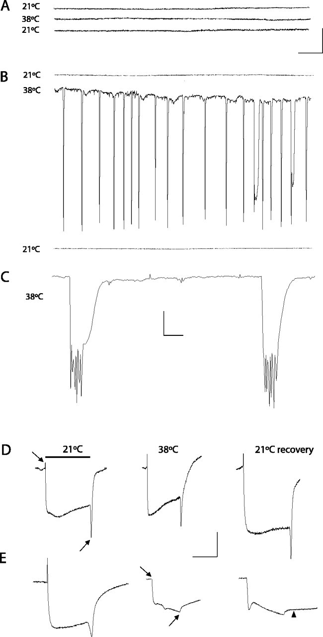
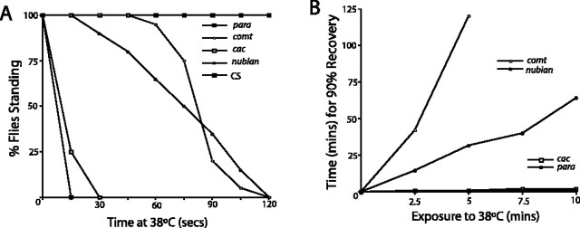
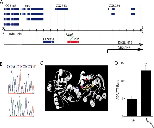
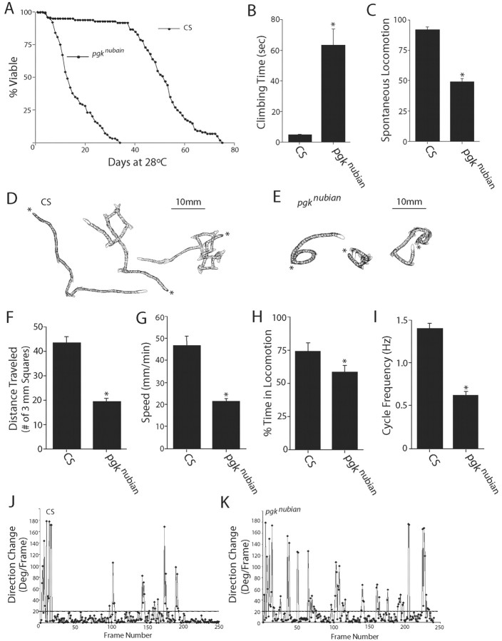
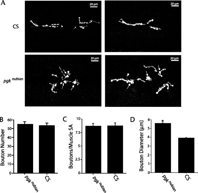
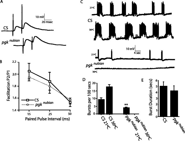
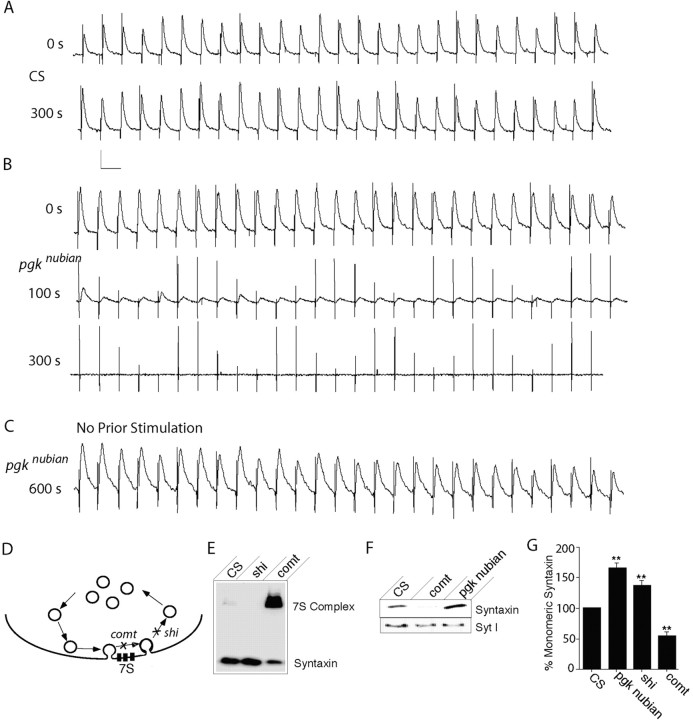
A novel paralytic mutant, nubian, was identified in a behavioral screen for conditional temperature-sensitive seizure mutants in Drosophila melanogaster. nubian mutants display reduced lifespan, abnormal motor behavior, altered synaptic structure, and defective neurotransmitter release. The nubian mutant disrupts phosphoglycerate kinase (PGK), an enzyme required for ATP generation in the terminal stage of the glycolytic pathway. Consistent with altered ATP generation in nubian animals, brain extracts show a threefold reduction in resting ATP levels compared with controls. Microarray analysis of nubian mutants reveals altered transcription of genes implicated in glucose and lipid metabolism. Disruption of ATP generation in nubian animals is accompanied by temperature-dependent defects in neuronal activity, with initial seizure activity, followed by an activity-dependent loss of synaptic transmission. nubian mutants also display structural defects at the synapse, with larger varicosity size but normal varicosity number, indicating that these synaptic parameters are regulated independently. Both exocytotic (NSF) and endocytotic (dynamin) ATPase/GTPase activity are required for normal synaptic transmission. Biochemical and physiological analyses indicate that synaptic defects in nubian animals are secondary to defective endocytosis, suggesting that endocytotic pathways may be generally more sensitive to altered ATP levels than those used for exocytosis. Alterations in ATP metabolism likely disrupt similar pathways in humans, because PGK deficiency is associated with mental retardation, seizures, and exercise intolerance. Given the behavioral similarities between disruptions of PGK function in Drosophila and humans, the analysis of nubian animals may reveal conserved neuronal responses associated with altered ATP generation within the brain.


Figures

Figure 1.
Figure 1.
Electrophysiological defects in nubian animals. A-C, Extracellular recordings from the DLM flight pathway from controls (CS; shown in A) or nubian adults (B, C) at 2-4 d of age. Recordings were taken at 21°C (top trace), at the nonpermissive 38°C (middle trace), and after recovery at 21°C (bottom trace). Control animals display no seizure activity when the temperature is raised to 38°C. In contrast, a rapid onset of spontaneous firing is observed in nubian animals at 38°C. After several minutes at 38°C the seizure activity in nubian animals ceases. Complete recovery was observed with return to the permissive temperature. C, An enlarged time scale trace for seizure activity in nubian at 38°C, revealing multiple bursting muscle action potentials. Calibration: A, B, 5 mV, 0.5 sec; C, 5 mV, 20 msec. D, E, Electroretinograms (ERGs) recorded from CS and nubian animals. Flies were heated rapidly from 21 to 38°C and maintained at 38°C for 4 min. Then the flies were cooled rapidly to 21°C and allowed to recover in darkness, with test light pulses given at regular intervals. Note the loss of on- and off-transients (arrows) in nubian flies. The on-/off-transients take ∼30 min to recover when shifted back to the permissive temperature. During this recovery period nubian photoreceptors show a delayed repolarization, indicating defects in membrane excitability as well. Of each genotype 5-10 individuals aged 2-4 d were tested with similar results to those shown. Calibration: D, E, 5 mV, 0.5 sec.
Figure 2.
Figure 2.
Temperature-sensitive nubian animals exhibit behavioral paralysis with slow recovery. A, The behavioral onset of motor uncoordination is plotted for nubian animals as well as for Dmca1ATS2 (Kawasaki et al., 2000), paraTS1, comtST17, and CS flies. Similar to comtST17 mutants, nubian animals require exposure to 38°C for several minutes before the onset of paralysis. Twenty flies of the indicated genotype were aged 3-5 d and placed in preheated glass vials at 38°C. The percentage of flies standing was measured in 15 sec intervals and plotted. B, The recovery time after exposure to 38°C is plotted versus the length of exposure to the nonpermissive temperature. Unlike Dmca1ATS2 and paraTS1 mutants that show a fast recovery, nubian mutants are affected more severely, requiring progressively longer recovery times, similar to comtST17.
Figure 3.
Figure 3.
The nubian locus corresponds to phosphoglycerate kinase. A, The nubian locus was mapped to the breakpoints defined by Df(2L)N6 and Df(2L)N19, which contains five loci based on breakpoint analysis. The blue diagrams represent predicted alternative splicing at each locus. The pgk locus is shown in red. nubian mutants failed to complement a P-element lethal mutation (pgkKG064431, green arrow) in the pgk locus. B, Sequence analysis of the pgk locus revealed a single base pair substitution (indicated by asterisk) that results in a V357E mutation in nubian. C, The location of the V357 amino acid (red) is shown in relation to the binding of ATP (yellow) and 1,3-diphosphoglycerate (blue). The V357E mutation introduces a negative charge near the catalytic binding pocket for ADP/ATP (Bernstein et al., 1997). D, To analyze ATP levels in pgknubian, we performed HPLC nucleotide analysis from head extracts of pgknubian and control adults maintained at room temperature. The ADP/ATP ratio is elevated significantly in pgknubian animals (**p < 0.05; Student's t test). Three separate experiments were performed with 30 flies in each sample.
Figure 4.
Figure 4.
Behavioral analysis of pgknubian animals. A, The pgknubian mutant shows a dramatic reduction in lifespan, with a decrease in the T50 (age at which 50% of the culture has died) by 75%. One hundred adult virgin males of each genotype were maintained at 28°C, and lethality was counted daily. B, C, Behavioral assays performed on 4- to 5-d-old pgknubian animals revealed a decrease in both climbing ability (B) and spontaneous locomotion (C). Climbing assays were performed by recording the time required for a fly placed at the bottom of a vial to climb 5 cm, with a maximum period of 5 min allowed. Spontaneous locomotion was assayed by introducing a fly in a cylindrical plastic chamber (diameter, 8 mm; height, 3 mm) and quantifying the number of times it crossed a straight line bisecting the chamber in a 4 min period. The data in B and C are presented as the mean ± SEM. D-K, Analysis of larval locomotion with the use of computer-assisted DIAS to examine the function of the central motor pattern generator. D, E, Representative crawling patterns of CS and pgknubian larvae. Images of the crawling larva were video recorded for a period of 2 min and digitized at 2 frames/sec; an outline or perimeter of the larva was drawn on each frame of a digital movie. Perimeters at different time points were summated to reproduce crawling patterns. The beginning of locomotion is marked by an asterisk. F, In a period of 2 min pgknubian larvae traveled ∼50% less distance than that of CS larvae. Distance traveled was measured by tracing the tracks created by a crawling larva on an agarose surface during a period of 2 min and then counting the number of 3 mm grid squares the trace traversed. G, The speed of pgknubian larvae was reduced ∼50% compared with control larvae. Locomotor speed was calculated by averaging the speed of the centroid (the center of the area of a perimeter) for each frame of digital movie, using the central difference method. H, pgknubian mutant larvae spend 20% less time in locomotion than controls. The frame-by-frame data for direction change (shown in J, K) were used to calculate the time spent in locomotion. Periods of locomotion were defined as a crawling segment of five or more consecutive frames in which direction change was <20° for each frame when the larval images were digitized at 2 frames/sec. At 2 frames/sec resolution the periods of pausing were defined as frames in which the direction of change per frame was >20°. I, The frequency of locomotor cycles was reduced significantly in pgknubian compared with that of CS larvae. The frequency of cycles was calculated by counting directly the number of the rhythmic waves of contraction from posterior to anterior while playing back the video recording of a crawling larva and by measuring the duration in a stretch of crawling that consisted of at least five consecutive cycles. J, K, Representative plots of direction change in pgknubian and CS larvae show the change of angle of the centroid for each computed frame via the central difference method. In comparisons with CS larvae the pgknubian larvae have more frames with a direction change that exceeds 20°, indicating increased pausing compared with that for controls. *p < 0.05.
Figure 5.
Figure 5.
Synaptic growth defects in pgknubian animals. A, Immunocytochemical analysis with anti-synaptotagmin antibodies at muscle fiber 6/7 in segment A3 of third instar larvae. Top panels, CS; bottom panels, pgknubian. pgknubian mutants have an increase in varicosity size (arrows) compared with controls. B, Quantification of varicosity number at muscle fiber 6/7 of segments A3-A5 of the indicated genotypes. For each genotype 8-10 larvae were examined, with a total of 39 6/7 NMJs quantified for CS and pgknubian in B-D. C, Quantification of varicosity number of the indicated genotypes at muscle fiber 6/7 of segments A3-A5 normalized to muscle surface area. D, Quantification of varicosity diameter for the five largest type 1B varicosities per 6/7 NMJ for CS and pgknubian. Varicosities were significantly larger in pgknubian animals (*p < 0.001; Student's t test), with an average diameter of 3.9 ± 0.07 μm ± SEM in CS versus 5.5 ± 0.08 μm ± SEM in pgknubian larvae. Error bars are SEM.
Figure 6.
Figure 6.
Paired pulse facilitation and central motor pattern generator activity in pgknubian mutants. A, Paired pulse facilitation in control and pgknubian mutant third instar larvae was recorded in saline containing 0.2 mm extracellular calcium. Calibration: 10 mV, 20 msec. B, Quantification of paired pulse facilitation (amplitude of pulse 2/amplitude of pulse 1) in control and pgknubian animals for 15, 25, and 50 msec intervals. No change in the amplitude of paired pulse facilitation was observed in pgknubian mutants. The number of preparations analyzed was 7 CS and 5 pgknubian. C, Voltage traces of spontaneous central pattern generator activity recorded from muscle fiber 6 in third instar preparations with an intact ventral nerve cord in 1.5 mm extracellular calcium. In control animals spontaneous bursts of motor neuron activity were observed, with increased frequency at 36°C. In pgknubian animals spontaneous activity was reduced at room temperature and abolished at 36°C. Calibration: 10 mV, 4 sec. The number of preparations analyzed was 22 CS at 21°C, 10 CS at 36°C, 23 pgknubian at 21°C, and 22 pgknubian at 36°C. D, Quantification of the number of bursts occurring in a 100 sec bin in control and pgknubian animals at 21 and 36°C. pgknubian mutants have a statistically significant (**p < 0.001; Student's t test) reduction in burst frequency compared with controls. At 36°C there was a complete absence of a central motor pattern in all 22 pgknubian animals that were analyzed. E, Quantification of burst duration in control and pgknubian animals. Although slightly reduced, there was no statistically significant change in burst duration. Ingeneral, burst duration was quite variable in both control and pgknubian animals.
Figure 7.
Figure 7.
Electrophysiological defects at pgknubian neuromuscular junctions. A-C, Voltage traces of evoked excitatory junction potentials recorded from muscle fiber 6 during 10 Hz stimulation at 36°C in either CS (A) or pgknubian third instar larvae (B, C) in 0.2 mm extracellular calcium. Calibration: 5 mV, 0.1 sec. pgknubian animals show a progressive reduction in EJP amplitude with high-frequency stimulation compared with controls. Maintaining the preparation at 36°C in the absence of stimulation did not result in EJP rundown. At room temperature the average initial EJP amplitude was 17.8 ± 0.8 mV (n = 16) in CS compared with 15.5 ± 0.7 (n = 20) in pgknubian, whereas resting membrane potential was -61.43 ± 1.4 (n = 16) in CS compared with -57.8 ± 1.2 (n = 20) in pgknubian. At 36°C the initial EJP amplitude was 8.8 ± 2.1 mV (n = 4) in CS compared with 8.0 ± 0.7 (n=32) in pgknubian, whereas resting membrane potential was -49.8 ± 2.1 (n=4) in CS compared with -41.8 ± 1.4 (n=32) in pgknubian. Spontaneous mEJP frequency was 1.4 ± 0.14 (n=16) in CS compared with 1.1 ± 0.09 (n=20) in pgknubian. All data are presented as the mean ± SEM. D, Model of SNARE complex (7S) generation during vesicle cycling. After vesicle fusion and SNARE complex formation the loss of NSF function in comt mutants blocks SNARE disassembly, leading to increased incorporation of monomeric syntaxin into 7S SNARE complexes. In shi mutants the vesicle recycling is blocked, leading to a reduction of SNARE complexes via NSF-mediated disassembly and an increase in monomeric syntaxin. E, Representative SNARE complex assay showing a reduction in the 7S complex with an increase in monomeric syntaxin in shi flies after exposure to 38°C, whereas comt mutants show an accumulation of 7S complex (Littleton et al., 2001). The distribution of syntaxin between monomeric and SNARE complexes was analyzed from 10 CS, comtST17, or shiTS1 flies after a 20 min, 38°C heat pulse. F, Western blots showing monomeric syntaxin accumulation in nubian animals compared with the depletion observed in comtST17 flies. Head homogenates were prepared from 10 CS, comtST17, or pgknubian flies after a 20 min, 38°C heat pulse and probed for syntaxin and synaptotagmin I. G, Quantification of monomeric syntaxin levels in pgknubian, shiTS1, and comtST17 compared with control CS. In each experiment the monomeric syntaxin levels measured by densitometry in CS were set to 100% and compared with each genotype. The number of experiments for quantification of each genotype was as follows: 4 pgknubian, 7 shiTS1, 10 comtST17, and 10 CS (**p < 0.001 by Student's t test analysis).

Similar articles

Cited by

References

    1. Akasu T, Tsurusaki M, Shoji S (1996) Depletion of glucose causes presynaptic inhibition of neuronal transmission in the rat dorsolateral septal nucleus. Synapse 24: 125-134. - PubMed
    1. Andrews J, Smith M, Merakovsky J, Coulson M, Hannan F, Kelly LE (1996) The stoned locus of Drosophila melanogaster produces a dicistronic transcript and encodes two distinct polypeptides. Genetics 143: 1699-1711. - PMC - PubMed
    1. Atkinson NS, Robertson GA, Ganetzky B (1991) A component of calcium-activated potassium channels encoded by the Drosophila slo locus. Science 253: 551-555. - PubMed
    1. Bernstein BE, Michels PA, Hol WG (1997) Synergistic effects of substrate-induced conformational changes in phosphoglycerate kinase activation. Nature 385: 275-278. - PubMed
    1. Davis GW, Bezprozvanny I (2001) Maintaining the stability of neural function: a homeostatic hypothesis. Annu Rev Physiol 63: 847-869. - PubMed

Publication types

MeSH terms