

Functional imaging of working memory after 24 hr of total sleep deprivation
- PMID: 15140927
- PMCID: PMC6729385
- DOI: 10.1523/JNEUROSCI.0007-04.2004
Functional imaging of working memory after 24 hr of total sleep deprivation
Abstract
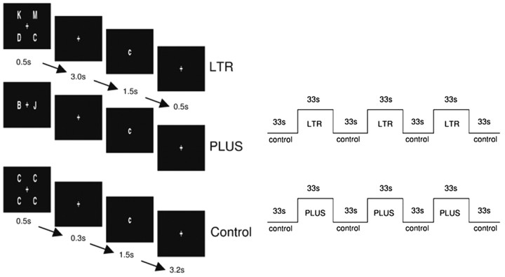
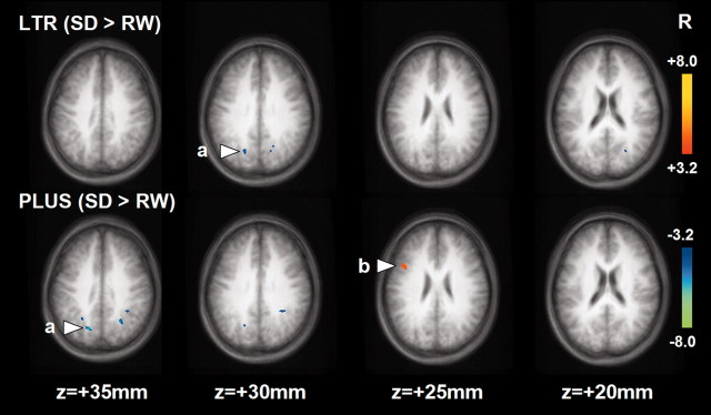
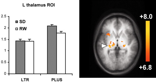
The neurobehavioral effects of 24 hr of total sleep deprivation (SD) on working memory in young healthy adults was studied using functional magnetic resonance imaging. Two tasks, one testing maintenance and the other manipulation and maintenance, were used. After SD, response times for both tasks were significantly slower. Performance was better preserved in the more complex task. Both tasks activated a bilateral, left hemisphere-dominant frontal-parietal network of brain regions reflecting the engagement of verbal working memory. In both states, manipulation elicited more extensive and bilateral (L>R) frontal, parietal, and thalamic activation. After SD, there was reduced blood oxygenation level-dependent signal response in the medial parietal region with both tasks. Reduced deactivation of the anterior medial frontal and posterior cingulate regions was observed with both tasks. Finally, there was disproportionately greater activation of the left dorsolateral prefrontal cortex and bilateral thalamus when manipulation was required. This pattern of changes in activation and deactivation bears similarity to that observed when healthy elderly adults perform similar tasks. Our data suggest that reduced activation and reduced deactivation could underlie cognitive impairment after SD and that increased prefrontal and thalamic activation may represent compensatory adaptations. The additional left frontal activation elicited after SD is postulated to be task dependent and contingent on task complexity. Our findings provide neural correlates to explain why task performance in relatively more complex tasks is better preserved relative to simpler ones after SD.
Figures


References
-
- Babkoff H, Caspy T, Mikulincer M (1991) Subjective sleepiness ratings: the effects of sleep deprivation, circadian rhythmicity and cognitive performance. Sleep 14: 534-539. - PubMed
-
- Barch DM, Braver TS, Nystrom LE, Forman SD, Noll DC, Cohen JD (1997) Dissociating working memory from task difficulty in human prefrontal cortex. Neuropsychologia 35: 1373-1380. - PubMed
-
- Cabeza R (2002) Hemispheric asymmetry reduction in older adults: the HAROLD model. Psychol Aging 17: 85-100. - PubMed
-
- Cabeza R, Dolcos F, Graham R, Nyberg L (2002) Similarities and differences in the neural correlates of episodic memory retrieval and working memory. NeuroImage 16: 317-330. - PubMed
Publication types
MeSH terms
LinkOut - more resources
Full Text Sources