. 2004 May 12;24(19):4614-24.
doi: 10.1523/JNEUROSCI.0765-04.2004.

KCNQ/M channels control spike afterdepolarization and burst generation in hippocampal neurons

Affiliations

KCNQ/M channels control spike afterdepolarization and burst generation in hippocampal neurons

Cuiyong Yue et al. J Neurosci. .

Abstract

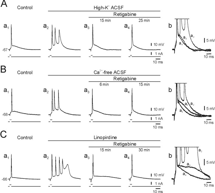
KCNQ channel subunits are widely expressed in peripheral and central neurons, where they give rise to a muscarinic-sensitive, subthreshold, and noninactivating K+ current (M-current). It is generally agreed that activation of KCNQ/M channels contributes to spike frequency adaptation during sustained depolarizations but is too slow to influence the repolarization of solitary spikes. This concept, however, is based mainly on experiments with muscarinic agonists, the multiple effects on membrane conductances of which may overshadow the distinctive effects of KCNQ/M channel block. Here, we have used selective modulators of KCNQ/M channels to investigate their role in spike electrogenesis in CA1 pyramidal cells. Solitary spikes were evoked by brief depolarizing current pulses injected into the neurons. The KCNQ/M channel blockers linopirdine and XE991 markedly enhanced the spike afterdepolarization (ADP) and, in most neurons, converted solitary ("simple") spikes to high-frequency bursts of three to seven spikes ("complex" spikes). Conversely, the KCNQ/M channel opener retigabine reduced the spike ADP and induced regular firing in bursting neurons. Selective block of BK or SK channels had no effect on the spike ADP or firing mode in these neurons. We conclude that KCNQ/M channels activate during the spike ADP and limit its duration, thereby precluding its escalation to a burst. Consequently, down-modulation of KCNQ/M channels converts the neuronal firing pattern from simple to complex spiking, whereas up-modulation of these channels exerts the opposite effect.


Figures

Figure 1.
Figure 1.
Effects of linopirdine on the spike ADP and firing pattern in a CA1 pyramidal cell. A, Intracellular recordings of the spikes evoked by brief (4 msec) and long (180 msec) threshold-straddling depolarizing current pulses. In each panel, the current stimulus is depicted below the voltage trace. Resting potential (in milli volts) is shown to the left of the voltage trace. In control, the neuron fired a solitary spike in response to a brief stimulus (a1). Adding 10 μm linopirdine to the ACSF caused a gradual depolarization of the neuron (from -70 to -66 mV) and facilitation of the spike ADP (a2, a3), ultimately converting regular firing to spontaneous bursting (a4; three superimposed traces). The ADP facilitation also occurred when membrane potential was maintained at its native value (-70 mV) by injecting steady negative current (b1-b4). Spike clustering was seen early during exposure to linopirdine when the neuron was stimulated with long depolarizing current pulses (c1-c4). B, Overlay of expanded portions of the voltage traces a1-a3, showing the facilitation of the spike ADP by linopirdine. C, Overlay of expanded portions of the voltage traces b1-b4, showing that even when membrane potential was maintained constant, linopirdine decreased the fAHP and augmented the spike ADP. D, Overlay of expanded portions of the voltage traces b1-b4, showing that linopirdine caused slowing of spike repolarization without affecting the rising phase of the spike.
Figure 2.
Figure 2.
Effects of different doses of linopirdine on the spike ADP and firing pattern in a CA1 pyramidal cell. A, Intracellular recordings of the spikes evoked by brief (a1-a5) and long (b1-b5) threshold-straddling depolarizing current pulses. In control, the neuron fired a solitary spike in response to these stimuli. Exposing the neuron to increasing concentrations of linopirdine (0.3, 1, 3, and 10 μm; exposure to each concentration lasted 30 min) caused a dose-dependent increase in the spike ADP and the propensity to burst fire. The resting potential was maintained at its native value (-68 mV) by injecting steady negative current. B, Overlay of expanded portions of the voltage traces a1-a5, showing that linopirdine decreased the fAHP and augmented the ADP in a dose-dependent manner. C, Bar diagram summarizing of the effects of different concentrations of linopirdine on the spike ADP size in CA1 pyramidal cells. The numbers of neurons averaged in each condition were 30 (control; no linopirdine), 9 (0.3 μm), 10 (1 μm), 10 (3 μm), and 20 (10 μm linopirdine).
Figure 3.
Figure 3.
The facilitatory action of linopirdine on the spike ADP depends on membrane potential. A, Intracellular recordings of spikes evoked by brief depolarizing current pulses at resting potential (-68 mV) and at depolarized (-63 mV) and hyperpolarized (-75 mV) potentials in control ACSF (a1, b1, c1) and during exposure to 3 μm linopirdine (a2, b2, c2). B, Overlay of expanded portions of the voltage traces in A, showing that linopirdine facilitated the spike ADP at resting potential (b1, b2). This effect was enhanced by depolarization (a1, a2) and abolished by hyperpolarization (c1, c2). C, Overlay of expanded portions of the voltage traces in A, showing that a modest slowing of spike repolarization after linopirdine exposure is apparent at the three membrane potentials examined.
Figure 4.
Figure 4.
Effects of XE991 on the spike ADP and firing pattern in a CA1 pyramidal cell. A, Intracellular recordings of the spikes evoked by brief and long threshold-straddling depolarizing current pulses. In control, the neuron fired a solitary spike in response to brief stimuli (a1). Adding 3 μm XE991 to the ACSF caused a gradual depolarization of the neuron (from -72 to -68 mV) and facilitation of the spike ADP (a1-a3), ultimately converting regular firing to spontaneous bursting (a4; two superimposed traces). The ADP facilitation also occurred when membrane potential was maintained at its native value (-72 mV) by injecting steady negative current (b1-b4). Spike clustering was seen early during exposure to linopirdine when the neuron was stimulated with long depolarizing current pulses (c1-c4). B, Overlay of expanded portions of the voltage traces a1-a3, showing the facilitation of the spike ADP by XE991. C, Overlay of expanded portions of the voltage traces b1-b4, showing that even when membrane potential was maintained constant, XE991 augmented the spike ADP. D, Overlay of expanded portions of the voltage traces b1-b4, showing that XE991 caused modest slowing of spike repolarization without affecting the rising phase of the spike.
Figure 5.
Figure 5.
Comparison of the effects of blockers of different K+ channels on spike waveform in CA1 pyramidal cells. A, Effects of paxilline. Top, Overlay of intracellular recordings of spikes evoked in control ASCF (solid line), 30 min after adding 10 μm paxilline (dotted line) and 20 min after adding 10 μm linopirdine to the paxilline-containing ACSF (dashed line). Paxilline had no detectible effect on the spike ADP, whereas linopirdine enhanced the ADP to the point of bursting. Bottom, Data are the same as above, but at an expanded time scale, showing spike broadening by paxilline. B, Effects of iberiotoxin. Recordings from another neuron in control ASCF (solid line), 30 min after adding 100 nm iberiotoxin to the ACSF (dotted line) and 25 min after adding 10 μm linopirdine to the iberiotoxin-containing ACSF (dashed line). Iberiotoxin blocked the fAHP but had no visible effect on the spike ADP, whereas linopirdine enhanced the ADP to the point of bursting. Bottom, Expanded traces showing attenuation of spike repolarization by iberiotoxin. C, Effects of apamin. Recordings from another neuron in control ASCF (solid line), 30 min after adding 50 nm apamin to the ACSF (dotted line) and 30 min after adding 10 μm linopirdine to the apamin-containing ACSF (dashed line). Apamin did not affect the spike ADP, whereas linopirdine induced an intense burst response. Bottom, Expanded traces showing that apamin does not affect spike repolarization. D, Effects of bicuculline. Recordings from another neuron in control ASCF (solid line),30 min after adding 10 μm bicuculline methiodide to the ACSF (dotted line) and 30 min after adding 10 μm linopirdine to the bicuculline-containing ACSF (dashed line). Bicuculline slightly augmented the spike ADP, whereas linopirdine facilitated it to the point of bursting. Bottom, Expanded traces showing that bicuculline markedly attenuates spike repolarization, and this effect was further enhanced by linopirdine. E, Effects of 4-AP. Recordings from another neuron in control ASCF (solid line), 30 min after adding 100 μm 4-AP to the ACSF (dotted line) and 20 min after adding 10 μm linopirdine to the 4-AP-containing ACSF (dashed line). The spike ADP was not affected by 4-AP, whereas linopirdine induced a burst response. Bottom, Expanded traces showing that no effect of 4-AP on fast spike repolarization. F-G, Bar diagrams summarizing the effects of the seven K+ channel blockers on spike width (expressed as percentage of control), fAHP (expressed as absolute change in millivolts), and spike ADP size (expressed as percentage of control), respectively. Each bar represents the change in spike parameter after 30 min of exposure to a drug. The number of neurons in each of the five experimental groups was 20 (linopirdine), 7 (XE991), 7 (paxilline), 7 (iberiotoxin), 5 (apamin), 5 (bicuculline), and 6 (4-AP). The asterisks above the bars denote that the observed changes were statistically significant.
Figure 6.
Figure 6.
Effects of retigabine on the spike ADP and repetitive firing in a CA1 pyramidal cell. A, Intracellular recordings of the spikes evoked by brief and long depolarizing current pulses. Exposing the neuron sequentially to increasing concentrations of retigabine (1, 3, and 10 μm; exposure to each concentration lasted 30 min) hyperpolarized the neuron by 4 mV, but the resting potential was maintained at its native value (-72 mV) by injecting steady positive current. Retigabine also caused a dose-dependent decrease in the spike ADP and increase in the mAHP, without affecting the fAHP (a1-a4). The number of spikes elicited by long, suprathreshold depolarizing current pulses decreased as the concentration of retigabine increased (b1-b4). B, Overlay of expanded portions of the voltage traces a1-a4, showing that retigabine dose-dependently suppresses the spike ADP and enhances the mAHP. C, Overlay of expanded portions of the voltage traces a1-a4, showing that 10 μm retigabine slightly broadens the spike ADP.
Figure 7.
Figure 7.
Retigabine suppresses intrinsic bursting in CA1 pyramidal cells. A, The effects of retigabine on intrinsic bursting induced by elevating K+ concentration in the ACSF to 7.5 mm. In normal ACSF, the neuron fired a solitary spike in response brief stimuli (a1). Changing to high-K+ ACSF converted it to burst mode (a2). Adding 10 μm retigabine to the latter ACSF suppressed the burst response by decreasing the underlying spike ADP (a3, a4). Portions of the traces in a2-a4 are expanded and overlaid in b. B, The effects of retigabine on intrinsic bursting induced by Ca2+-free ACSF. In normal ACSF, the neuron fired a solitary spike in response brief stimuli (a1). Changing to Ca2+-free ACSF converted it to the burst mode (a2). Adding 10 μm retigabine to the latter ACSF suppressed the burst response by decreasing the underlying spike ADP (a3, a4). Portions of the traces in a2-a4 are expanded and overlaid in b. C, The effects of retigabine on intrinsic bursting induced by linopirdine. In normal ACSF, the neuron fired a solitary spike in response to brief stimuli (a1). Exposure to 10 μm linopirdine converted it to the burst mode (a2). Adding 10 μm retigabine to the linopirdine-containing ACSF suppressed the burst response by decreasing the underlying spike ADP (a3, a4). Portions of the traces in a2-a4 are expanded and overlaid in b.
Figure 8.
Figure 8.
Effects of linopirdine, XE991, and retigabine on subthreshold ADPs. A, Effects of linopirdine. The neuron was stimulated with brief (4 msec) threshold-straddling depolarizing current pulses that evoked spikes in approximately half of the trials (a1, top trace) and subthreshold responses in the other trials (a1, middle trace). Adding 10 μm linopirdine to the ACSF facilitated the spike ADP (a2, top solid trace) until it elicited a burst (a2, top dashed trace) and also facilitated the subthreshold ADP (a2, middle trace). Portions of the top traces in a1 and a2 are expanded and overlaid in b to facilitate comparison of spike ADPs. Likewise, portions of the middle traces in a1 and a2 are expanded and overlaid in c to facilitate comparison of subthreshold ADPs. B, Effects of XE991. This neuron was also stimulated with brief (4 msec) threshold-straddling depolarizing current pulses that evoked spikes in approximately half of the trials (a1, top trace) and subthreshold responses in the other trials (a1, middle trace). Adding 3 μm XE991 to the ACSF facilitated the spike ADP (a2, top solid trace) until it elicited a burst (a2, top dashed trace) and also facilitated the subthreshold ADP (a2, middle trace). Portions of the top traces in a1 and a2 are expanded and overlaid in b to facilitate comparison of spike ADPs. Likewise, portions of the middle traces in a1 and a2 are expanded and overlaid in c to facilitate comparison of subthreshold ADPs. C, Effects of retigabine. This neuron also was stimulated with brief (4 msec) threshold-straddling depolarizing current pulses that evoked spikes in approximately half of the trials (a1, top trace) and subthreshold responses in the other trials (a1, middle trace). Adding 10 μm retigabine to the ACSF suppressed the spike ADP (a2, top trace) and the subthreshold ADP (a2, middle trace). Portions of the top traces in a1 and a2 are expanded and overlaid in b to facilitate comparison of spike ADPs. Likewise, portions of the middle traces in a1 and a2 are expanded and overlaid in c to facilitate comparison of subthreshold ADPs.

Similar articles

Cited by

References

    1. Aiken SP, Lampe BJ, Murphy PA, Brown BS (1995) Reduction of spike frequency adaptation and blockade of M-current in rat CA1 pyramidal neurones by linopirdine (DuP 996), a neurotransmitter release enhancer. Br J Pharmacol 11: 1163-1168. - PMC - PubMed
    1. Alroy G, Su H, Yaari Y (1999) Protein kinase C mediates muscarinic block of intrinsic bursting in rat hippocampal neurons. J Physiol (Lond) 518: 71-79. - PMC - PubMed
    1. Andreasen M, Lambert JDC (1999) Somatic amplification of distally generated subthreshold EPSPs in rat hippocampal pyramidal neurones. J Physiol (Lond) 519: 85-100. - PMC - PubMed
    1. Azouz R, Jensen AM, Yaari Y (1994) Muscarinic modulation of intrinsic burst firing in rat hippocampal neurons. Eur J Neurosci 6: 961-966. - PubMed
    1. Azouz R, Jensen MS, Yaari Y (1996) Ionic basis of spike afterdepolarization and burst generation in adult rat hippocampal CA1 pyramidal cells. J Physiol (Lond) 492: 211-223. - PMC - PubMed

Publication types

MeSH terms

LinkOut - more resources