. 2004 May 12;24(19):4657-67.
doi: 10.1523/JNEUROSCI.0797-04.2004.

Retarded axonal transport of R406W mutant tau in transgenic mice with a neurodegenerative tauopathy

Affiliations

Retarded axonal transport of R406W mutant tau in transgenic mice with a neurodegenerative tauopathy

Bin Zhang et al. J Neurosci. .

Abstract

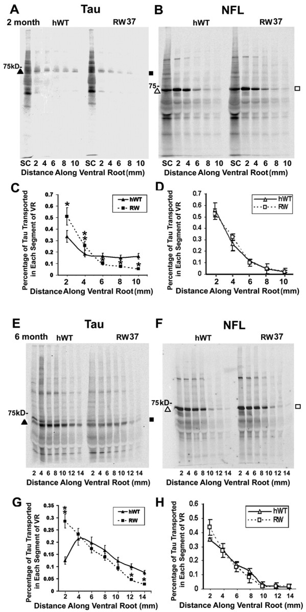
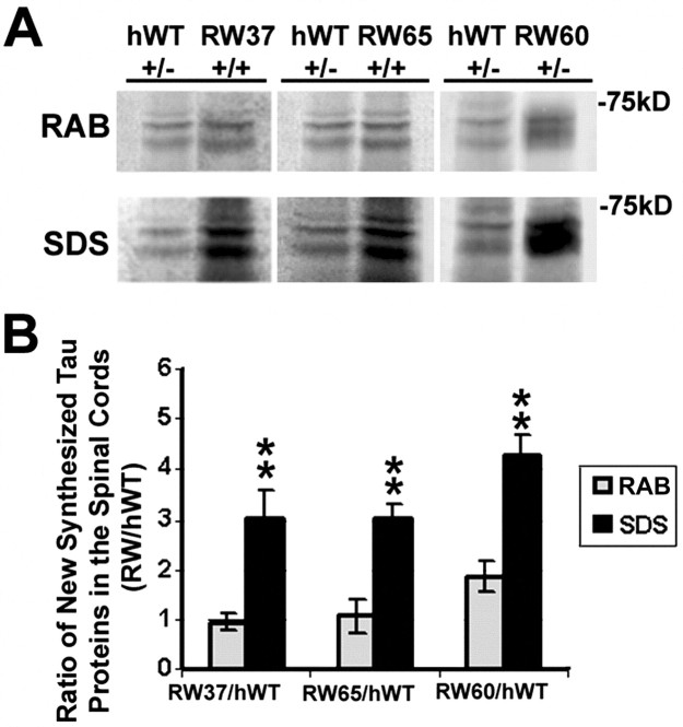
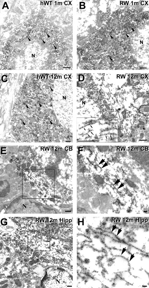
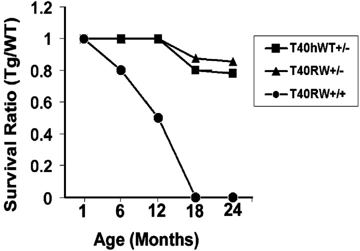
Intracellular accumulations of filamentous tau inclusions are neuropathological hallmarks of neurodegenerative diseases known as tauopathies. The discovery of multiple pathogenic tau gene mutations in many kindreds with familial frontotemporal dementia with parkinsonism linked to chromosome 17 (FTDP-17) unequivocally confirmed the central role of tau abnormalities in the etiology of neurodegenerative disorders. To examine the effects of tau gene mutations and the role of tau abnormalities in neurodegenerative tauopathies, transgenic (Tg) mice were engineered to express the longest human tau isoform (T40) with or without the R406W mutation (RW and hWT Tg mice, respectively) that is pathogenic for FTDP-17 in several kindreds. RW but not hWT tau Tg mice developed an age-dependent accumulation of insoluble filamentous tau aggregates in neuronal perikarya of the cerebral cortex, hippocampus, cerebellum, and spinal cord. Significantly, CNS axons in RW mice contained reduced levels of tau when compared with hWT mice, and this was linked to retarded axonal transport and increased accumulation of an insoluble pool of RW but not hWT tau. Furthermore, RW but not hWT mice demonstrated neurodegeneration and a reduced lifespan. These data indicate that the R406W mutation causes reduced binding of this mutant tau to microtubules, resulting in slower axonal transport. This altered tau function caused by the RW mutation leads to increased accumulation and reduced solubility of RW tau in an age-dependent manner, culminating in the formation of filamentous intraneuronal tau aggregates similar to that observed in tauopathy patients.


Figures

Figure 1.
Figure 1.
RW mutant tau in Tg mice accumulates as an insoluble pool with advancing age and shows reduced binding to MTs. A-C, To compare the levels of hWT and RW tau expression in several lines of Tg mice, Western blot analysis of transgenic human tau and total tau (human plus mouse) levels were estimated using T14, a human-specific anti-tau monoclonal antibody, and 17026, a polyclonal anti-tau antibody that recognizes both human and mouse tau. A, Total tau in brains from the different Tg mice were recovered after direct extraction with 2% SDS buffer and resolved on 7.5% SDS-PAGE gels. Equal amounts of total proteins (60 μg) were loaded for non-Tg (N-Tg) and heterozygous hWT (line 23+/-) and RW (line 60+/- and 65+/+) and homozygous RW tau Tg mice (line 37+/+). Heterozygous hWT and homozygous RW tau Tg mice (line 37) show similar expression levels of tau in the brain and the spinal cord at 6 month of age (A). Although no obvious tau was clearly detected in RW tau Tg line 65 when 60 μg of total CNS proteins were loaded (A), the loading of 10-fold more total proteins (600 μg) resulted in the visualization of a protein band that was recognized by the human tau-specific T14 monoclonal antibody (B). Quantification of the relative tau levels in hWT and all three lines of RW mice are shown in C. To demonstrate the presence of increase level of less soluble tau protein, brains from 1-, 6-, and 12-month-old non-Tg and hWT and RW Tg mice were sequentially extracted using RAB hi-salt buffer, RIPA buffers, and 70% FA (D). Tau protein levels were determined by quantitative Western blotting using the 17026 polyclonal antibody, followed by [125I]Protein A. Human transgenic T40 in the RIPA and FA fractions progressively accumulate in the brains of both RW and hWT tau Tg mice but not in the brains of non-Tg mice. However, the age-related increase of the FA-extractable tau species is larger in the RW mice than the hWT mice (E). **p < 0.01. To compare the binding of hWT and RW tau to endogenous mouse MTs, MT-binding assays were conducted (F). RW mutant tau showed reduced binding to MTs when compared with hWT tau from Tg mice (G). Total tau (T) from hWT and RW mouse brains were used for MT binding assays, and quantification of tau bound to MTs in the pellet (P) versus those that remained in the supernatant (S) is shown in G. n = 3. **p < 0.001.
Figure 2.
Figure 2.
RW tau Tg mice develop somato-dendritic NFT-like tau pathology. A-F, Immunostaining using the polyclonal anti-tau antibody 17026 in the spinal cord (A, B), neocortex (C, D), and cerebellum (E, F) of hWT and RW tau Tg mice at 12 months of age. Neurons in RW tau Tg mice progressively accumulated tau in their somato-dendritic compartment, where as neurons in the hWT tau Tg mice showed little or no perikaryal tau immunoreactivity. G, H, High-power photomicrographs demonstrate intense perikaryal tau immunoreactivity in hippocampal hilar neurons of an 18-month-old RW Tg mouse using the 17026 antibody (G), which resemble the filamentous neuronal tau inclusions in the hippocampus of a patient with FTDP-17 caused by a R406W tau gene mutation (H). Some of the tau-positive neurons in the RW tau Tg mice are thioflavin S positive (I), similar to the NFTs in an R406W FTDP-17 brain (J). Scale bars: A-F, 100 μm; G-J, insets, 10 μm.
Figure 3.
Figure 3.
The slow axonal transport of RW mutant tau is retarded in the ventral roots of RW tau Tg mice. SDS-PAGE shows progressive retardation of the slow axonal transport for tau in the ventral roots (VR) and contiguous spinal nerves (SN) of RW line 37 tau Tg mice at 2 months of age (A, C), at 6 months of age (E, G), and at 12 months of age (I, K) compared with age-matched hWT tau Tg mice, but the slow axonal transport of NFL remains the same in the RW and hWT tau Tg mice at 2 months (B, D), 6 months (F, H), and 12 months of age (J, L). Similar retardation of RW tau transport was also detected in a second RW line, i.e., line 60 at 6 months of age (M, O), with no obvious differences in NFL transport (N, P). tau or NFL protein samples were collected by immunoprecipitation with the anti-tau antibody 17026 (A, C, E, G, I, K, M, O) or the anti-NFL antibody (B, D, F, H, J, L, N, P) 7 d after injection of [35S]methionine into spinal cord (SC). The graphs illustrate quantitative measurements of tau (C, G, K, O) and NFL (D, H, L, P) proteins conveyed by slow axonal transport in pairs of age-matched RW and hWT Tg mice (n = 6 per age group). At all ages studies, slow axonal transport of tau in both lines of RW Tg mice is significantly reduced compared with hWT, but no differences were found in the slow axonal transport of NFL between the RW lines and hWT tau Tg mice. *p < 0.05; **p < 0.01.
Figure 3.
Figure 3.
The slow axonal transport of RW mutant tau is retarded in the ventral roots of RW tau Tg mice. SDS-PAGE shows progressive retardation of the slow axonal transport for tau in the ventral roots (VR) and contiguous spinal nerves (SN) of RW line 37 tau Tg mice at 2 months of age (A, C), at 6 months of age (E, G), and at 12 months of age (I, K) compared with age-matched hWT tau Tg mice, but the slow axonal transport of NFL remains the same in the RW and hWT tau Tg mice at 2 months (B, D), 6 months (F, H), and 12 months of age (J, L). Similar retardation of RW tau transport was also detected in a second RW line, i.e., line 60 at 6 months of age (M, O), with no obvious differences in NFL transport (N, P). tau or NFL protein samples were collected by immunoprecipitation with the anti-tau antibody 17026 (A, C, E, G, I, K, M, O) or the anti-NFL antibody (B, D, F, H, J, L, N, P) 7 d after injection of [35S]methionine into spinal cord (SC). The graphs illustrate quantitative measurements of tau (C, G, K, O) and NFL (D, H, L, P) proteins conveyed by slow axonal transport in pairs of age-matched RW and hWT Tg mice (n = 6 per age group). At all ages studies, slow axonal transport of tau in both lines of RW Tg mice is significantly reduced compared with hWT, but no differences were found in the slow axonal transport of NFL between the RW lines and hWT tau Tg mice. *p < 0.05; **p < 0.01.
Figure 4.
Figure 4.
Nerve ligation of L5 ventral roots in RW mice showed retardation of slow axonal transport of RW mutant tau (A, B). Quantitative Western blot analysis of tau (bottom panels) and NFM (top panels) protein levels in the ligated L5 ventral roots (L) compared with nonligated L5 ventral roots (U) of non-Tg as well as the hWT and RW mice at 2 (A) and 12 (B) months of age. Samples were resolved on 7.5% SDS-PAGE and blotted with the 17026 antibody. Equal volumes of sample (10 μl) were loaded in all lanes. Non-Tg mice showed no detectable signals in the ventral roots (A, B). C, A histogram demonstrates the signal intensity of tau proteins from Western blots in the ligated and nonligated ventral roots of the non-Tg, hWT, and RW Tg mice. D, E, These histograms show the ratio of tau (D) or NFM (E) proteins in the ligated verse nonligated ventral roots. The ratio of tau in RW Tg mice was significantly lower than that in hWT Tg mice at both 2 and 12 months of age (D). In contrast, there was no significant difference in the ratio of NFM among non-Tg and hWT and RW Tg mice at 2 and 12 months of age (E). *p < 0.05; **p < 0.01.
Figure 5.
Figure 5.
Increased accumulation of newly synthesized tau as a less soluble pool in the spinal cords of the RW Tg mice. A, Sequential extraction of tau with RAB hi-salt buffer, followed by 2% SDS 7 d after [35S]methionine injection into ventral horns of the spinal cords of three different RW lines and age-matched hWT mice. Radiolabeled tau was collected by immunoprecipitation with the anti-tau antibody 17026. B, Bar graph shows significant increases in newly synthesized tau proteins in the SDS fractions of ventral horn of the spinal cord in all three lines of RW Tg mice compared with age matched hWT mice. n = 18. **p < 0.01.
Figure 6.
Figure 6.
Intraneuronal tau inclusions in the RW tau Tg mice are composed of straight filaments. A-H, Preembedding immuno-EM using 17026 antibodies show tau immunostaining in neuronal perikarya of the hWT and RW tau Tg mice. Neocortical neurons of a 1-month-old hWT mouse (A), a 1-month-old RW mouse (B), and a 12-month-old hWT mouse (C) demonstrate diffuse tau staining (arrowheads) but no obvious tau filaments. D, Randomly oriented tau-positive straight filaments are demonstrated in the perikaryon of a neocortical neuron of a 12-month-old RW tau Tg mouse, and a higher-power view of these filaments (small arrows) is shown in the inset. E-H, Preembedding immuno-EM using the 17026 antibody shows tau-immunoreactive straight filaments in neurons from several CNS areas of a 12-month-old RW mouse. Higher-power views of the tau filaments (large arrows in F, H) are from the boxed areas in the adjacent panels showing lower-power views (E, G). The tau-immunoreactive filaments were found in the cerebellum (E, F) and hippocampus (G, H). N, Nucleus. Scale bars: A-E, G, 500 nm; F, H, 100 nm.
Figure 7.
Figure 7.
RW tau Tg mice show reduced longevity. Longevity of hWT and RWL37 tau Tg mice was studied in cohorts of pups weaned at 1 month of age, and survival ratio of mice in each cohort was assessed at 6, 12, 18, and 24 months of age (n = 20 mice per age group). Approximately 90 and 55% of hWT+/- and RWL37+/- tau Tg mice survived up to 24 months of age, respectively, and 40% of RWL37+/+ tau Tg mice died by 12 months of age.

Similar articles

Cited by

References

    1. Barghorn S, Zheng-Fischhofer Q, Ackmann M, Biernat J, von Bergen M, Mandelkow E, Mandelkow E (2000) Structure, microtubule interactions, and paired helical filament aggregation by tau mutants of frontotemporal dementias. Biochemistry 39: 11714-11721. - PubMed
    1. Bugiani O, Murrell JR, Giaccone G, Hasegawa M, Ghigo G, Tabaton M, Morbin M, Primavera A, Carella F, Solaro C, Grisoli M, Savoiardo MG, Spillantini F, Tagliavini M, Goedert M, Ghetti B (1999) Frontotemporal dementia and corticobasal degeneration in a family with a P301S mutation in tau. J Neuropathol Exp Neurol 58: 667-677. - PubMed
    1. Clark LN, Poorkaj P, Wszolek Z, Geschwind DH, Nasreddine ZS, Miller B, Li D, Payami H, Awert F, Markopoulou K, Andreadis A, D'Souza I, Lee VM-Y, Reed L, Trojanowski JQ, Zhukareva V, Bird T, Schellenberg G, Wilhelmsen KC (1998) Pathogenic implications of mutations in the tau gene in pallidopontonigral degeneration and related neurodegenerative disorders linked to chromosome 17. Proc Natl Acad Sci USA 95: 13103-13107. - PMC - PubMed
    1. Connell JW, Gibb GM, Betts JC, Blackstock JC, Gallo J, Lovestone S, Hutton M, Anderton BH (2001) Effects of FTDP-17 mutations on the in vitro phosphorylation of tau by glycogen synthase kinase 3β identified by mass spectrometry demonstrate certain mutations exert long-range conformational changes. FEBS Lett 493: 40-44. - PubMed
    1. Crowther RA, Goedert M (2000) Abnormal tau-containing filaments in neurodegenerative diseases. J Struct Biol 130: 271-279. - PubMed

Publication types

MeSH terms