. 2004 May 12;24(19):4668-82.
doi: 10.1523/JNEUROSCI.0580-04.2004.

The function of neurotrophic factor receptors expressed by the developing adductor motor pool in vivo

Affiliations

The function of neurotrophic factor receptors expressed by the developing adductor motor pool in vivo

Thomas W Gould et al. J Neurosci. .

Abstract

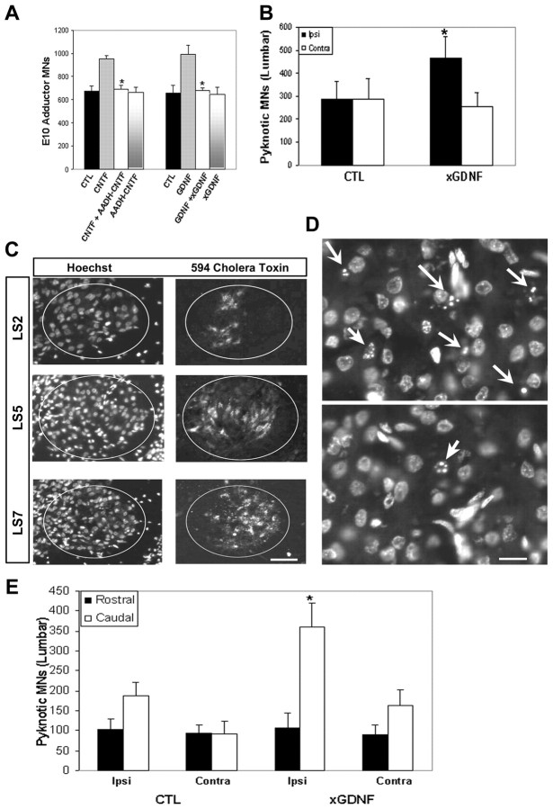
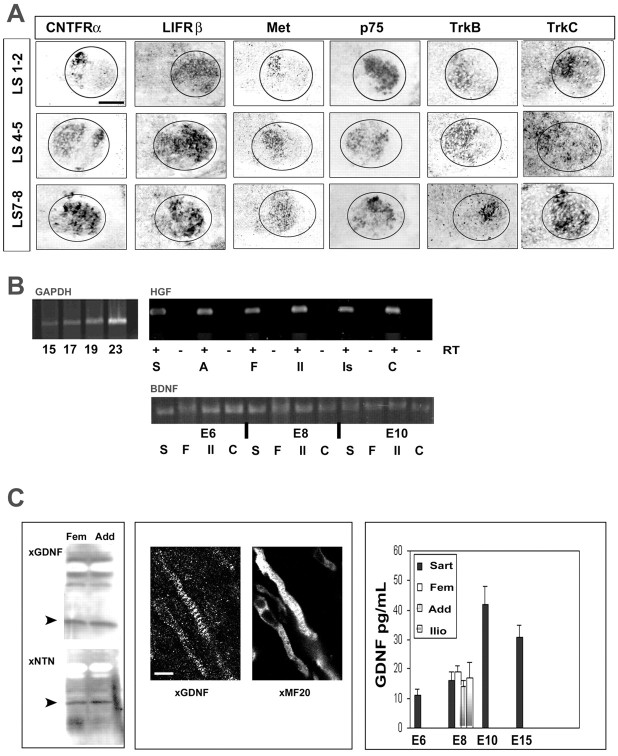
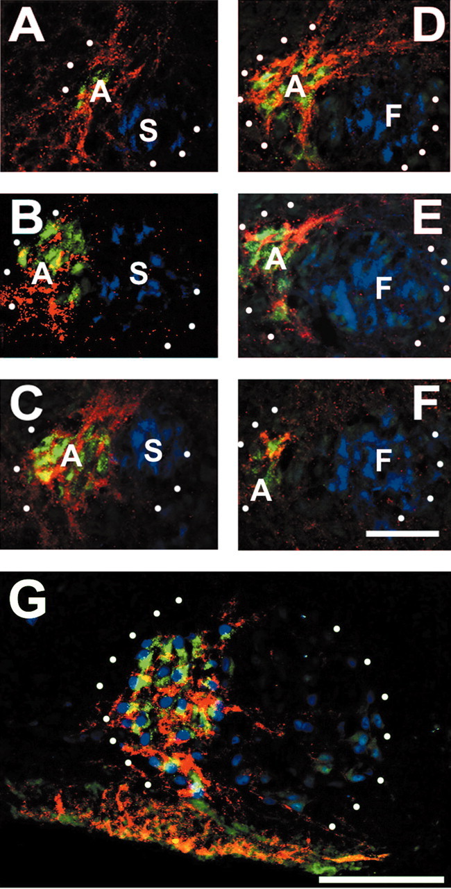
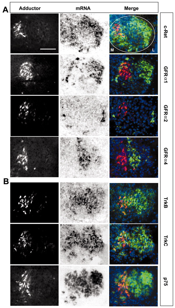
We examined the spatio-temporal relationship between neurotrophic factor receptor (NTF-R) expression and motoneuron (MN) survival in the developing avian spinal cord and observed heterogeneity in the expression of NTF-Rs between, but not within, pools of MNs projecting to individual muscles. We then focused on the role of NTFs in regulating the survival of one motor pool of MNs, all of which innervate a pair of adductor muscles in the thigh and hence compete for survival during the period of programmed cell death (PCD). The complete NTF-R complement of these MNs was analyzed and found to include many, but not all, NTF-Rs. Treatment with exogenous individual NTFs rescued some, but not all, adductor MNs expressing appropriate NTF-Rs. In contrast, administration of multiple NTFs completely rescued adductor MNs from PCD. Additionally, adductor MNs were partially rescued from PCD by NTFs for which they failed to express receptors. NTF-Rs expressed by the nerve but not in the muscle target were capable of mediating survival signals to MNs in trans. Finally, the expression of some NTF-Rs by adductor MNs was not required for MN survival. These studies demonstrate the complexity in NTF regulation of a defined subset of competing MNs and suggest that properties other than NTF-R expression itself can play a role in mediating trophic responses to NTFs.


Figures

Figure 7.
Figure 7.
Neither CNTF nor GDNF signaling is necessary for adductor MN survival during the period of PCD. A, Embryos were treated with control vehicle (CTL), CNTF (5 μg, E6-E9), AADH-CNTF (100 μg, E6-E9), AADH-CNTF together with CNTF, GDNF (5 μg, E6-E9), neutralizing antibodies against human GDNF (anti-GDNF, 20 μg, E6 and E8), or anti-GDNF together with GDNF, and adductor MNs were retrogradely labeled at E10. Data in A, B, and E represent the means ± SD; n = 4 per group. *p < 0.05 for AADH-CNTF + CNTF versus CNTF alone, or for anti-GDNF + GDNF versus GDNF alone; Student's t test. B, GDNF signaling is required for lumbar MN survival. Unilateral injections of 5 μg/μl anti-GDNF (also containing 0.5 μg/μl Alexa-Fluor 594-conjugated cholera toxin β) were made into the hindlimb at E7, and pyknotic MNs were counted on Nissl-stained sections throughout the entire lumbar spinal cord at E8. Data represent the means ± SD; n = 4 for anti-GDNF, and n = 10 for control, which included 5 μg/μl IgG-treated, sham-operated, and windowed-only embryos. *p < 0.02 for ipsilateral anti-GDNF-injected versus ipsilateral control; Student's t test. C, The distribution of lumbar MNs exposed to anti-GDNF injections made into the hindlimb at E7 was measured by examining transverse sections through the lumbar spinal cord at E8 for Alexa-Fluor 594-conjugated cholera toxin β-positive cells. The ovals represent the LMC; medial is to the left. The adductor motor nucleus resides medially from lumbar (LS) 1-3, which is retrogradely labeled after anti-GDNF hindlimb injection (top right). D, Hindlimb anti-GDNF injection (top) increases the number of dying lumbar MNs (white arrows) versus control in Hoechst-labeled transverse sections. E, Hindlimb anti-GDNF injection increases dying lumbar MNs only in the caudal half of the lumbar spinal cord. Counts of pyknotic MNs generated from B were subdivided into rostral and caudal halves of the lumbar spinal cord. The caudal, but not rostral, lumbar spinal cord ipsilateral to the injection of anti-GDNF (ipsi) exhibits an increase in pyknotic MNs versus control ipsilateral caudal and rostral lumbar spinal cord, respectively. *p < 0.01; Student's t test. Scale bars: C, 50 μm; D, 20 μm.
Figure 1.
Figure 1.
Heterogeneous expression of NTF-R mRNAs in the lumbar spinal cord versus homogeneous expression of NTFs in hindlimb muscles at E8, the peak of MN PCD. A, Transverse sections of three different rostrocaudal levels of the lumbar spinal cord reveal spatial differences in the expression of different NTF-RmRNAs. Ellipses encircle the LMC. Medial is to the left, and lateral to the right. Scale bar, 100 μm. B, RNA was extracted from individual muscles at E6, E8, or E10; reverse transcribed; and subjected to semiquantitative PCR with primers designed against chick BDNF, HGF, and GAPDH. The intensity of the GAPDH PCR product increases linearly between 15 and 23 cycles (left). Similar amounts of HGF PCR product are present in six hindlimb muscles at E8 (top right). Equal amounts of BDNF PCR product are present in four hindlimb muscles at E6, E8, and E10 (bottom right). S, Sartorius; A, adductor; F, femerotibialis; Il, iliofibularis; Is, ischioflexorius; C, caudilioflexorius; RT, reverse transcriptase. C, Left, Immunoblot analysis reveals similar amounts of GDNF and NTN protein in the adductor and femerotibialis hindlimb muscles at E8. Molecular weight of GDNF and NTN are 19 and 25 kDa, respectively (arrowheads). Middle, Immunohistochemical detection of GDNF (left) in muscle cells identified by sarcomeric myosin (MF20) immunoreactivity (right). Scale bar, 5 μm. Right, GDNF protein quantitation by ELISA demonstrates equivalent amounts of GDNF in various hindlimb muscles at E8, the peak of MN PCD.
Figure 3.
Figure 3.
Virtually all MNs within the adductor motor pool express CNTFRα. Adductor MNs were retrogradely labeled at E8 with HRP, and both sartorius and femerotibialis MNs were also retrogradely labeled at E8 with Alexa-Fluor 488-conjugated β-subunit cholera toxin (blue channel). Sections were immunolabeled with Cy2- and Cy5-conjugated antibodies against HRP (green) and CNTFRα (red). The images in A-F are every 15th 10 μm section, proceeding from the rostralmost (LS1) to the caudalmost (LS3) portion of the adductor pool. All adductor MNs (green), but neither sartorius nor femerotibialis MNs (blue), express CNTFRα protein (red). A, Adductor; S, sartorius; F, femerotibialis. G, Islet1/2-positive MNs (blue) in LS2 in the adductor motor pool (green) are immunoreactive for CNTFRα (red). The white dots in indicate the LMC. Scale bars: A-F, 50 μm; G, 75 μm.
Figure 2.
Figure 2.
NTF-R expression by the adductor motor nucleus. The adductor muscles of the chick were injected with Alexa-Fluor 594-conjugated β-subunit cholera toxin, and retrogradely labeled MNs were identified and imaged on fresh-frozen spinal cord transverse sections (left). Sections were then processed for in situ hybridization analysis of specific NTF-Rs (middle). Images could be compared with the aid of Hoechst-labeled nuclei (blue in right column), which were captured before and after in situ hybridization. A, The adductor motor pool expresses the GDNF family receptor cRet and GFRα1 but not GFRα2 or GFRα4. B, All MNs within the adductor motor pool express the NTF-Rs TrkB, TrkC, and p75. C, All adductor MNs express the neuropoietic cytokine receptors LIFRβ, gp130, and CNTFRα and the HGF receptor cMet. D, Lateral LMC MNs fail to express GFRα1. The medial adductor from an E8 embryo was surgically removed, and the lateral adductor was retrogradely labeled. The top row shows GFRαl mRNA expression in the medial portion of the LMC, where medial adductor MNs are usually detected. The bottom row shows that GFRαl expression fails to colocalize with the more laterally situated lateral adductor MNs. Large dots indicate the LMC, whereas small dots in the bottom row indicate lateral adductor MNs. Medial is to the left, and lateral to the right. Scale bar (in A), 50 μm.
Figure 2.
Figure 2.
NTF-R expression by the adductor motor nucleus. The adductor muscles of the chick were injected with Alexa-Fluor 594-conjugated β-subunit cholera toxin, and retrogradely labeled MNs were identified and imaged on fresh-frozen spinal cord transverse sections (left). Sections were then processed for in situ hybridization analysis of specific NTF-Rs (middle). Images could be compared with the aid of Hoechst-labeled nuclei (blue in right column), which were captured before and after in situ hybridization. A, The adductor motor pool expresses the GDNF family receptor cRet and GFRα1 but not GFRα2 or GFRα4. B, All MNs within the adductor motor pool express the NTF-Rs TrkB, TrkC, and p75. C, All adductor MNs express the neuropoietic cytokine receptors LIFRβ, gp130, and CNTFRα and the HGF receptor cMet. D, Lateral LMC MNs fail to express GFRα1. The medial adductor from an E8 embryo was surgically removed, and the lateral adductor was retrogradely labeled. The top row shows GFRαl mRNA expression in the medial portion of the LMC, where medial adductor MNs are usually detected. The bottom row shows that GFRαl expression fails to colocalize with the more laterally situated lateral adductor MNs. Large dots indicate the LMC, whereas small dots in the bottom row indicate lateral adductor MNs. Medial is to the left, and lateral to the right. Scale bar (in A), 50 μm.
Figure 5.
Figure 5.
NTF-R expression by cells in the peripheral nerve mediates survival signaling in trans to MNs in vivo. A, In ovo treatment with 5 μg of NTN or PSP from E6 to E9 rescues adductor MNs, which fail to express either GFRα2 and GFRα4, from PCD. B, GFRα2 and GFRα4, but not GFRα1 or cRet, are expressed by putative Schwann cells (SCs) in the peripheral nerve. DRG, Dorsal root ganglion; SG, sympathetic ganglion. Scale bar, 250 μm. C, D, MN-derived expression of cRet is required for the rescue effects of GDNF family ligands. C, In ovo treatment with 5 μg of CNTF but not GDNF, PSP, or NTN from E6 to E9 rescues MNs innervating the medial gastrocnemius (MG). D, MG-innervating MNs retrogradely labeled at E8 with Alexa-Fluor 594-conjugated β-subunit cholera toxin (left) express CNTFRα and GFRα1 mRNA (middle) but not cRet mRNA. Hoechst-stained nuclei are in the right panel. Scale bar, 50 μm. Data represent the means ± SD; n = 3 for all conditions. *p < 0.05 versus control; Student's t test.
Figure 4.
Figure 4.
In ovo treatment with multiple, but not individual, NTFs rescues all adductor MNs from PCD. A, Adductor muscles were injected with HRP at different times during the period of MN PCD (E6-E10). MNs innervating the adductor are reduced by about one-half during the normal period of PCD. B, Treatment with individual NTFs (5 μg daily from E6 to E9) partially rescues adductor MNs from PCD, whereas combined treatment with BDNF, GDNF, CNTF, and HGF (B/G/C/H) completely prevents adductor MN PCD (compare BCGH with E6.5 in A). Data represent the mean ± SD; n = 5 for all conditions. *p < 0.05 versus control; **p < 0.01 versus control and versus single-factor, Student's t test with Bonferroni corrections. C, Representative images of cross sections at E10 of the adductor motor pool in embryos treated from E6 to E9 with multiple NTFs (top four images) and in control embryos (bottom four images). The right column contains the same images as the left column at higher magnification. Retrogradely labeled MNs were quantified by counting the number of HRP (green) and islet1/2 (red)-immunoreactive nuclei (blue). The white dots in the left column denote the medial and lateral borders of the LMC. The white arrows in the right column depict the individual countable adductor MNs in these two conditions, the totals of which are presented in the bottom right corner for each condition. The top and bottom rows of each treatment condition are the same images at different focal planes to illustrate all of the adductor MNs within the 10 μm section. D, Differences in subcellular patterns of immunoreactivity for specific NTF-Rs (red) at E8, counter-stained with Hoechst (blue). The LMC is enclosed with dots. Medial is to the left, and lateral (L) to the right. Hoechst counterstain is omitted for TrkB to better visualize immunoreactivity. Scale bar: C, D, 50 μm.
Figure 6.
Figure 6.
NTF-R expression by non-muscle cells in the limb does not mediate survival signaling in trans to MNs in vivo. A, CNTFRα immunoreactivity (top; red in bottom panel) is detected in transverse sections of the chick hindlimb at E8 but does not colocalize with sarcomeric myosin (MF20) immunoreactivity (middle; green in bottom panel). B, CNTFRα immunoreactivity (top; red in bottom panel) colocalizes with fibronectin immunoreactivity (middle; green in bottom panel) in the chick hindlimb at E8. C, CNTFRα mRNA (top; green in bottom panel) is expressed by non-muscle cells in the chick hindlimb at E8, both within the muscle (arrowheads) and in the connective tissue surrounding the muscle (arrow), but does not colocalize with sarcomeric myosin (MF20) immunoreactivity (middle; red in bottom panel). Hoechst-stained nuclei are blue in the bottom panel in A-C. Scale bars: A-C, 50 μm. D, In ovo treatment with 5 μg of CNTF from E6 to E9 rescues adductor but not sartorius MNs from PCD. In contrast, treatment with CNTF and soluble CNTFRα rescues sartorius MNs. Data represent the means ± SD; n = 4 for all conditions. *p < 0.05 versus control; Student's t test. E, Diagram of constructs used to overexpress chicken CNTFRα in MNs during the period of PCD. Two plasmids, one containing the tetracycline repressor protein (TetR) cDNA driven by the cytomegalovirus (CMV) promoter, the other containing chicken CNTFRα and GFP cDNAs expressed as a single bicistronic transcript under the control of the tetracycline repressor response element (TRE), were electroporated at E3 into the chicken neural tube. Doxycycline (100 mg) was added at E3 and E4 to initiate gene expression, and the embryo was killed at E4.5 and examined for CNTFRα mRNA (left) or protein (right). Note the expansion of CNTFRα mRNA and protein to the electroporated half of the spinal cord (ipsi), compared with the restriction of CNTFRα mRNA to MNs (LMC) and CNTFRα protein to the dorsal roots (arrowhead) in the control half (contra). The arrowhead in the right panel indicates ectopic CNTFRα immunoreactivity exhibited by crossing commissural interneuronal fibers. Scale bar, 200 μm. F, Expression of CNTFRα at E10 in the rostral lumbar spinal cord after electroporation of chicken CNTFRα constructs at E3 and doxycycline treatment from E6 to E9. Ectopic CNTFRα-positive MNs, detected by examining GFP expression (green in the middle and right panels), are observed lateral to their usual adductor motor pool domain (red in the left and right panels). The arrowheads indicate CNTFRα-overexpressing MNs within the adductor pool, and arrows indicate ectopic CNTFRα-overexpressing MNs in presumptive sartorius and femerotibialis MNs. The white ellipse in the right panel encloses the LMC; medial is to the right. Hoechst-stained nuclei are blue in the right panel. Scale bar, 50 μm.

Similar articles

Cited by

References

    1. Airaksinen MS, Saarma M (2002) The GDNF family: signaling, biological functions and therapeutic value. Nat Rev Neurosci 3: 383-394. - PubMed
    1. Alexander WS, Rakar S, Robb L, Farley A, Willson TA, Zhang JG, Hartley L, Kikuchi Y, Kojima T, Nomura H, Hasegawa M, Maeda M, Fabri L, Jachno K, Nash A, Metcalf D, Nicola NA, Hilton DJ (1999) Suckling defect in mice lacking the soluble haemopoietin receptor NR6. Curr Biol 9: 605-608. - PubMed
    1. Arce V, Pollock RA, Philippe JM, Pennica D, Henderson CE, deLapeyrière O (1998) Synergistic effects of Schwann- and muscle-derived factors on motoneuron survival involve GDNF and cardiotrophin-1 (CT-1). J Neurosci 18: 1440-1448. - PMC - PubMed
    1. Baloh RH, Tansey MG, Golden JP, Creedon DJ, Heuckeroth RO, Keck CL, Zimonjic DB, Popescu NC, Johnson Jr EM, Milbrandt J (1997) TrnR2, a novel receptor that mediates neurturin and GDNF signaling through Ret. Neuron 18: 793-802. - PubMed
    1. Bottaro DP, Rubin JS, Faletto DL, Chan AM, Kmiecik TE, Vande Woude GF, Aaronson SA (1991) Identification of the hepatocyte growth factor receptor as the c-met proto-oncogene product. Science 251: 802-804. - PubMed

Publication types

MeSH terms

Substances