. 2004 May 12;24(19):4698-708.
doi: 10.1523/JNEUROSCI.5523-03.2004.

Ca2+-binding protein-1 facilitates and forms a postsynaptic complex with Cav1.2 (L-type) Ca2+ channels

Affiliations

Ca2+-binding protein-1 facilitates and forms a postsynaptic complex with Cav1.2 (L-type) Ca2+ channels

Hong Zhou et al. J Neurosci. .

Abstract

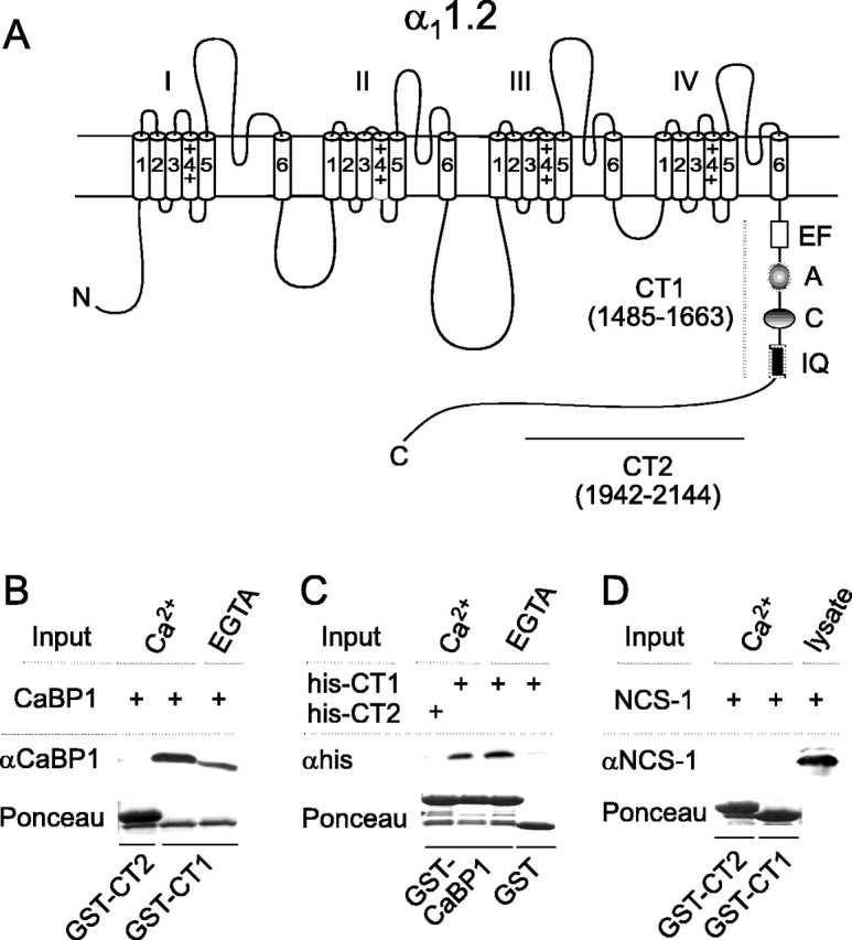
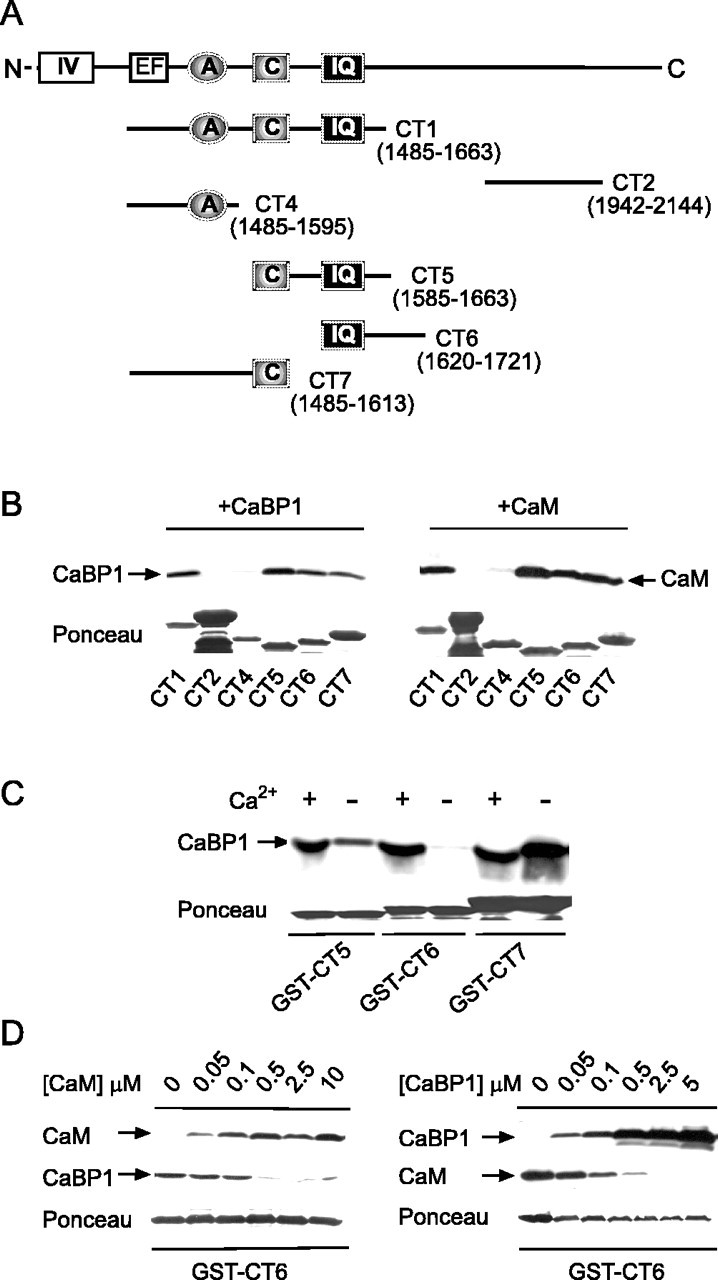
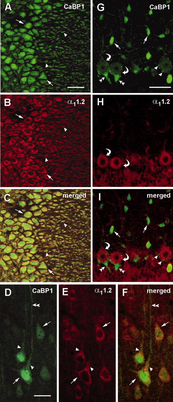
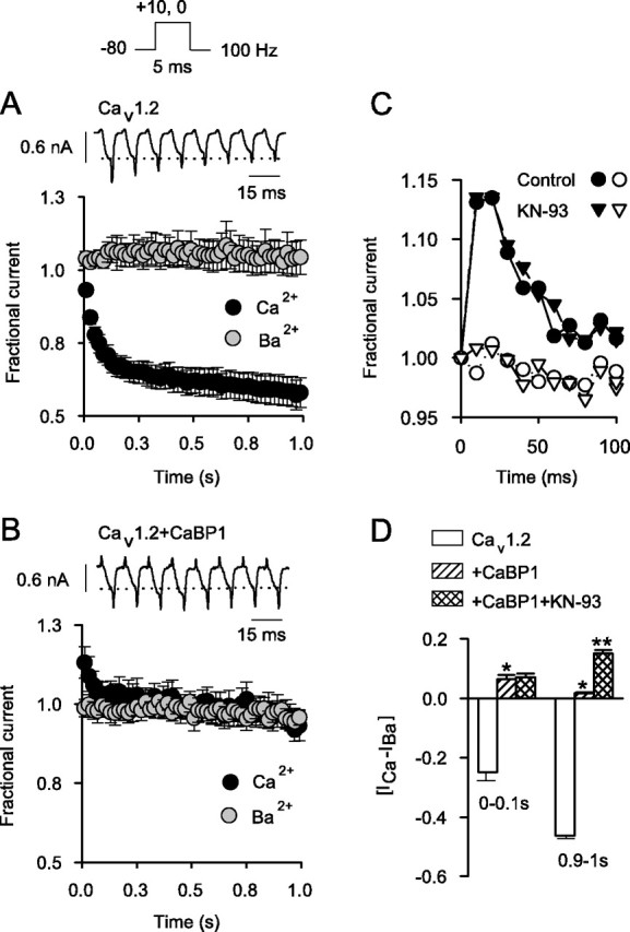
Ca2+-binding protein-1 (CaBP1) is a Ca2+-binding protein that is closely related to calmodulin (CaM) and localized in somatodendritic regions of principal neurons throughout the brain, but how CaBP1 participates in postsynaptic Ca2+ signaling is not known. Here, we describe a novel role for CaBP1 in the regulation of Ca2+ influx through Ca(v)1.2 (L-type) Ca2+ channels. CaBP1 interacts directly with the alpha1 subunit of Ca(v)1.2 at sites that also bind CaM. CaBP1 binding to one of these sites, the IQ domain, is Ca2+ dependent and competitive with CaM binding. The physiological significance of this interaction is supported by the association of Ca(v)1.2 and CaBP1 in postsynaptic density fractions purified from rat brain. Moreover, in double-label immunofluorescence experiments, CaBP1 and Ca(v)1.2 colocalize in numerous cell bodies and dendrites of neurons, particularly in pyramidal cells in the CA3 region of the hippocampus and in the dorsal cortex. In electrophysiological recordings of cells transfected with Ca(v)1.2, CaBP1 greatly prolonged Ca2+ currents, prevented Ca2+-dependent inactivation, and caused Ca2+-dependent facilitation of currents evoked by step depolarizations and repetitive stimuli. These effects contrast with those of CaM, which promoted strong Ca2+-dependent inactivation of Ca(v)1.2 with these same voltage protocols. Our findings reveal how Ca2+-binding proteins, such as CaM and CaBP1, differentially adjust Ca2+ influx through Ca(v)1.2 channels, which may specify diverse modes of Ca2+ signaling in neurons.


Figures

Figure 1.
Figure 1.
CaBP1 interacts with the C-terminal domain of the α1 subunit of Cav1.2. A, Schematic of α11.2 with C-terminal domain showing CT1 and CT2 regions used for GST fusion constructs in binding experiments. Amino acid boundaries are indicated in parentheses. CT1 included sequences involved in Ca2+-dependent inactivation of Cav1.2: the EF-hand (EF) and CaM-interacting sites A, C, and the IQ domain (IQ). N, N terminus of α11.2. B, Binding of CaBP1 to CT1 but not CT2. GST-CT1 or CT2 fusion proteins were immobilized on glutathione-agarose beads and incubated with lysates from cells transfected with CaBP1 in the presence of 2 mm Ca2+ or 10 mm EGTA. Bound CaBP1 was detected by Western blot. Ponceau staining of the blot indicated levels of GST fusion protein used in each binding assay. C, Binding of His-tagged CT1 to GST-tagged CaBP1. GST or GST-CaBP1 was immobilized on glutathione-agarose beads and in cubated with purified His-CT1 or His-CT2. Bound fusion proteins were detected by Western blot with anti-His antibodies. D, NCS-1 does not bind to CT1. Binding assay was performed as in B, except that lysates from cells transfected with NCS-1 were used and immunoblotting was done with rabbit polyclonal antibodies against NCS-1. NCS-1 was detected in transfected cell lysates (last lane) but not in samples pulled down by GST-CT1 or CT2.
Figure 2.
Figure 2.
CaBP1 binds to putative CaM-binding sites in the C-terminal domain of α11.2. A, Schematic of C-terminal domain of α11.2 showing regions tested for binding CaM or CaBP1 in binding assays. B, Binding of CaM and CaBP1 to multiple sites in α11.2. GST-tagged α11.2 fragments shown in A were incubated with CaBP1 from transfected cells (left) or purified CaM (right) in the presence of 2 mm Ca2+. Bound proteins were detected by Western blotting with specific antibodies. Integrity and levels of GST-tagged proteins were confirmed by Ponceau staining. C, Ca2+-dependent and -independent binding of CaBP1. Binding of CaBP1 to GST-CT5, GST-CT6, or GST-CT7 was performed as in B and compared when assay was done with 2 mm Ca2+ (+) or 10 mm EGTA (-). D, Competitive binding of CaM and CaBP1 to CT6. Binding of CaBP1 or CaM to GST-CT6 was performed as in B with 2 mm Ca2+ and compared in the presence of increasing concentrations of purified CaM (left) or CaBP1 (right).
Figure 3.
Figure 3.
Coimmunoprecipitation of CaBP1 and α11.2 from transfected cells and rat brain. A, Cells transfected with Cav1.2 (α11.2-FLAG, β2A, and α2δ), Cav1.2 plus CaBP1, or CaBP1 alone were subject to lysis and immunoprecipitation using anti-FLAG antibodies. Experiments were done in the presence of 2 mm Ca2+ or 10 mm EGTA. Immunoprecipitated proteins were detected by Western blotting with antibodies recognizing the FLAG epitope (top) or CaBP1 (bottom). B, Immunopurification of a complex containing both α11.2 and CaBP1 from PSD fractions of rat brain. PSD proteins were isolated on sucrose gradients, solubilized, and incubated with α11.2 antibodies. Immunoprecipitated proteins were detected by Western blotting with antibodies against α11.2 (top) or CaBP1 (bottom). Immunoblotting of the PSD fraction was also performed with antibodies against PSD-95 (right).
Figure 4.
Figure 4.
Colocalization of CaBP1 and α11.2 in rat brain. A-I, Confocal images of rat brain sections sequentially double labeled with antibodies against CaBP1 and α11.2. Immunofluorescence was viewed under optics for fluorescein(for CaBP1; A,D,G) or rhodamine (for α11.2;B, E, H). Regions of colocalization appear yellow in the merged images (C, F, I). Extensive colocalization of CaBP1 and α11.2 was detected in cell bodies (arrows) and dendrites (arrowheads) of neurons in the CA3 region of the hippocampus (A-C). In layer V of the cerebral cortex (D-F), CaBP1 and α11.2 colocalized in pyramidal cell soma (arrows) and proximal dendrites (arrowheads) but not more distal dendrites (double arrowheads). In the cerebellum (G-I), CaBP1 and α11.2 were colocalized in Purkinje cell bodies (curved arrows), but only CaBP1 was found in interneurons in the molecular layer (arrows) and in small perisomatic synapses (arrowheads) and large Pinceaux synapses (double arrowheads) onto Purkinje cell bodies. Scale bars: A-C, G-I, 100 μm; D-F, 50 μm.
Figure 5.
Figure 5.
CaBP1 prolongs Ca2+ currents through Cav1.2 channels and does not support Ca2+-dependent inactivation. A, Effect of CaBP1 on Cav1.2 Ca2+ currents evoked by step depolarizations. Traces represent Ca2+ currents evoked by a 1 sec step from -80 to +10 mV in whole-cell patch-clamp recordings of HEK293T cells transfected with Cav1.2 subunits with or without CaBP1. Ires /Ipk was determined by dividing the residual current amplitude at the end of the pulse by the peak current amplitude. Extracellular solution contained 10 mm Ca2+, and intracellular solution contained 5 mm EGTA. Data represent mean ± SEM for Cav1.2 (n = 17) and Cav1.2 plus CaBP1 (n = 11) (*p < 0.001). B, Impact of CaBP1 on fast and slow inactivation. Ca2+ currents obtained in A were fit with a double-exponential function. The fast and slow time constants (τfastslow) and fraction of channels showing fast and slow components of inactivation (Ffast, Fslow) were averaged and shown for cells transfected with Cav1.2 alone or Cav1.2 plus CaBP1 (*p < 0.001). C, Comparison of Ca2+-dependent inactivation in cells transfected with Cav1.2 alone or cotransfected with CaBP1. Traces show currents evoked by 1 sec depolarizing step from -80 to +10 mV for ICa (black trace) or 0 mV for IBa (gray trace) to compensate for -10 mV voltage shift for IBa. Extracellular solution contained either 10 mm Ca2+ or Ba2+.[ICa - IBa] represents the difference between Ires /Ipk for ICa and the mean Ires /Ipk for IBa for Cav1.2 channels alone (n = 17; open bar) or Cav1.2 plus CaBP1 (n = 11; hatched bar) (*p < 0.001). D, U-Shaped voltage dependence of Ires /Ipk for ICa in cells transfected with Cav1.2 alone but not in cells cotransfected with CaBP1. Ires /Ipk was determined for ICa evoked by 1 sec test pulses from -80 mV to various voltages, averaged, and plotted against test voltage for Cav1.2 alone (open circles; n = 9) or Cav1.2 plus CaBP1 (filled circles; n = 8).
Figure 6.
Figure 6.
CaBP1 blocks Ca2+-dependent inactivation and causes facilitation during repetitive stimuli. A, Ca2+-dependent inactivation of Cav1.2 during trains of depolarizations. ICa and IBa were evoked by 5 msec test pulses from -80 to +10 mV for ICa or 0 mV for IBa as shown in voltage protocol above. Shown are representative traces of ICa during the first eight pulses. Dotted line indicates initial current amplitude. Fractional current, plotted below, represents test current amplitude normalized to that for the first pulse in the train and plotted against time for ICa(black circles; n = 11) and IBa (gray circles; n=5). Points represent the mean ± SEM, and every other point is plotted. B, Loss of Ca2+-dependent inactivation but gain of ICa facilitation with CaBP1. Current traces (top) and fractional current (bottom) were obtained as in A, except that recordings were from cells cotransfected with CaBP1 (n = 5 for ICa; n = 4 for IBa). C, Ca2+-dependent facilitation in cells cotransfected with CaBP1 is not prevented by KN-93. Facilitation of ICa (filled symbols) but not IBa (open symbols) is shown for cells cotransfected with Cav1.2 plus CaBP1 recorded with control intracellular solution (circles) or that containing the CaM kinase II inhibitor KN-93 (2 μm; triangles). Results were obtained as in A; and data from single representative cells were plotted for the first 100 msec of the train. D, Effects of CaBP1 on Ca2+-dependent modulation of Cav1.2. [ICa - IBa] represents the difference between the average fractional current for ICa and IBa for the first 10 (0-0.1 sec) or last 10 (0.9-1 sec) pulses. Shown are results obtained from cells transfected with Cav1.2 alone (n = 11), Cav1.2 plus CaBP1 (n = 5), and cells cotransfected with Cav1.2 plus CaBP1 that were treated with KN-93 (n = 5) (*p < 0.05 compared with Cav1.2 alone; **p < 0.01 compared with Cav1.2 plus CaBP1).
Figure 7.
Figure 7.
Essential role for the IQ in CaBP1 binding and modulation of Cav1.2 during repetitive stimuli. A, Effect of alanine substitutions in the IQ on binding of CaBP1 and modulation of Cav1.2. Top panels show CaBP1 binding to GST-tagged fragments containing the IQ with or without IQ-AA substitutions (CT6, CT6IQ-AA). Binding was done with 2 mm Ca2+ as described in Figure 2B. Bottom panel shows pattern of ICa evoked by repetitive stimuli in cells transfected with Cav1.2IQ-AA alone (open circles) or cotransfected with CaBP1 (filled circles). Data were obtained with voltage protocols and plotted as in Figure 6. Points represent mean ± SEM (n = 4-6), with every second point plotted. B, Fractional current obtained in A was averaged for the first 10 (0-0.1 sec) and last 10 (0.9-1 sec) pulses in the train and shown for Cav1.2IQ-AA transfected alone (open bars) or cotransfected with CaBP1 (filled bars) (*p < 0.005 compared with Cav1.2IQ-AA). C, IQ to EE substitutions prevent binding and inhibit functional effects of CaBP1. Top panel shows loss of CaBP1 binding to GST-tagged CT6 with IQ-EE substitutions. Bottom panel shows ICa, obtained as in A, in cells transfected with Cav1.2 channels containing the IQ-EE mutation alone (n = 5; open circles) or cotransfected with CaBP1 (n = 6; filled circles). D, Fractional current for data plotted in C was averaged as in B for Cav 1.2IQ-EE alone (open bars) or Cav1.2IQ-EE plus CaBP1 (filled bars) (* p < 0.05 compared with Cav1.2IQ-EE).

Similar articles

Cited by

References

    1. Avery RB, Johnston D (1996) Multiple channel types contribute to the low-voltage-activated calcium current in hippocampal CA3 pyramidal neurons. J Neurosci 16: 5567-5582. - PMC - PubMed
    1. Bading H, Ginty DD, Greenberg ME (1993) Regulation of gene expression in hippocampal neurons by distinct calcium signaling pathways. Science 260: 181-186. - PubMed
    1. Beck H, Steffens R, Heinemann U, Elger CE (1997) Properties of voltage-activated Ca2+ currents in acutely isolated human hippocampal granule cells. J Neurophysiol 77: 1526-1537. - PubMed
    1. Brehm P, Eckert R, Tillotson D (1980) Calcium-mediated inactivation of calcium current in Paramecium. J Physiol (Lond) 306: 193-203. - PMC - PubMed
    1. Brown AM, Schwindt PC, Crill WE (1993) Voltage dependence and activation kinetics of pharmacologically defined components of the high-threshold calcium current in rat neocortical neurons. J Neurophysiol 70: 1530-1543. - PubMed

Publication types

MeSH terms