. 2004 Jun;78(11):5633-41.
doi: 10.1128/JVI.78.11.5633-5641.2004.

Nipah virus V and W proteins have a common STAT1-binding domain yet inhibit STAT1 activation from the cytoplasmic and nuclear compartments, respectively

Affiliations

Nipah virus V and W proteins have a common STAT1-binding domain yet inhibit STAT1 activation from the cytoplasmic and nuclear compartments, respectively

Megan L Shaw et al. J Virol. 2004 Jun.

Abstract

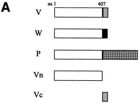
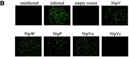
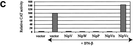
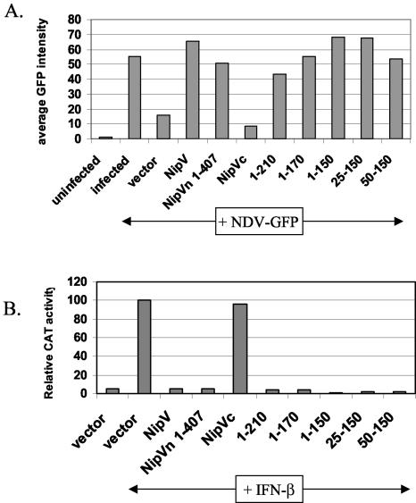
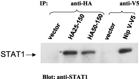
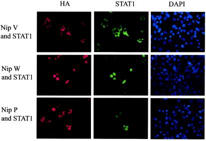
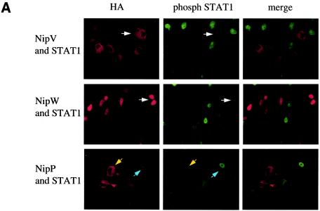
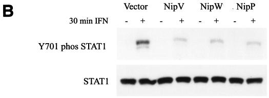
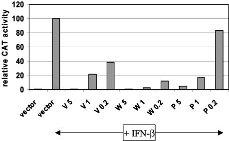
In previous reports it was demonstrated that the Nipah virus V and W proteins have interferon (IFN) antagonist activity due to their ability to block signaling from the IFN-alpha/beta receptor (J. J. Rodriguez, J. P. Parisien, and C. M. Horvath, J. Virol. 76:11476-11483, 2002; M. S. Park et al., J. Virol. 77:1501-1511, 2003). The V, W, and P proteins are all encoded by the same viral gene and share an identical 407-amino-acid N-terminal region but have distinct C-terminal sequences. We now show that the P protein also has anti-IFN function, confirming that the common N-terminal domain is responsible for the antagonist activity. Truncation of this N-terminal domain revealed that amino acids 50 to 150 retain the ability to block IFN and to bind STAT1, a key component of the IFN signaling pathway. Subcellular localization studies demonstrate that the V and P proteins are predominantly cytoplasmic whereas the W protein is localized to the nucleus. In all cases, STAT1 colocalizes with the corresponding Nipah virus protein. These interactions are sufficient to inhibit STAT1 activation, as demonstrated by the lack of STAT1 phosphorylation on tyrosine 701 in IFN-stimulated cells expressing P, V, or W. Therefore, despite their common STAT1-binding domain, the Nipah virus V and P proteins act by retaining STAT1 in the cytoplasm while the W protein sequesters STAT1 in the nucleus, creating both a cytoplasmic and a nuclear block for STAT1. We also show that the IFN antagonist activity of the P protein is not as strong as that of V or W, perhaps explaining why Nipah virus has evolved to express these two edited products.


Figures

FIG. 1.
FIG. 1.
The Nipah virus P protein has IFN antagonist activity. (A) Schematic representation of the Nipah virus V, W, and P proteins, their common N-terminal domain (Vn), and the unique C-terminal domain of V (Vc). Open box, N-terminal domain shared by all three proteins; shaded, solid, and checked boxes, unique C-terminal domains of V, W, and P, respectively, which arise due to the insertion of 1, 2, or 0 extra nontemplate G residues. (B) GFP expression in A549 cells following NDV-GFP infection. First panel, untransfected, uninfected cells; second panel, untransfected, NDV-GFP-infected cells. For the remaining panels, cells were transfected with the indicated expression plasmid and subsequently infected with NDV-GFP. (C) Inhibition of IFN-β-induced reporter gene expression in the presence of V, W, P, or Vn. Vero cells were transfected with an ISRE-CAT reporter plasmid and the indicated expression plasmid and were then either mock treated or treated with 1,000 IU of IFN-β/ml (+IFN-β) for 24 h. Data are expressed as the relative CAT activity determined 1 day posttreatment, normalized to a constitutively expressed Renilla luciferase control.
FIG. 1.
FIG. 1.
The Nipah virus P protein has IFN antagonist activity. (A) Schematic representation of the Nipah virus V, W, and P proteins, their common N-terminal domain (Vn), and the unique C-terminal domain of V (Vc). Open box, N-terminal domain shared by all three proteins; shaded, solid, and checked boxes, unique C-terminal domains of V, W, and P, respectively, which arise due to the insertion of 1, 2, or 0 extra nontemplate G residues. (B) GFP expression in A549 cells following NDV-GFP infection. First panel, untransfected, uninfected cells; second panel, untransfected, NDV-GFP-infected cells. For the remaining panels, cells were transfected with the indicated expression plasmid and subsequently infected with NDV-GFP. (C) Inhibition of IFN-β-induced reporter gene expression in the presence of V, W, P, or Vn. Vero cells were transfected with an ISRE-CAT reporter plasmid and the indicated expression plasmid and were then either mock treated or treated with 1,000 IU of IFN-β/ml (+IFN-β) for 24 h. Data are expressed as the relative CAT activity determined 1 day posttreatment, normalized to a constitutively expressed Renilla luciferase control.
FIG. 2.
FIG. 2.
The minimal domain that retains IFN antagonist activity is contained within amino acids 50 to 150. (A) Amino acids 50 to 150 constitute the smallest domain that rescues GFP expression in the NDV-GFP complementation assay. A549 cells were transfected with the indicated plasmid followed by NDV-GFP infection. Average GFP expression, as determined by FACS analysis, is shown. IFN antagonist activity is indicated by an increase in GFP expression compared to that with the vector control. (B) Amino acids 50 to 150 constitute the smallest domain that shows complete inhibition of IFN-β-inducible reporter gene expression. Vero cells were transfected with an ISRE-CAT reporter construct and the indicated expression construct and were then either mock treated or treated with 1,000 IU of IFN-β/ml (+IFN-β) for 24 h. Data are expressed as the relative CAT activity determined 1 day posttreatment, normalized to a constitutively expressed Renilla luciferase control.
FIG. 3.
FIG. 3.
Amino acids 50 to 150 constitute the smallest domain that is able to interact with STAT1. 293T cells were transfected with a STAT1 expression construct and the indicated Nipah virus construct. Protein complexes were coimmunoprecipitated with an antibody against the HA or V5 epitope, and the interacting protein was analyzed by SDS-PAGE and immunoblotting with an anti-STAT1 antibody. The position of STAT1 is indicated.
FIG. 4.
FIG. 4.
Nipah virus W is localized to the nucleus and causes redistribution of STAT1 to the nucleus, whereas V and P localize to the cytoplasm and retain STAT1 in the cytoplasm. HeLa cells were transfected with the STAT1 expression plasmid and HA-tagged V, W, or P. Cells were fixed and permeabilized 1 day later. Proteins were stained with antibodies against the HA epitope (left panels) or STAT1 (center panels), and nuclei were stained with 4′,6-diamidino-2-phenylindole (DAPI) (right panels).
FIG. 5.
FIG. 5.
Nipah virus V, W, and P all inhibit IFN-induced phosphorylation of STAT1. (A) HeLa cells were transfected with a STAT1 plasmid and an HA-tagged V, W, or P plasmid. STAT1 phosphorylation was induced by treatment with 100 ng of IFN-α/ml for 30 min. Proteins were stained with antibodies against the HA epitope (left panels) or pY701-STAT1 (center panels). In cells expressing V or W, there was no evidence of phosphorylated STAT1 in the nucleus (white arrows). Among cells expressing P, some had activated STAT1 in the nucleus (blue arrows), while others did not (yellow arrows). Merged images are shown in the panels on the right. (B) 293T cells were transfected with the indicated plasmids. One day later, STAT1 phosphorylation was induced by treatment with 1,000 IU of IFN-β/ml for 30 min. Cells were lysed, and levels of pY701-STAT1 (upper panel) and bulk STAT1 (lower panel) were determined by Western blot analysis.
FIG. 5.
FIG. 5.
Nipah virus V, W, and P all inhibit IFN-induced phosphorylation of STAT1. (A) HeLa cells were transfected with a STAT1 plasmid and an HA-tagged V, W, or P plasmid. STAT1 phosphorylation was induced by treatment with 100 ng of IFN-α/ml for 30 min. Proteins were stained with antibodies against the HA epitope (left panels) or pY701-STAT1 (center panels). In cells expressing V or W, there was no evidence of phosphorylated STAT1 in the nucleus (white arrows). Among cells expressing P, some had activated STAT1 in the nucleus (blue arrows), while others did not (yellow arrows). Merged images are shown in the panels on the right. (B) 293T cells were transfected with the indicated plasmids. One day later, STAT1 phosphorylation was induced by treatment with 1,000 IU of IFN-β/ml for 30 min. Cells were lysed, and levels of pY701-STAT1 (upper panel) and bulk STAT1 (lower panel) were determined by Western blot analysis.
FIG. 6.
FIG. 6.
At limiting concentrations, Nipah virus P has a weaker ability to block IFN signaling than the V and W proteins. Vero cells were transfected with an ISRE-CAT reporter plasmid and either an empty vector or 5, 1, or 0.2 μg of an HA-tagged V, W, or P plasmid. In all cases, the total DNA was made up to 5 μg with the empty vector. Cells were either mock treated or treated with 1,000 IU of IFN-β/ml for 24 h, lysed, and analyzed for CAT activity. Data are expressed as relative levels of CAT activity, normalized to a constitutively expressed luciferase control.

Similar articles

Cited by

References

    1. Andrejeva, J., E. Poole, D. F. Young, S. Goodbourn, and R. E. Randall. 2002. The p127 subunit (DDB1) of the UV-DNA damage repair binding protein is essential for the targeted degradation of STAT1 by the V protein of the paramyxovirus simian virus 5. J. Virol. 76:11379-11386. - PMC - PubMed
    1. Basler, C. F., A. Mikulasova, L. Martinez-Sobrido, J. Paragas, E. Muhlberger, M. Bray, H. D. Klenk, P. Palese, and A. Garcia-Sastre. 2003. The Ebola virus VP35 protein inhibits activation of interferon regulatory factor 3. J. Virol. 77:7945-7956. - PMC - PubMed
    1. Basler, C. F., A. H. Reid, J. K. Dybing, T. A. Janczewski, T. G. Fanning, H. Zheng, M. Salvatore, M. L. Perdue, D. E. Swayne, A. Garcia-Sastre, P. Palese, and J. K. Taubenberger. 2001. Sequence of the 1918 pandemic influenza virus nonstructural gene (NS) segment and characterization of recombinant viruses bearing the 1918 NS genes. Proc. Natl. Acad. Sci. USA 98:2746-2751. - PMC - PubMed
    1. Basler, C. F., X. Wang, E. Muhlberger, V. Volchkov, J. Paragas, H. D. Klenk, A. Garcia-Sastre, and P. Palese. 2000. The Ebola virus VP35 protein functions as a type I IFN antagonist. Proc. Natl. Acad. Sci. USA 97:12289-12294. - PMC - PubMed
    1. Bossert, B., and K. K. Conzelmann. 2002. Respiratory syncytial virus (RSV) nonstructural (NS) proteins as host range determinants: a chimeric bovine RSV with NS genes from human RSV is attenuated in interferon-competent bovine cells. J. Virol. 76:4287-4293. - PMC - PubMed

Publication types