. 2004 Jun;78(11):5658-69.
doi: 10.1128/JVI.78.11.5658-5669.2004.

Murine coronavirus replication induces cell cycle arrest in G0/G1 phase

Affiliations

Murine coronavirus replication induces cell cycle arrest in G0/G1 phase

Chun-Jen Chen et al. J Virol. 2004 Jun.

Abstract

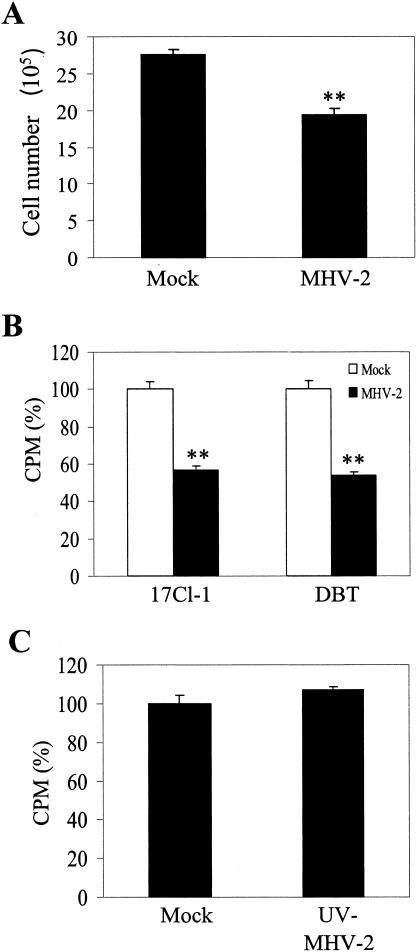
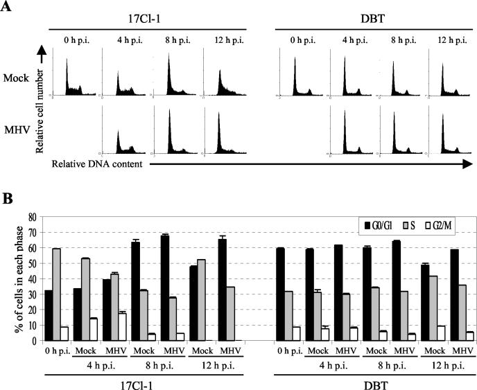
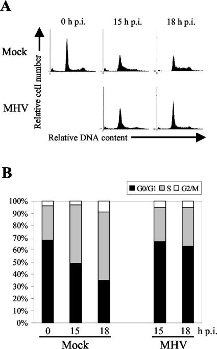
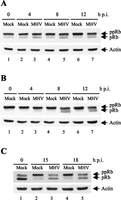
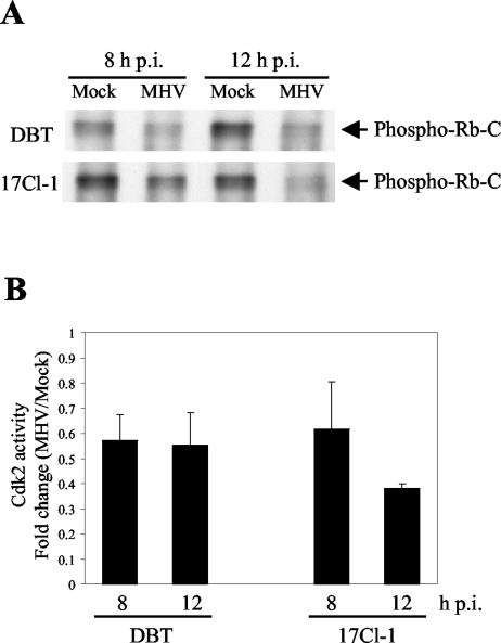
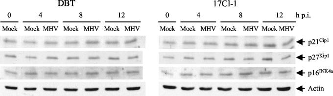
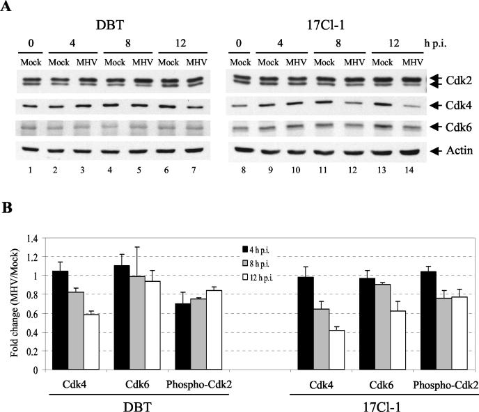
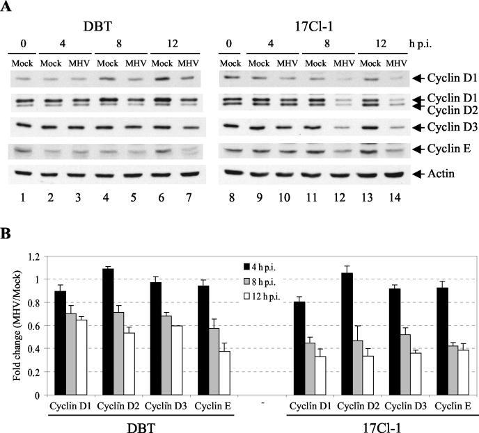
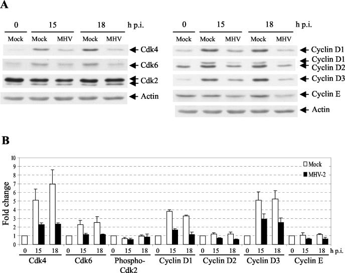
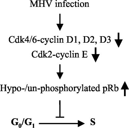
Mouse hepatitis virus (MHV) replication in actively growing DBT and 17Cl-1 cells resulted in the inhibition of host cellular DNA synthesis and the accumulation of infected cells in the G(0)/G(1) phase of the cell cycle. UV-irradiated MHV failed to inhibit host cellular DNA synthesis. MHV infection in quiescent 17Cl-1 cells that had been synchronized in the G(0) phase by serum deprivation prevented infected cells from entering the S phase after serum stimulation. MHV replication inhibited hyperphosphorylation of the retinoblastoma protein (pRb), the event that is necessary for cell cycle progression through late G(1) and into the S phase. While the amounts of the cellular cyclin-dependent kinase (Cdk) inhibitors p21(Cip1), p27(Kip1), and p16(INK4a) did not change in infected cells, MHV infection in asynchronous cultures induced a clear reduction in the amounts of Cdk4 and G(1) cyclins (cyclins D1, D2, D3, and E) in both DBT and 17Cl-1 cells and a reduction in Cdk6 levels in 17Cl-1 cells. Infection also resulted in a decrease in Cdk2 activity in both cell lines. MHV infection in quiescent 17Cl-1 cells prevented normal increases in Cdk4, Cdk6, cyclin D1, and cyclin D3 levels after serum stimulation. The amounts of cyclin D2 and cyclin E were not increased significantly after serum stimulation in mock-infected cells, whereas they were decreased in MHV-infected cells, suggesting the possibility that MHV infection may induce cyclin D2 and cyclin E degradation. Our data suggested that a reduction in the amounts of G(1) cyclin-Cdk complexes in MHV-infected cells led to a reduction in Cdk activities and insufficient hyperphosphorylation of pRb, resulting in inhibition of the cell cycle in the G(0)/G(1) phase.


Figures

FIG. 1.
FIG. 1.
MHV infection inhibits cell proliferation and cellular DNA synthesis. (A) 17Cl-1 cells at 30% confluence in a 6-cm dish were mock infected or infected with MHV-2 at an MOI of 10. At 18 h p.i., cell numbers were counted in a hemocytometer. The data are presented as means and SEs (n = 5). (B) 17Cl-1 cells and DBT cells were mock infected or infected with MHV-2 at an MOI of 20. Cellular DNA synthesis was measured by [3H]thymidine incorporation from 4 to 11 h p.i. The results are presented as the mean and SE counts per minute (CPM) for six independent experiments. The CPM for MHV-infected samples were normalized to the CPM for mock-infected samples (100%). (C) 17Cl-1 cells were mock infected or infected with UV-inactivated MHV-2, and cellular DNA synthesis was measured as described for panel B. Double asterisks indicate a P value of < 0.001 in comparisons with mock-infected samples.
FIG. 2.
FIG. 2.
MHV infection in asynchronously growing cells induces the accumulation of cells in the G0/G1 phase of the cell cycle. (A) DBT and 17Cl-1 cells were mock infected (Mock) or infected with MHV-2 (MHV) at an MOI of 10. At the indicated times p.i., cells were collected and stained with propidium iodide for FACS analysis. The data are from one of three experiments. (B) The histograms in panel A were analyzed by the ModFit LT program to determine the percentage of cells in each phase of the cell cycle. The results are presented as means and SEs for three experiments. The S- and G2/M-phase populations in mock- and MHV-infected 17Cl-1 cells at 12 h p.i. were combined and presented as the S-phase population because the G2/M-phase population in these histograms could not be identified accurately by the ModFit LT program.
FIG. 3.
FIG. 3.
MHV infection of quiescent 17Cl-1 cells prevents cell cycle reentry. (A) Serum-starved 17Cl-1 cells were mock infected (Mock) or infected with MHV-2 (MHV) at an MOI of 10. After 1 h of virus adsorption, medium containing 10% FCS was added to the cells, and cell cycle profiles at the indicated times p.i. were determined by FACS analysis. The data are from one of three experiments. (B) The histograms in panel A were analyzed by the Synch Wizard algorithm of the ModFit LT program to determine the percentage of cells in each phase of the cell cycle.
FIG. 4.
FIG. 4.
MHV infection induces the accumulation of nonphosphorylated and/or hypophosphorylated pRb. Asynchronously growing DBT cells (A) and 17Cl-1 cells (B) were mock infected (Mock) or infected with MHV-2 (MHV) at an MOI of 10. At the indicated times p.i., cells were lysed with SDS sample buffer, and equal amounts of protein from the samples were subjected to Western blot analysis for pRb and actin. Nonphosphorylated and hypophosphorylated forms of pRb (pRb) appeared as rapidly migrating bands, and hyperphosphorylated pRb (ppRb) appeared as slowly migrating bands. (C) Serum-starved 17Cl-1 cells were mock infected (Mock) or infected with MHV-2 (MHV) at an MOI of 10. After 1 h of virus adsorption, medium containing 10% FCS was added to the cells. At the indicated times p.i., cell lysates were collected and analyzed as described above. Similar results were obtained in three independent experiments.
FIG. 5.
FIG. 5.
Effect of MHV infection on Cdk2 activity. (A) Cdk2 was immunoprecipitated from mock-infected (Mock) or MHV-2-infected (MHV) DBT and 17Cl-1 cell lysates, and kinase activities were determined by an immunocomplex kinase assay with Rb-C as a substrate followed by Western blot analysis for Rb-C phosphorylation at Ser807 and Ser811 (phospho-Rb-C). The data are from one of three independent experiments. (B) Cdk2 activities in panel A were quantified by densitometric analysis, and the ratio of activity in MHV-infected samples to that in mock-infected samples is presented as the mean and SE (n = 3).
FIG. 6.
FIG. 6.
Effect of MHV infection on the levels of expression of the cellular CKIs p21Cip1, p27Kip1, and p16INK4a. Asynchronously growing DBT cells and 17Cl-1 cells were mock infected (Mock) or infected with MHV-2 (MHV) at an MOI of 10. At the indicated times p.i., cells were lysed with SDS sample buffer, and equal amounts of protein from the samples were tested by Western blot analysis against probes for p21Cip1, p27Kip1, p16INK4a, and actin. The data are from one of three independent experiments.
FIG. 7.
FIG. 7.
Effect of MHV infection on levels of G1 Cdks in asynchronously growing cells. (A) DBT and 17Cl-1 cells were mock infected (Mock) or infected with MHV-2 (MHV) at an MOI of 10. At the indicated times p.i., cells were lysed with SDS sample buffer, and equal amounts of protein from the samples were tested by Western blot analysis against probes for Cdk2, Cdk4, Cdk6, and actin. The data are from one of three independent experiments. (B) Cdk amounts in panel A were quantified by densitometric analysis and normalized against an internal control (actin). Bars indicate the ratio of Cdk amounts in MHV-infected samples to those in mock-infected samples. The results are presented as means and SEs (n = 3).
FIG. 8.
FIG. 8.
Effect of MHV infection on levels of G1 cyclins in asynchronously growing cells. (A) DBT and 17Cl-1 cells were mock infected (Mock) or infected with MHV-2 (MHV) at an MOI of 10. At the indicated times p.i., cells were lysed with SDS sample buffer, and equal amounts of protein from the samples were tested by Western blot analysis against probes for cyclin D1, cyclin D2, cyclin D3, cyclin E, and actin. The data are from one of three independent experiments. (B) Cyclin amounts in panel A were quantified by densitometric analysis and normalized against an internal control (actin). Bars indicate the ratio of cyclin amounts in MHV-infected samples to those in mock-infected samples. The results are presented as means and SEs (n = 3).
FIG. 9.
FIG. 9.
Effect of MHV infection on levels of G1 Cdks and G1 cyclins in cells released from quiescence. (A) Serum-starved 17Cl-1 cells were mock infected (Mock) or infected with MHV-2 (MHV) at an MOI of 10. After 1 h of virus adsorption, medium containing 10% FCS was added to the cells. At the indicated times p.i., cells were lysed with SDS sample buffer, and equal amounts of protein from the samples were tested by Western blot analysis against probes for Cdk2, Cdk4, Cdk6, cyclin D1, cyclin D2, cyclin D3, cyclin E, and actin. The data are from one of three independent experiments. (B) Cdk and cyclin amounts in panel A were quantified by densitometric analysis and normalized against an internal control (actin). The amounts of each Cdk and each cyclin at different times p.i. were further normalized to the amounts at 0 h p.i., which were arbitrarily set to a value of 1.0. The results are presented as means and SEs (n = 3).
FIG. 10.
FIG. 10.
Proposed mechanism for MHV-induced G0/G1 cell cycle arrest. MHV infection causes decreases in the amounts of G1 cyclins and Cdks, resulting in a reduction in Cdk2/4/6 activities and the accumulation of hypophosphorylated and/or nonphosphorylated pRb, which blocks cell cycle progression from the G0/G1 phase to the S phase.

Similar articles

Cited by

References

    1. Aktas, H., H. Cai, and G. M. Cooper. 1997. Ras links growth factor signaling to the cell cycle machinery via regulation of cyclin D1 and the Cdk inhibitor p27KIP1. Mol. Cell. Biol. 17:3850-3857. - PMC - PubMed
    1. An, S., C. J. Chen, X. Yu, J. L. Leibowitz, and S. Makino. 1999. Induction of apoptosis in murine coronavirus-infected cultured cells and demonstration of E protein as an apoptosis inducer. J. Virol. 73:7853-7859. - PMC - PubMed
    1. Banerjee, S., S. An, A. Zhou, R. H. Silverman, and S. Makino. 2000. RNase L-independent specific 28S rRNA cleavage in murine coronavirus-infected cells. J. Virol. 74:8793-8802. - PMC - PubMed
    1. Belyavsky, M., E. Belyavskaya, G. A. Levy, and J. L. Leibowitz. 1998. Coronavirus MHV-3-induced apoptosis in macrophages. Virology 250:41-49. - PMC - PubMed
    1. Bonneau, A. M., and N. Sonenberg. 1987. Involvement of the 24-kDa cap-binding protein in regulation of protein synthesis in mitosis. J. Biol. Chem. 262:11134-11139. - PubMed

Publication types