. 2004 Jun;78(11):5686-97.
doi: 10.1128/JVI.78.11.5686-5697.2004.

Passive and active inclusion of host proteins in human immunodeficiency virus type 1 gag particles during budding at the plasma membrane

Affiliations

Passive and active inclusion of host proteins in human immunodeficiency virus type 1 gag particles during budding at the plasma membrane

Maria Hammarstedt et al. J Virol. 2004 Jun.

Abstract

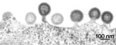
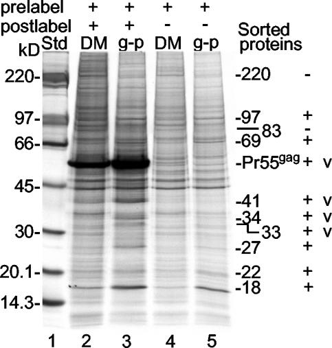
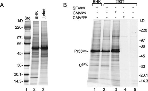
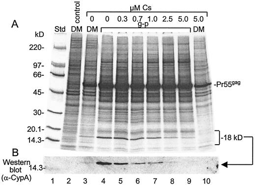
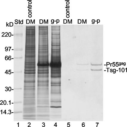
Human immunodeficiency virus type 1 particles form by budding at the surface of most cell types. In this process, a piece of the plasma membrane is modified into an enveloped virus particle. The process is driven by the internal viral protein Pr55(gag). We have studied how host proteins in the membrane are dealt with by Pr55(gag) during budding. Are they included in or excluded from the particle? The question was approached by measuring the relative concentrations of host and viral proteins in the envelope of Pr55(gag) particles and in their donor membranes in the cell. We observed that the bulk of the host proteins, including actin and clathrin, were passively included into the virus-like Gag particles. This result suggests that budding by Pr55(gag) proceeds without significant alteration of the original host protein composition at the cell membrane. Nevertheless, some proteins were concentrated in the particles, and a few were excluded. The concentrated proteins included cyclophilin A and Tsg-101. These were recruited to the plasma membrane by Pr55(gag). The membrane-bound cyclophilin A was concentrated into particles as efficiently as Pr55(gag), whereas Tsg-101 was concentrated more efficiently. The latter finding is consistent with a role for Tsg-101 in Gag particle release.


Figures

FIG. 1.
FIG. 1.
Budding of Gag particles at the cell surface. BHK-21 cells were infected with SFV-C/Pr55gag vectors and fixed after 6 h. Thin sections of the cells were prepared and analyzed by EM.
FIG. 2.
FIG. 2.
Purification of 35S-labeled Gag particles. (A) Sedimentation analyses of 35S-labeled particles released from HIV-1 gag-expressing cells. BHK-21 cells were labeled with [35S]Met before and after infection with SFV-C/Pr55gag vectors. Gag particles in the medium were isolated by sedimentation in a 5 to 20% iodixanol gradient. After fractionation and dilution of the iodixanol, particles were recovered from each fraction by centrifugation and analyzed by SDS-PAGE (6 to 15%). Autoradiographs of the gels are shown. Lanes: 1, top fraction; P, pellet in the tube with the iodixanol gradient; L, NP-40 extract of vector-infected cells. (B) Sedimentation analyses of 35S-labeled particles released from NPflu-expressing cells. Labeling of cells that had been infected with SFV-C/NPflu vectors and analyses of particles were as described for panel A.
FIG. 3.
FIG. 3.
Purification of 32P-labeled Gag particles. (A) Sedimentation analyses of 32P-labeled particles released from HIV-1 gag-expressing cells. BHK-21 cells were labeled with [32P]orthophosphate for 40 h and then infected with SFV-C/Pr55gag vectors. After these steps, labeling was continued, and released particles were collected and separated as described in the legend to Fig. 2A. The isolated particles were solubilized in hot SDS and analyzed for 32P-labeled SDS-phospholipid (SDS-PL) mixed micelles by SDS-PAGE (20%). Labeled RNA at the top of the gels and free orthophoshate (Pi) at the bottom are indicated. (B) Sedimentation analyses of 32P-labeled particles released from NPflu-expressing cells. Labeling of cells and analyses of particles were as described for panel A. (C) Quantification of 32P-labeled SDS-lipid mixed micelles from panels A and B. PL, phospholipids; PSL, photostimulated luminescence. (D) EM analysis of isolated particles. Particles in fractions 13 to 15 of the separation procedure shown in Fig. 2A were pooled, concentrated by centrifugation at the tip of a centrifuge tube, embedded in gelatin, processed for sectioning, and analyzed by EM. Note the ∼100-nm particles with an apparent Gag layer.
FIG. 4.
FIG. 4.
Purification of donor membranes. (A) Flotation centrifugation analyses of microsomes from HIV-1 gag-expressing cells. BHK-21 cells were labeled with [35S]Met and infected with SFV-C/Pr55gag vectors as described in the legend to Fig. 2A. The cells were homogenized, and a postnuclear supernatant was prepared and subjected to flotation centrifugation in a 10, 30, 35, 40, 50, and 55% sucrose step gradient. The gradient was fractionated, and membranes were recovered from each fraction, after dilution, by centrifugation and analyzed by SDS-PAGE (6 to 15%). Autoradiographs of the gels are shown. Std, standard. (B) Comparison of protein profiles of Gag-enriched membranes (donor membranes [DM]) from SFV-C/Pr55gag vector-infected cells and corresponding membranes from uninfected cells. Analyses were done as described for panel A. Lanes show protein analyses for pooled fractions 6 and 7. (C) Quantification of Pr55gag in the floating microsomes (M) from panel A. Included is a quantification of Pr55gag in Gag particles (g-p) that were subjected to a similar flotation analysis. PSL, photostimulated luminescence. (D) Morphological analysis of donor membranes. Membranes in fractions 6 and 7 were pooled, concentrated by centrifugation at the tip of a centrifuge tube, sectioned, and analyzed by EM. Note the apparent budding Gag particles in some of the vesicles.
FIG. 5.
FIG. 5.
Host protein sorting in the donor membrane during the budding of Gag particles. Samples of 35S-labeled Gag particles (g-p) and donor membranes (DM) were adjusted to contain equal amounts of membranes and then analyzed by SDS-PAGE (6 to 15%). The donor membrane and Gag particle preparations were made in cells that were either labeled both before (prelabeled) and after (postlabeled) vector infection (lanes 2 and 3) or only prelabeled (lanes 4 and 5). In the latter case, only host proteins were labeled and detected. Std, standard. The molecular masses of vector proteins (v) as well as those of host proteins that were either concentrated or diluted during budding are indicated. The latter proteins are marked as positively (+) or negatively (−) sorted proteins.
FIG. 6.
FIG. 6.
Inclusion of host proteins in Gag particles made in Jurkat T lymphocytes by SFV expression and in 293T cells by nuclear gene expression. (A) BHK-21 cells and Jurkat cells were prelabeled with [35S]Met and infected with SFV-C/Pr55gag vectors. The cells then were postlabeled, and Gag particles were collected between 5 and 6 h postinfection. Samples of iodixanol gradient-purified Gag particles were adjusted to contain equal amounts of membranes and then analyzed by SDS-PAGE (6 to 15%). Particles produced in BHK-21 cells are shown in lane 2, and particles produced in Jurkat cells are shown in lane 3. Std, standard. (B) BHK-21 cells were infected with SFV-C/Pr55gag vectors (lane 1), and 293T cells were infected with SFV-C/Pr55gag vectors (lane 2). 293T cells were additionally transfected with pCMV-HIVgag DNA (lane 3) or pRRLsin.cPPT.CMV.GFP.Wpre DNA (lane 4) or mock transfected (lane 5). The infected BHK-21 and 293T cells were labeled with [35S]Met before and after infection for 17 and 6 h, respectively. Particles produced during the last 1 h of the labeling period were collected and purified by sedimentation in iodixanol gradients. The transfected and mock-transfected cells were incubated for 25.5 h and then labeled for 21 h. In these cases, the particles produced during the last 4 h of the labeling period were collected and purified. SDS-PAGE (6 to 15%) analysis of the various particle preparations is shown. Note that 1.25 and 1.07 times more Gag particles were analyzed in lanes 3 and 1, respectively, than in lane 2. Note also the apparent SFV capsid protein that is seen only in the particles produced by SFV-C vector-infected cells.
FIG. 7.
FIG. 7.
Concentration of CypA into Gag particles. Donor membranes (DM) and Gag particles (g-p) were produced by using SFV-C/Pr55gag vector-infected BHK-21 cells that were pre- and postlabeled with [35S]Met but with cyclosporine (Cs) added at the indicated concentrations at 2 h postinfection. Gag particles (lanes 4 to 9) and donor membranes (lanes 3 and 10) were adjusted to contain equal amounts of membranes and then analyzed by SDS-PAGE (6 to 15%) in duplicate. One gel was processed for autoradiography (A), and the other one was used for the detection of CypA by Western blotting (B). Donor membranes from uninfected cells were analyzed in parallel (lane 2). Std, standard.
FIG. 8.
FIG. 8.
Concentration of Tsg-101 into Gag particles. Donor membranes (DM) and Gag particles (g-p) from BHK-21 cells infected with SFV-C/Pr55gag vectors and pre- and postlabeled with [35S]Met as well as donor membranes from uninfected control cells were analyzed by SDS-PAGE on an equal-lipid basis. The proteins in the gel were transferred to a filter, and this was used for the detection of Tsg-101 by Western blotting (lanes 5 to 7). Lanes 2 to 4 represent an autoradiograph of 35S-labeled proteins separated on a duplicate gel. Note that abundant Pr55gag was stained nonspecifically in the blot. Std, standard.
FIG. 9.
FIG. 9.
Passive inclusion of actin and clathrin in Gag particles. (A and B) Donor membranes (DM) from SFV-C/Pr55gag vector-infected BHK-21 cells were analyzed by multiple SDS-PAGE (6 to 15%) analyses. The proteins in the gels were transferred to filters, and these were used for the detection of actin (panel A, lane 2) and clathrin (panel B, lane 2). Lanes 1 in panels A and B represent autoradiographs of the respective filters. Note that Pr55gag was recognized nonspecifically in all Western blot analyses. (C) Actin and clathrin proteins from earlier protein analyses of membrane-equalized donor membrane and Gag particle (g-p) samples derived from prelabeled SFV-C/Pr55gag vector-infected BHK-21 cells (Fig. 5, lanes 4 and 5). Note that actin and clathrin are present at corresponding concentrations in the donor membranes and the Gag particles. Std, standard.

Similar articles

Cited by

References

    1. Arthur, L. O., J. W. Bess, Jr., R. C. Sowder II, R. E. Benveniste, D. L. Mann, J. C. Chermann, and L. E. Henderson. 1992. Cellular proteins bound to immunodeficiency viruses: implications for pathogenesis and vaccines. Science 258:1935-1938. - PubMed
    1. Bess, J. W., Jr., R. J. Gorelick, W. J. Bosche, L. E. Henderson, and L. O. Arthur. 1997. Microvesicles are a source of contaminating cellular proteins found in purified HIV-1 preparations. Virology 230:134-144. - PubMed
    1. Braaten, D., E. K. Franke, and J. Luban. 1996. Cyclophilin A is required for an early step in the life cycle of human immunodeficiency virus type 1 before the initiation of reverse transcription. J. Virol. 70:3551-3560. - PMC - PubMed
    1. Braaten, D., and J. Luban. 2001. Cyclophilin A regulates HIV-1 infectivity, as demonstrated by gene targeting in human T cells. EMBO J. 20:1300-1309. - PMC - PubMed
    1. Cheng, R. H., R. J. Kuhn, N. H. Olson, M. G. Rossmann, H. K. Choi, T. J. Smith, and T. S. Baker. 1995. Nucleocapsid and glycoprotein organization in an enveloped virus. Cell 80:621-630. - PMC - PubMed

Publication types

LinkOut - more resources