. 2004 Jun;78(11):5707-19.
doi: 10.1128/JVI.78.11.5707-5719.2004.

Longitudinal analysis of CD8+ T cells specific for structural and nonstructural hepatitis B virus proteins in patients with chronic hepatitis B: implications for immunotherapy

Affiliations

Longitudinal analysis of CD8+ T cells specific for structural and nonstructural hepatitis B virus proteins in patients with chronic hepatitis B: implications for immunotherapy

George J M Webster et al. J Virol. 2004 Jun.

Abstract

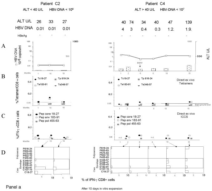
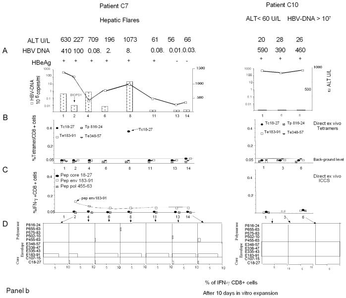
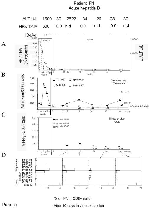
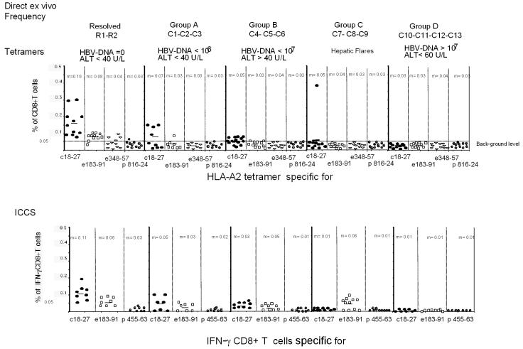
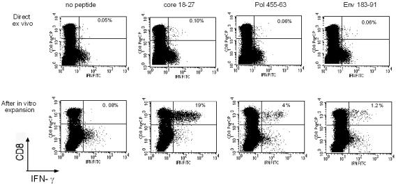
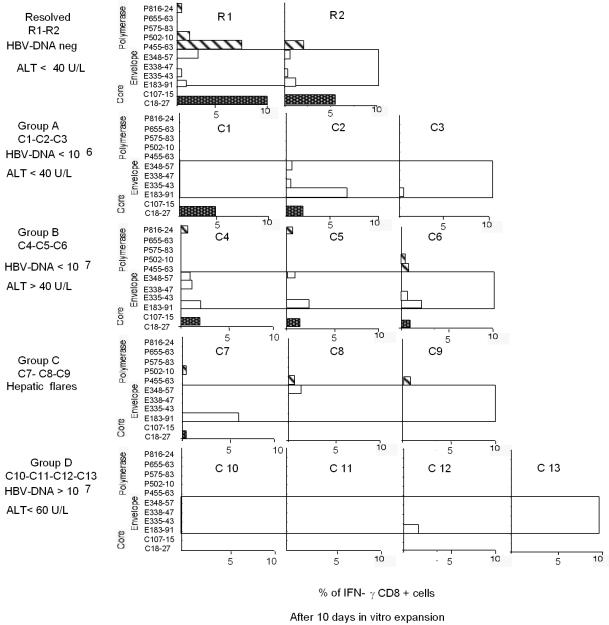
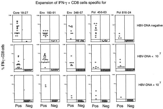
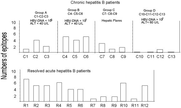
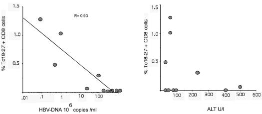
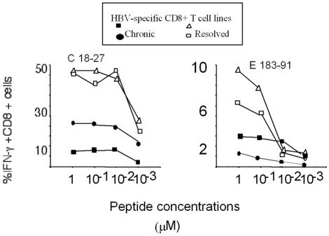
The cytotoxic T-cell response in chronic hepatitis B virus (HBV) infection has been described as weak and mono- or oligospecific in comparison to the more robust virus-specific T-cell response present in resolved infection. However, chronic hepatitis B is a heterogeneous disease with markedly variable levels of virus replication and liver disease activity. Here we analyzed (both directly ex vivo and after in vitro stimulation) the HBV-specific CD8 T-cell responses against structural and nonstructural HBV proteins longitudinally in patients with different patterns of chronic infections. We found that the profiles of virus-specific CD8(+)-T-cell responses during chronic infections are highly heterogeneous and influenced more by the level of HBV replication than by the activity of liver disease. An HBV DNA load of <10(7) copies/ml appears to be the threshold below which circulating multispecific HBV-specific CD8(+) T cells are consistently detected. Furthermore, CD8(+) T cells with different specificities are differentially regulated during chronic infections. HBV core-specific CD8(+) T cells are associated with viral control, while CD8(+) T cells specific for envelope and polymerase epitopes can occasionally be found in the setting of high levels (>10(7) copies) of HBV replication. These findings have implications for the design of immunotherapy for chronic HBV infections.


Figures

FIG. 1.
FIG. 1.
Longitudinal analysis of HBV-specific CD8+-T-cell responses. Patients with different clinical profiles of HBV infection were studied longitudinally for ALT and HBV DNA levels and the frequencies of HBV-specific CD8+ T cells directly ex vivo and after in vitro expansion. A representative patient for each clinical group is shown: chronically infected patients C2, C4, C7, and C10 in panels a and b and patient R1, with resolved acute hepatitis B, in panel c. (A) Time course analysis of HBV DNA and ALT levels. Serum ALT levels (units per liter [U/L]) (bars) and HBV DNA levels (copies per milliliter) (squares) were analyzed longitudinally. Time zero in patient R1 is the day of clinical onset. n.d., not determined. (B) Percentages of CD8+ T cells that were tetramer positive at each time point. PBMC were tested directly ex vivo with the indicated tetramers (Tc18-27, core 18-27 tetramer; Te183-91, env 183-191 tetramer; Tp816-24, pol 816-824 tetramer; Te348-57, env 348-357 tetramer). The background level of direct ex vivo tetramer staining (0.05%, indicated by a horizontal line) was calculated in HLA-A2-positive, noninfected subjects and in HLA-A2-negative, HBV-infected patients (21). (C) Percentages of peptide-specific IFN-γ-producing CD8+ T cells determined directly ex vivo at different time points. PBMC were stimulated directly ex vivo with the indicated peptides (Pep). The percentages of IFN-γ-producing CD8+ T cells were calculated after subtraction of doubly positive cells obtained from nonstimulated PBMC. (D) Percentages of peptide-specific IFN-γ-producing CD8+ T cells after in vitro expansion (bars). PBMC were stimulated with the indicated peptides. After 10 days of in vitro expansion, the percentages of peptide-specific IFN-γ-producing CD8+ T cells were calculated.
FIG. 1.
FIG. 1.
Longitudinal analysis of HBV-specific CD8+-T-cell responses. Patients with different clinical profiles of HBV infection were studied longitudinally for ALT and HBV DNA levels and the frequencies of HBV-specific CD8+ T cells directly ex vivo and after in vitro expansion. A representative patient for each clinical group is shown: chronically infected patients C2, C4, C7, and C10 in panels a and b and patient R1, with resolved acute hepatitis B, in panel c. (A) Time course analysis of HBV DNA and ALT levels. Serum ALT levels (units per liter [U/L]) (bars) and HBV DNA levels (copies per milliliter) (squares) were analyzed longitudinally. Time zero in patient R1 is the day of clinical onset. n.d., not determined. (B) Percentages of CD8+ T cells that were tetramer positive at each time point. PBMC were tested directly ex vivo with the indicated tetramers (Tc18-27, core 18-27 tetramer; Te183-91, env 183-191 tetramer; Tp816-24, pol 816-824 tetramer; Te348-57, env 348-357 tetramer). The background level of direct ex vivo tetramer staining (0.05%, indicated by a horizontal line) was calculated in HLA-A2-positive, noninfected subjects and in HLA-A2-negative, HBV-infected patients (21). (C) Percentages of peptide-specific IFN-γ-producing CD8+ T cells determined directly ex vivo at different time points. PBMC were stimulated directly ex vivo with the indicated peptides (Pep). The percentages of IFN-γ-producing CD8+ T cells were calculated after subtraction of doubly positive cells obtained from nonstimulated PBMC. (D) Percentages of peptide-specific IFN-γ-producing CD8+ T cells after in vitro expansion (bars). PBMC were stimulated with the indicated peptides. After 10 days of in vitro expansion, the percentages of peptide-specific IFN-γ-producing CD8+ T cells were calculated.
FIG. 2.
FIG. 2.
Direct ex vivo frequencies of HBV-specific CD8+ T cells. The direct ex vivo frequencies of CD8+ T cells stained with HLA tetramers (Tc18-27, Te183-191, Te348-357, and Tp816-824) or producing IFN-γ after stimulation with core 18-27, env 183-191, and pol 455-463 peptides are shown for the indicated groups of patients. The frequency of IFN-γ-producing CD8+ T cells stimulated by the single peptides was subtracted from the frequency obtained in non-peptide-stimulated PBMC. All of the data obtained at different time points for each patient are shown. m, mean. HBV DNA levels are given in copies per milliliter; ALT levels are given in units per liter (U/L).
FIG. 3.
FIG. 3.
Analysis of IFN-γ-producing CD8+ T cells directly ex vivo or after in vitro expansion. IFN-γ-producing CD8+ T cells were tested directly ex vivo in PBMC (upper dot plots) or after 10 days of in vitro expansion with the corresponding peptides (lower dot plots). The results shown are from experiments performed with PBMC from patient R1. PBMC were stimulated with the indicated individual peptides either for direct ex vivo analysis or for experiments performed after 10 days of in vitro expansion. In the experiments performed after in vitro expansion, the growing cells were restimulated either with no peptide or with the initial stimulatory HBV peptide. Subdominant peptides (pol 455-463 and env 183-191) tested positive only after 10 days of in vitro expansion.
FIG. 4.
FIG. 4.
Profiles of HBV-specific CD8+ responses obtained after in vitro expansion in various patients. Peptide-specific IFN-γ-producing CD8+ T cells were quantified after 10 days of in vitro expansion with the indicated peptides. One representative experiment for each patient is shown. Results for patients R1 and R2 were obtained, respectively, at 12 (R1) and 18 (R2) months after the resolution of acute hepatitis. Results for patients C2, C4, C7, and C10 represent the results shown in Fig. 1a and b at month 10 (C2 and C4), month 8 (C7), and month 3 (C10). HBV DNA levels are given in copies per milliliter; ALT levels are given in units per liter (U/L).
FIG. 5.
FIG. 5.
Expansion of epitope-specific IFN-γ-producing CD8+ T cells in relation to HBV DNA levels. The percentages of IFN-γ-producing CD8+ T cells specific for the indicated HBV epitopes present in the T-cell lines generated after in vitro peptide stimulation of PBMC were grouped in relation to the HBV DNA levels present at the time of the assays. Data include “positive” (Pos) experiments (presence of peptide-specific IFN-γ-producing CD8+ T cells) and “negative” (Neg) experiments (absence of peptide-specific IFN-γ-producing CD8+ T cells) performed for patients studied longitudinally. HBV DNA levels are given in copies per milliliter.
FIG. 6.
FIG. 6.
Numbers of HBV epitopes recognized by patients with chronic or resolved HBV infections. Bars indicate the numbers of peptides recognized by each patient for the 11 HBV epitopes tested. PBMC from the indicated patients with chronic or resolved HBV infections were stimulated with 11 HBV peptides representing known HLA-A2-restricted epitopes (Table 2). After 8 to 10 days of in vitro expansion, the short-term cell lines were tested for the presence of peptide-specific IFN-γ-producing CD8+ T cells. HBV DNA levels are given in copies per milliliter; ALT levels are given in units per liter (U/L).
FIG. 7.
FIG. 7.
Frequencies of intrahepatic core 18-27-specific CD8+ T cells in relation to HBV DNA and ALT levels. Percentages of tetramer core 18-27-specific (Tc18-27) CD8+ T cells present in lymphocytes purified from liver biopsy specimens were plotted in relation to the HBV DNA and ALT levels (units per liter [U/l]) present at the time of liver biopsy. Results obtained for 10 different patients are shown. There was a strong negative correlation between HBV DNA and core 18-27-specific CD8+ T cells (r value determined by the Spearman correlation test, −0.93; P = 0.0001) but no correlation between ALT and core 18-27-specific CD8+ T cells (r = 0.23; P = 0.53).
FIG. 8.
FIG. 8.
Functional T-cell avidity of HBV epitope-specific CD8+ T cells. Various T-cell lines specific for the indicated HBV epitopes and produced in patients with resolved infections or chronic infections were stimulated with various concentrations of peptides. Core 18-27- and env 183-191-specific CD8+ T cells were derived from patients R1 and R2 (with resolved infections). Core 18-27-specific CD8+ T cells were derived from chronically infected patients C4 and C5; env 183-191-specific CD8+ T cells were derived from patients C5 and C7. Frequencies of IFN-γ-producing CD8+ T cells activated by the indicated peptide concentrations are shown.

Similar articles

Cited by

References

    1. Alexander-Miller, M. A., G. R. Leggatt, A. Sarin, and J. A. Berzofsky. 1996. Role of antigen, CD8, and CTL avidity in high dose antigen induction of apoptosis of effector CTL. J. Exp. Med. 184:485-492. - PMC - PubMed
    1. Bertoletti, A., A. Costanzo, F. V. Chisari, M. Levrero, M. Artini, A. Sette, A. Penna, T. Giuberti, F. Fiaccadori, and C. Ferrari. 1994. Cytotoxic T lymphocyte response to a wild-type hepatitis B virus epitope in patients chronically infected by variant viruses carrying substitutions within the epitope. J. Exp. Med. 180:933-943. - PMC - PubMed
    1. Bertoletti, A., and N. V. Naoumov. 2003. Translation of immunological knowledge into better treatments of chronic hepatitis B. J. Hepatol. 39:115-124. - PubMed
    1. Bertoni, R., J. Sidney, P. Fowler, R. Chesnut, F. Chisari, and A. Sette. 1997. Human histocompatibility leukocyte antigen-binding supermotifs predict broadly cross-reactive cytotoxic T lymphocyte responses in patients with acute hepatitis. J. Clin. Investig. 100:503-513. - PMC - PubMed
    1. Bocher, W., S. Herzog-Hauff, J. Schlaak, K. Meyer zum Buschenfelde, and H. Lohr. 1998. Kinetics of hepatitis B surface antigen-specific immune responses in acute and chronic hepatitis B or after HBs vaccination: stimulation of the in vitro antibody response by interferon gamma. Hepatology 29:238-244. - PubMed

Publication types