. 2004 Jun;78(11):5720-7.
doi: 10.1128/JVI.78.11.5720-5727.2004.

IGFBP-3, a marker of cellular senescence, is overexpressed in human papillomavirus-immortalized cervical cells and enhances IGF-1-induced mitogenesis

Affiliations

IGFBP-3, a marker of cellular senescence, is overexpressed in human papillomavirus-immortalized cervical cells and enhances IGF-1-induced mitogenesis

Astrid C Baege et al. J Virol. 2004 Jun.

Abstract

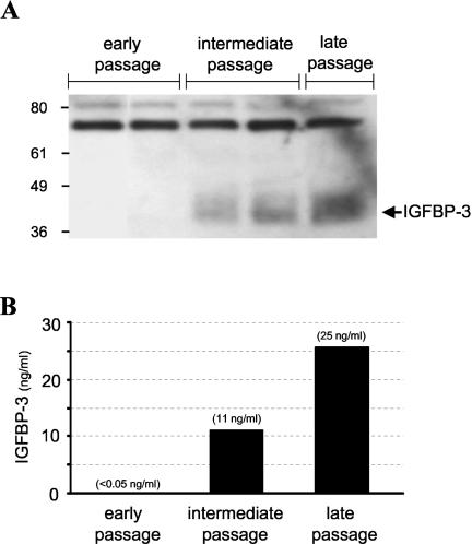
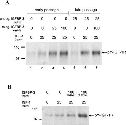
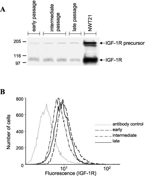
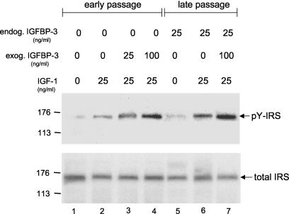
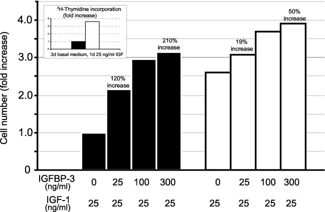
Human ectocervical cells, following retroviral transduction with the human papillomavirus type 16 E6/E7 oncogenes, are altered in their array of transcribed cellular genes, including increased mRNA for the insulin-like growth factor binding protein 3 (IGFBP-3). IGFBP-3 expression is associated with cellular senescence, and its addition to many cell types inhibits growth or induces apoptosis. By immunoblotting and enzyme-linked immunosorbent assay methods, we demonstrate that late-passage, immortalized E6/E7-transduced cells secrete high levels of IGFBP-3 (25 ng/ml), which represent a 500-fold increase compared to levels in early-passage, nonimmortalized transduced cells (<0.05 ng/ml). Concomitantly, these late-passage cervical cells exhibit an increase in sensitivity to IGF-1, including enhanced phosphorylation of the IGF receptor (IGF-R) and insulin receptor substrate as well as increased DNA synthesis (5-fold) and cell proliferation (3.7-fold). However, there was no change in the level of IGF-R in these cells (surface or total), and the cells did not synthesize IGF-1, indicating that these arms of the IGF pathway were independently regulated and not responsible for the augmented signaling. Consistent with a causal relationship between IGFBP-3 expression and enhanced IGF-1 responses, we found that early-passage cells could be converted to the late-passage, IGF-1-responsive phenotype by preincubation with IGFBP-3. Thus, in contrast to findings with some cell types, IGFBP-3 expression in cervical cells is associated with augmented IGF-1 signaling and cell proliferation and correlates with the timing of cellular immortalization.


Figures

FIG. 1.
FIG. 1.
IGFBP-3 secretion increases during in vitro passaging of E6/E7-transduced cervical epithelial cells. (A) Detection of secreted IGFBP-3 protein in cell culture medium. Culture medium supernatants were assayed for the presence of IGFBP-3 by immunoblotting as described in Materials and Methods. Measurements were performed at different passages after retroviral transduction (early, passage 40). The arrow indicates the position of the IGFBP-3 protein. (B) Quantification of secreted IGFBP-3 protein. The levels of secreted IGFBP-3 were determined by ELISA of conditioned cell culture medium at the indicated passages. Early-passage, E6/E7-transduced cells did not secrete detectable protein levels of IGFBP-3, whereas intermediate- and late-passage cultures expressed the binding protein at 11 and 25 ng/ml, respectively. The sensitivity of the ELISA was 0.04 ng/ml.
FIG. 2.
FIG. 2.
IGFBP-3 augments ligand activation of the IGF-1R. (A) Dose response of IGFBP-3 effects on IGF-1R activation. Cultured cervical cells were maintained for 3 days in basal medium containing various concentrations of IGFBP-3. The cells were then treated with 25 ng of IGF-1/ml for 10 min, lysed, and analyzed for tyrosine phosphorylation of the IGF-1R by immunoblotting as described in the text. The arrow indicates the position of the IGF-1R β-subunit. (B) Effects of immediate and prolonged IGFBP-3 exposure on IGF-1R activation. Early-passage cervical cells were exposed to IGFBP-3 at the same time as they received IGF-1 for 0 days or, alternatively, they were exposed to IGFBP-3 for 3 days prior to addition of IGF-1 (3 days). Late-passage cells responded to exogenous IGF-1 with enhanced IGF-1R phosphorylation. In early-passage cells, a similar increase in tyrosine-phosphorylated (pY) IGF-1R was observed after preincubation with IGFBP-3, indicating their conversion to the IGF-1-responsive phenotype of late-passage cells. Simultaneous IGFBP-3-IGF-1 incubation of early-passage cells diminished this effect, suggesting a necessity for preincubation with IGFBP-3.
FIG. 3.
FIG. 3.
Total and cell surface IGF-1R levels remain stable during cell passaging. (A) Detection of total cell IGF-1R. Total IGF-1R protein levels were detected by Western blotting at the indicated passages using an antibody that specifically binds to the IGF-1R β-chain. NWT21 cells (a mouse 3T3 fibroblast cell line overexpressing the IGF-1R) served as a positive control. The positions of the IGF-1R β-subunit and the IGF-1R precursor are indicated. (B) Detection of cell surface IGF-1R. Cell surface IGF-1R levels were evaluated on live cervical cells by flow cytometry using the indicated antibody. The number of cells versus relative fluorescence intensity is plotted. The levels of total and surface IGF-1R showed no significant changes during cell passaging.
FIG. 4.
FIG. 4.
IGFBP-3 enhances IGF-1R downstream signaling. Cells were incubated for 3 days in basal medium containing various concentrations of IGFBP-3. The cultures were then treated with IGF-1 for 10 min, and cell lysates were analyzed by immunoprecipitation and immunoblotting for the amount of tyrosine-phosphorylated (pY) IRS. After detection of (pY) IRS levels, the same membrane was stripped and reprobed for total IRS levels as described in the text.
FIG. 5.
FIG. 5.
IGFBP-3 enhances IGF-induced DNA synthesis and cell division. (Inset) Cellular DNA synthesis (measured by [H3]thymidine incorporation) was enhanced 3.5-fold in late-passage cells, consistent with their production of 25 ng of IGFBP-3/ml. Early-passage (black bars) and late-passage (white bars) cells (2.5 × 10⁵ per plate) were maintained for 3 days in basal growth medium followed by a 3-day period in the same medium supplemented with 25 ng of IGF-1/ml and increasing amounts of IGFBP-3. The number of cells per plate was quantified with a Coulter Counter, and the results are expressed as the fold increase in the number of cells. IGFBP-3 at 25 and 300 ng/ml induced a 120 and 210% increase in cell number, respectively. In contrast, there was only a 19 and 50% increase in the number of late-passage cells treated with the same concentrations of IGFBP-3.

Similar articles

Cited by

References

    1. Andreatta-Van Leyen, S., J. R. Hembree, and R. L. Eckert. 1994. Regulation of insulin-like growth factor 1 binding protein 3 levels by epidermal growth factor and retinoic acid in cervical epithelial cells. J. Cell. Physiol. 160:265-274. - PubMed
    1. Baege, A. C., A. Berger, R. Schlegel, T. Veldman, and R. Schlegel. 2002. Cervical epithelial cells transduced with the papillomavirus E6/E7 oncogenes maintain stable levels of oncoprotein expression but exhibit progressive, major increases in hTERT gene expression and telomerase activity. Am. J. Pathol. 160:1251-1257. - PMC - PubMed
    1. Barreca, A., M. De Luca, P. Del Monte, S. Bondanza, G. Damonte, G. Cariola, E. Di Marco, G. Giordano, R. Cancedda, and F. Minuto. 1992. In vitro paracrine regulation of human keratinocyte growth by fibroblast-derived insulin-like growth factors. J. Cell. Physiol. 151:262-268. - PubMed
    1. Baserga, R. 1995. The insulin-like growth factor 1 receptor: a key to tumor growth? Cancer Res. 55:249-252. - PubMed
    1. Baserga, R., A. Hongo, M. Rubin, M. Prisco, and B. Valentinis. 1997. The IGF-1 receptor in cell growth, transformation and apoptosis. Biochem. Biophys. Acta 1332:105-126. - PubMed

Publication types

MeSH terms

Substances

LinkOut - more resources