

Reduced sensitivity to human serum inactivation of enveloped viruses produced by pig cells transgenic for human CD55 or deficient for the galactosyl-alpha(1-3) galactosyl epitope
- PMID: 15140979
- PMCID: PMC415822
- DOI: 10.1128/JVI.78.11.5812-5819.2004
Reduced sensitivity to human serum inactivation of enveloped viruses produced by pig cells transgenic for human CD55 or deficient for the galactosyl-alpha(1-3) galactosyl epitope
Abstract
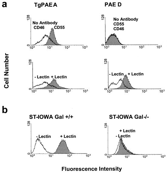
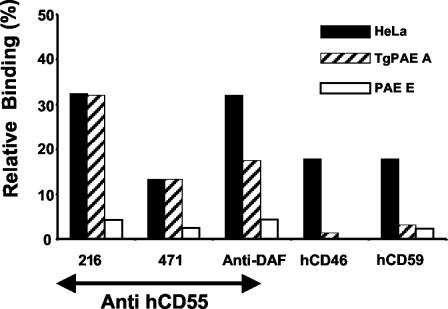
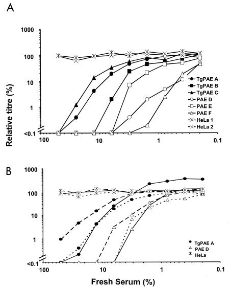
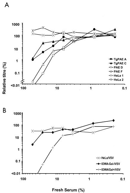
Complement activation mediated by the major xenogeneic epitope in the pig, galactosyl-alpha(1-3) galactosyl sugar structure (alpha-Gal), and human natural antibodies could cause hyperacute rejection (HAR) in pig-to-human xenotransplantation. The same reaction on viruses bearing alpha-Gal may serve as a barrier to zoonotic infection. Expressing human complement regulatory proteins or knocking out alpha-Gal epitopes in pig in order to overcome HAR may therefore pose an increased risk in xenotransplantation with regard to zoonosis. We investigated whether amphotropic murine leukemia virus, porcine endogenous retrovirus, and vesicular stomatitis virus (VSV) budding from primary transgenic pig aortic endothelial (TgPAE) cells expressing human CD55 (hCD55 or hDAF) was protected from human-complement-mediated inactivation. VSV propagated through the ST-IOWA pig cell line, in which alpha-galactosyl-transferase genes were disrupted (Gal null), was also tested for sensitivity to human complement. The TgPAE cells were positive for hCD55, and all pig cells except the Gal-null ST-IOWA expressed alpha-Gal epitopes. Through antibody binding, we were able to demonstrate the incorporation of hCD55 onto VSV particles. Viruses harvested from TgPAE cells were relatively resistant to complement-mediated inactivation by the three sources of human sera tested. Additionally, VSV from Gal-null pig cells was resistant to human complement inactivation. Such protection of enveloped viruses may increase the risk of zoonosis from pigs genetically modified for pig-to-human xenotransplantation.
Figures
Similar articles
-
A desirable transgenic strategy using GGTA1 endogenous promoter-mediated knock-in for xenotransplantation model.Sci Rep. 2022 Jun 10;12(1):9611. doi: 10.1038/s41598-022-13536-z. Sci Rep. 2022. PMID: 35688851 Free PMC article.
-
A novel mechanism of retrovirus inactivation in human serum mediated by anti-alpha-galactosyl natural antibody.J Exp Med. 1995 Nov 1;182(5):1345-55. doi: 10.1084/jem.182.5.1345. J Exp Med. 1995. PMID: 7595205 Free PMC article.
-
Sensitivity to human serum of gammaretroviruses produced from pig endothelial cells transduced with glycosyltransferase genes.Xenotransplantation. 2003 Nov;10(6):562-8. doi: 10.1034/j.1399-3089.2003.00056.x. Xenotransplantation. 2003. PMID: 14708522
-
Gal alpha (1,3)Gal, the major xenoantigen(s) recognised in pigs by human natural antibodies.Immunol Rev. 1994 Oct;141:169-90. doi: 10.1111/j.1600-065x.1994.tb00877.x. Immunol Rev. 1994. PMID: 7532618 Review.
-
Complement activation, its consequences, and blockade by gene transfer.World J Surg. 1997 Nov-Dec;21(9):907-12. doi: 10.1007/s002689900325. World J Surg. 1997. PMID: 9361503 Review.
Cited by
-
Engineering a serum-resistant and thermostable vesicular stomatitis virus G glycoprotein for pseudotyping retroviral and lentiviral vectors.Gene Ther. 2013 Aug;20(8):807-15. doi: 10.1038/gt.2013.1. Epub 2013 Jan 31. Gene Ther. 2013. PMID: 23364315 Free PMC article.
-
Incorporation of host complement regulatory proteins into Newcastle disease virus enhances complement evasion.J Virol. 2012 Dec;86(23):12708-16. doi: 10.1128/JVI.00886-12. Epub 2012 Sep 12. J Virol. 2012. PMID: 22973037 Free PMC article.
-
Social and environmental risk factors in the emergence of infectious diseases.Nat Med. 2004 Dec;10(12 Suppl):S70-6. doi: 10.1038/nm1150. Nat Med. 2004. PMID: 15577934 Free PMC article.
-
Vibrio vulnificus biotype 2 serovar E gne but not galE is essential for lipopolysaccharide biosynthesis and virulence.Infect Immun. 2008 Apr;76(4):1628-38. doi: 10.1128/IAI.01393-07. Epub 2008 Jan 28. Infect Immun. 2008. PMID: 18227162 Free PMC article.
-
A comprehensive microbiological safety approach for agarose encapsulated porcine islets intended for clinical trials.Xenotransplantation. 2016 Nov;23(6):444-463. doi: 10.1111/xen.12277. Epub 2016 Nov 11. Xenotransplantation. 2016. PMID: 27862363 Free PMC article.
References
-
- Auchincloss, H., Jr., and D. H. Sachs. 1998. Xenogeneic transplantation. Annu. Rev. Immunol. 16:433-470. - PubMed
-
- Bartosch, B., R. A. Weiss, and Y. Takeuchi. 2002. PCR-based cloning and immunocytological titration of infectious porcine endogenous retrovirus subgroup A and B. J. Gen. Virol. 83:2231-2240. - PubMed
-
- Byrne, G. W., K. R. McCurry, M. J. Martin, S. M. McClellan, J. L. Platt, and J. S. Logan. 1997. Transgenic pigs expressing human CD59 and decay-accelerating factor produce an intrinsic barrier to complement-mediated damage. Transplantation 63:149-155. - PubMed
-
- Carrington, C. A., A. C. Richards, E. Cozzi, G. Langford, N. Yannoutsos, and D. J. White. 1995. Expression of human DAF and MCP on pig endothelial cells protects from human complement. Transplant. Proc. 27:321-323. - PubMed
-
- Carrington, C. A., A. C. Richards, A. W. Tucker, A. L. Peters, and D. J. G. White. 1996. A line of transgenic pigs in which the expression of human decay-accelerating factor by endothelial cells increased in the presence of inflammatory stimuli. Xenotransplantation 3:87-91.
Publication types
MeSH terms
Substances
LinkOut - more resources
Full Text Sources
Miscellaneous
