. 2004 Jun;78(11):5848-55.
doi: 10.1128/JVI.78.11.5848-5855.2004.

Integrase-specific enhancement and suppression of retroviral DNA integration by compacted chromatin structure in vitro

Affiliations

Integrase-specific enhancement and suppression of retroviral DNA integration by compacted chromatin structure in vitro

Konstantin D Taganov et al. J Virol. 2004 Jun.

Abstract

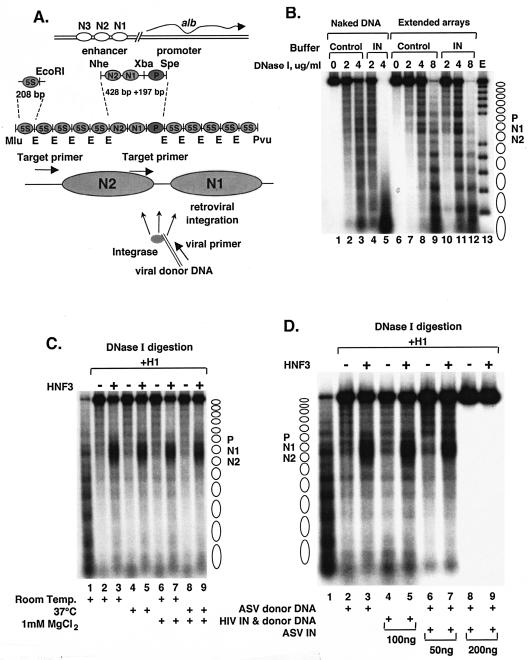
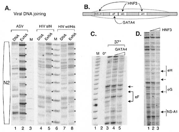
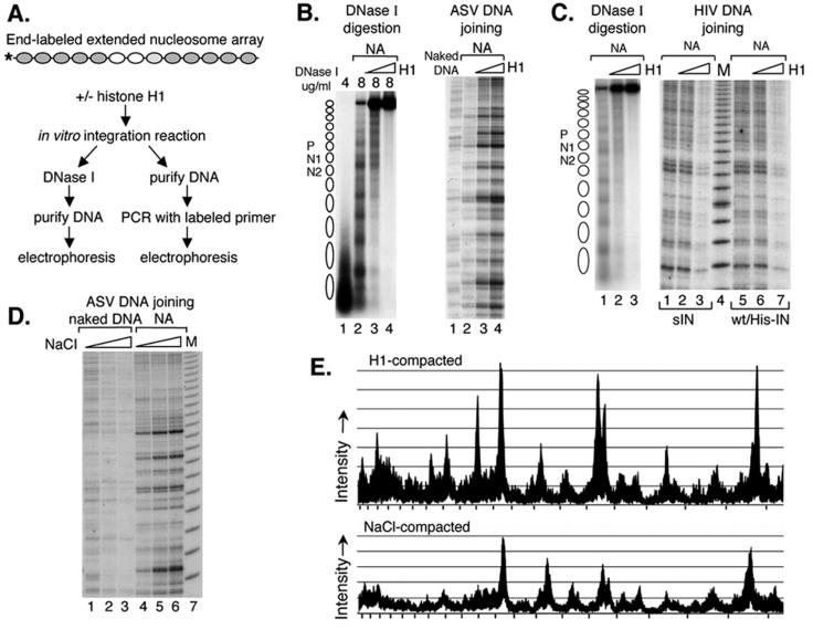
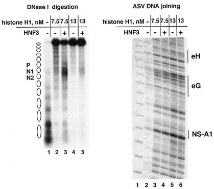
Integration of viral DNA into the host chromosome is an obligatory step in retroviral replication and is dependent on the activity of the viral enzyme integrase. To examine the influence of chromatin structure on retroviral DNA integration in vitro, we used a model target comprising a 13-nucleosome extended array that includes binding sites for specific transcription factors and can be compacted into a higher-ordered structure. We found that the efficiency of in vitro integration catalyzed by human immunodeficiency virus type 1 (HIV-1) integrase was decreased after compaction of this target with histone H1. In contrast, integration by avian sarcoma virus (ASV) integrase was more efficient after compaction by either histone H1 or a high salt concentration, suggesting that the compacted structure enhances this reaction. Furthermore, although site-specific binding of transcription factors HNF3 and GATA4 blocked ASV DNA integration in extended nucleosome arrays, local opening of H1-compacted chromatin by HNF3 had no detectable effect on integration, underscoring the preference of ASV for compacted chromatin. Our results indicate that chromatin structure affects integration site selection of the HIV-1 and ASV integrases in opposite ways. These distinct properties of integrases may also affect target site selection in vivo, resulting in an important bias against or in favor of integration into actively transcribed host DNA.


Figures

FIG. 1.
FIG. 1.
Control experiments to examine the influence of retroviral integration reaction conditions and components on nucleosome arrays. (A) Diagram of the MluI-PvuII DNA fragment used to reconstitute the albumin (alb)-5S nucleosome arrays. The locations of the PCR target and viral DNA primers are shown by arrows at the bottom. (B) Effects of integrase and integration reaction components of nucleosome arrays. DNase I digestion analysis of extended nucleosome arrays in the presence of array buffer with (IN lanes) or without (Control lanes) ASV integration reaction components. Lane E, partial EcoRI digest of a DNA end-labeled, extended nucleosome array. Positions of nucleosomes on reconstituted arrays are indicated to right. (C) Effect of temperature and MgCl2 on nucleosome compaction and accessibility of HNF3. Lanes: 1, extended nucleosome array; 2 to 9, array compacted with histone H1 in the absence (−) or presence (+) of HNF3; 2 and 3, incubation of the compacted nucleosome array for 1 h at room temperature; 4 and 5, incubation at 37°C; 6 and 7, incubation with 1 mM MgCl2 at room temperature; 8 and 9, incubation with 1 mM MgCl2 at 37°C. The locations of the nucleosomes corresponding to the albumin enhancer and promoter N1, N2, and P are indicated at the right. DNase I concentrations used: 4 μg/ml for lane 1 and 8 μg/ml for lanes 2 to 9. (D) Effects of viral DNA substrates and viral integrases on nucleosome compaction and accessibility of HNF3. Lanes: 1, extended nucleosome array; 2 to 9, array compacted with histone H1 in the absence (−) or presence (+) of HNF3 (the compacted nucleosome array was incubated for 1 h in lanes 2 and 3 with 1 mM MgCl2 and ASV donor DNA at 37°C); 4 and 5, array compacted with 100 ng of soluble HIV-1 (sIN) integrase protein and HIV-1 donor DNA; 6 and 7, array compacted with 50 ng of ASV integrase protein and ASV donor DNA; 8 and 9, array compacted with 200 ng of ASV integrase protein and ASV donor DNA amounts similar to those in lanes 6 and 7. DNase I concentrations used: 4 μg/ml for lane 1 and 8 μg/ml for lanes 2 to 11.
FIG. 2.
FIG. 2.
Retroviral integrase-mediated joining to the extended nucleosome array. (A) Controls for the retroviral integration reaction. Lanes: 1 and 2, ASV integrase-mediated joining of the cognate viral donor DNA to the naked target (DNA) and to the extended nucleosome array (ExNA) (dots mark the 10-bp periodicity for joining); 4 and 5, HIV-1sIN-mediated joining of the cognate viral donor DNA to the naked target and extended nucleosome array; 7 and 8, HIV-1wt/His integrase-mediated joining of the cognate viral donor DNA to the naked target and extended nucleosome array; 3 and 6, 10-bp DNA molecular weight marker (M). The position of the N2 nucleosome is shown at the left. (B) Sites of HNF3 and GATA4 binding to the nucleosome N1 region in the albumin enhancer region. The position of the nucleosome is indicated by the oval. Labeled boxes indicate transcription factor binding sites. (C) ASV DNA joining is blocked at sites at which transcription factor GATA4 is bound to the nucleosome array. Lanes: 1, 10-bp DNA molecular weight marker (M); 2, ASV DNA joining after incubation at 0°C; 3, ASV DNA joining after incubation at 37°C; 4, ASV DNA joining after incubation at 37°C after preincubation in 20 nM GATA4; 5, ASV DNA joining after incubation after preincubation in 40 nM GATA4. The GATA4 binding site is designated eF. Arrows show sites of target DNA protection (footprinting) from ASV DNA joining. (D) ASV DNA joining is blocked at sites at which transcription factor HNF3 is bound to the nucleosome array. Lanes: 1, ASV DNA joining to a nucleosome array at 37°C; 2, ASV DNA joining to a nucleosome array preincubated with 20 nM HNF3; 3, ASV DNA joining to a nucleosome array preincubated with 40 nM HNF3. HNF3 binding sites are designated NS-A1, eG, and eH. Arrows show sites of target DNA protection (footprinting) from ASV DNA joining.
FIG. 3.
FIG. 3.
Influence of target nucleosome array compaction on integrase-mediated joining of viral DNAs. (A) Experimental design. The extended nucleosome array was incubated with increasing amounts of histone H1 for 1 h, followed by addition of the ASV or HIV-1 integration components. One hour later, the mixture was analyzed by DNase I assay and by PCR with the N2-specific target DNA primer and an HIV-1 or ASV donor DNA primer. (B) DNase I analysis of the target DNA after incubation with the ASV DNA joining components (on the left) and PCR detection of the ASV DNA joining pattern (on the right). NA, extended nucleosome array. On the left side, DNase I concentrations are shown above the gel. Right side: lane 1, naked DNA target; lane 2, extended nucleosome array without histone H1; lane 3, array compacted by addition of 7.5 nM histone H1; lane 4, extended array compacted by addition of 13 nM histone H1. (C) DNase I analysis of the DNA used as a target for HIV-1 DNA joining (on the left) and PCR detection of the HIV-1 DNA joining pattern (on the right). DNase I concentrations used (left side, 4 μg/ml for lane 1 and 8 μg/ml for lanes 2 and 3). Right side: lanes 1 to 3, HIV-1sIN-mediated DNA joining; lanes 5 to 7, HIV-1wt/His IN-mediated DNA joining; lanes 1 and 5, extended nucleosome array; lanes 2 and 6, extended array compacted by addition of 7.5 nM histone H1; lanes 3 and 7, extended array compacted by addition of 13 nM histone H1; lane 4, 10-bp molecular weight marker (M). (D) ASV integrase-mediated joining into NaCl-mediated compacted targets. Naked DNA or the extended nucleosome array was incubated with 100 mM NaCl (lanes 2 and 5, respectively) or 120 mM NaCl (lanes 3 and 6, respectively) and then incubated with the ASV integration components. Lanes 1 and 4, ASV DNA joining to naked DNA and the extended nucleosome array in the presence of control buffer; lane 7, 10-bp molecular weight marker (M). (E) Comparison of ASV integration patterns in the region of nucleosome N1 with extended and compacted nucleosomal arrays. The sequencing gels of panels B, right side, lanes 2 and 4 (top tracing), and D, lanes 4 and 6 (bottom tracing), were scanned with a Fuji BAS-250 Bio-imaging analyzer and analyzed with Image Gauge 4.0 software. Tick marks on the x axis correspond to the 10-bp DNA molecular weight marker.
FIG. 4.
FIG. 4.
ASV integrase-mediated joining of viral DNA to the compacted array opened locally by the HNF3 transcription factor. The histone H1-compacted nucleosome array was incubated for 2 h with transcription factor HNF3, and then the ASV integration components were added. One hour later, the mixture was analyzed by DNase I assay and by PCR with N1-specific target DNA and ASV donor DNA primers. DNase I analysis is shown in the left-side DNA. Lanes: 1, extended array; 2, extended array compacted by 7.5 nM histone H1; 3, 7.5 nM histone H1-compacted array opened locally by HNF3; 4, extended array compacted by 13 nM histone H1; 5, 13 nM histone H1-compacted array opened locally by HNF3. The DNase I concentrations used were 4 μg/ml for lane 1 and 8 μg/ml for lanes 2 to 5. Results from the joining assay are on the right side. Lanes:1, 10-bp marker; 2, viral DNA joining to the extended nucleosome array; 3 and 4, DNA joining to the extended nucleosome array compacted with 7.5 nM histone H1 in the absence (−) or presence (+) of HNF3; 5 and 6, with 13 nM histone H1 in the absence (−) or presence (+) of HNF3.

Similar articles

Cited by

References

    1. Bor, Y. C., F. D. Bushman, and L. E. Orgel. 1995. In vitro integration of human immunodeficiency virus type 1 cDNA into targets containing protein-induced bends. Proc. Natl. Acad. Sci. USA 92:10334-10338. - PMC - PubMed
    1. Carruthers, L. M., J. Bednar, C. L. Woodcock, and J. C. Hansen. 1998. Linker histones stabilize the intrinsic salt-dependent folding of nucleosomal arrays: mechanistic ramifications for higher-order chromatin folding. Biochemistry 37:14776-14787. - PubMed
    1. Chaya, D., T. Hayamizu, M. Bustin, and K. S. Zaret. 2001. Transcription factor FoxA (HNF3) on a nucleosome at an enhancer complex in liver chromatin. J. Biol. Chem. 276:44385-44389. - PubMed
    1. Check, E. 2003. Second cancer case halts gene-therapy trials. Nature 421:305. - PubMed
    1. Check, E. 2002. A tragic setback. Nature 420:116-118. - PubMed

Publication types

LinkOut - more resources