

Hepatitis C virus persistence after spontaneous or treatment-induced resolution of hepatitis C
- PMID: 15140984
- PMCID: PMC415836
- DOI: 10.1128/JVI.78.11.5867-5874.2004
Hepatitis C virus persistence after spontaneous or treatment-induced resolution of hepatitis C
Abstract
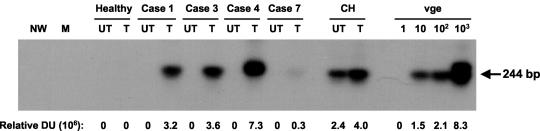
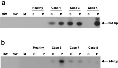
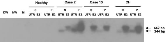
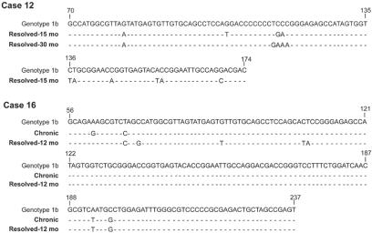
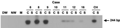
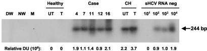
It is presumed that resolution of hepatitis C, as evidenced by normalization of liver function tests and disappearance of hepatitis C virus (HCV) RNA from serum, as determined by conventional laboratory assays, reflects virus eradication. In this study, we examined the expression of the HCV genome in the sera, peripheral blood mononuclear cells (PBMC), and, on some occasions, monocyte-derived dendritic cells (DC) long after resolution of hepatitis C by using a highly sensitive reverse transcription (RT)-PCR-nucleic acid hybridization (RT-PCR-NAH) assay. The samples obtained from 16 randomly selected patients (5 with spontaneous and 11 with treatment-induced resolution), monitored for up to 5 years, were studied by qualitative and semiquantitative RT-PCR-NAH and by real-time RT-PCR to detect the HCV RNA positive strand. The replicative HCV RNA negative strand was examined in PBMC after culture with a T-cell proliferation stimulating mitogen. The findings show that HCV RNA was carried in the convalescent-phase sera and/or PBMC in all 16 individuals investigated. Also, DC from six of seven patients were reactive for the HCV genome. Importantly, traces of the HCV RNA negative strand, suggesting progressing virus replication, were detected in the majority of mitogen-stimulated PBMC, including four samples collected 5 years after recovery. Sequencing of the HCV 5' untranslated region fragment revealed genotype 1b in four of nine individuals examined and genotypes 1a and 2a in three and two patients, respectively. These results imply that HCV RNA can persist at very low levels in the serum and peripheral lymphoid cells and that an intermediate replicative form of the HCV genome can persist in PBMC for many years after apparently complete spontaneous or antiviral therapy-induced resolution of chronic hepatitis C.
Figures
Similar articles
-
Persistence of Hepatitis C Virus Traces after Spontaneous Resolution of Hepatitis C.PLoS One. 2015 Oct 16;10(10):e0140312. doi: 10.1371/journal.pone.0140312. eCollection 2015. PLoS One. 2015. PMID: 26473969 Free PMC article. Clinical Trial.
-
Clearance of HCV RNA in peripheral blood mononuclear cell as a predictor of response to antiviral therapy in patients with chronic hepatitis C.Hepatobiliary Pancreat Dis Int. 2005 Nov;4(4):550-3. Hepatobiliary Pancreat Dis Int. 2005. PMID: 16286260
-
Positive and negative strand of hepatitis C virus RNA sequences in peripheral blood mononuclear cells in patients with chronic hepatitis C: no correlation with viral genotypes 1b, 2a, and 2b.J Med Virol. 1997 Jul;52(3):270-4. J Med Virol. 1997. PMID: 9210035
-
Hepatitis C virus lymphotropism: lessons from a decade of studies.Dig Liver Dis. 2007 Sep;39 Suppl 1:S38-45. doi: 10.1016/s1590-8658(07)80009-0. Dig Liver Dis. 2007. PMID: 17936221 Review.
-
The yin and yang of hepatitis C: synthesis and decay of hepatitis C virus RNA.Nat Rev Microbiol. 2015 Sep;13(9):544-58. doi: 10.1038/nrmicro3506. Epub 2015 Aug 10. Nat Rev Microbiol. 2015. PMID: 26256788 Free PMC article. Review.
Cited by
-
Patient-derived hepatitis C virus inhibits CD4⁺ but not CD8⁺ T lymphocyte proliferation in primary T cells.Virol J. 2015 Jun 19;12:93. doi: 10.1186/s12985-015-0322-4. Virol J. 2015. PMID: 26084511 Free PMC article.
-
Cell culture-produced hepatitis C virus does not infect peripheral blood mononuclear cells.Hepatology. 2008 Dec;48(6):1843-50. doi: 10.1002/hep.22550. Hepatology. 2008. PMID: 19003912 Free PMC article.
-
Prevalence and follow-up of occult HCV infection in an Italian population free of clinically detectable infectious liver disease.PLoS One. 2012;7(8):e43541. doi: 10.1371/journal.pone.0043541. Epub 2012 Aug 22. PLoS One. 2012. PMID: 22927986 Free PMC article.
-
Antibody-dependent enhancement of hepatitis C virus infection.J Virol. 2008 Mar;82(5):2140-9. doi: 10.1128/JVI.01867-07. Epub 2007 Dec 19. J Virol. 2008. PMID: 18094180 Free PMC article.
-
Hepatitis C virus upregulates B-cell receptor signaling: a novel mechanism for HCV-associated B-cell lymphoproliferative disorders.Oncogene. 2016 Jun 9;35(23):2979-90. doi: 10.1038/onc.2015.364. Epub 2015 Oct 5. Oncogene. 2016. PMID: 26434584 Free PMC article.
References
-
- Auffermann-Gretzinger, S., E. B. Keeffe, and S. Levy. 2001. Impaired dendritic cell maturation in patients with chronic, but not resolved, hepatitis C virus infection. Blood 97:3171-3176. - PubMed
-
- Bain, C., A. Fatmi, F. Zoulim, J. P. Zarski, C. Trepo, and G. Inchauspe. 2001. Impaired allostimulatory function of dendritic cells in chronic hepatitis infection. Gastroenterology 120:512-524. - PubMed
-
- Cohen, J. 1999. The scientific challenge of hepatitis C virus. Science 285:26-30. - PubMed
Publication types
MeSH terms
Substances
LinkOut - more resources
Full Text Sources
Medical
