. 2004 Jun;78(11):5900-12.
doi: 10.1128/JVI.78.11.5900-5912.2004.

Processing of a pestivirus protein by a cellular protease specific for light chain 3 of microtubule-associated proteins

Affiliations

Processing of a pestivirus protein by a cellular protease specific for light chain 3 of microtubule-associated proteins

Jens Fricke et al. J Virol. 2004 Jun.

Abstract

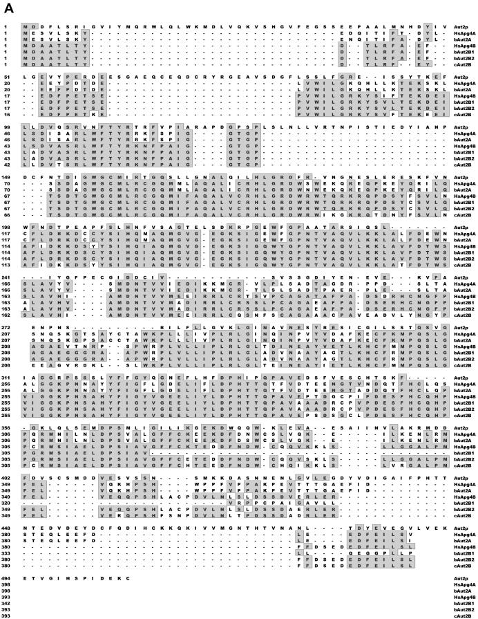
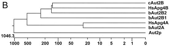
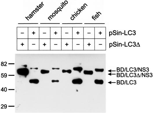
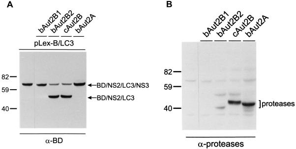
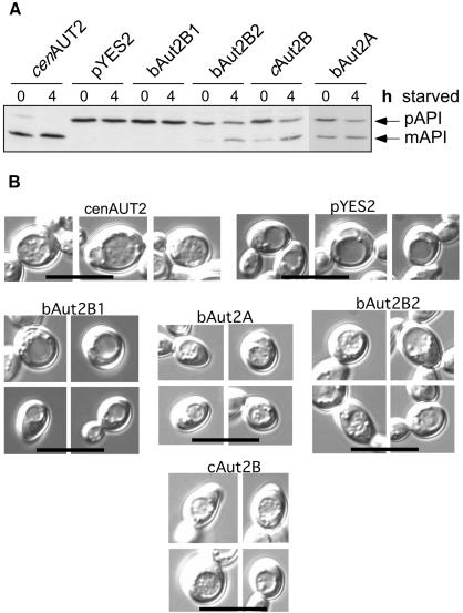
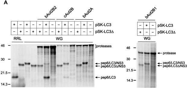
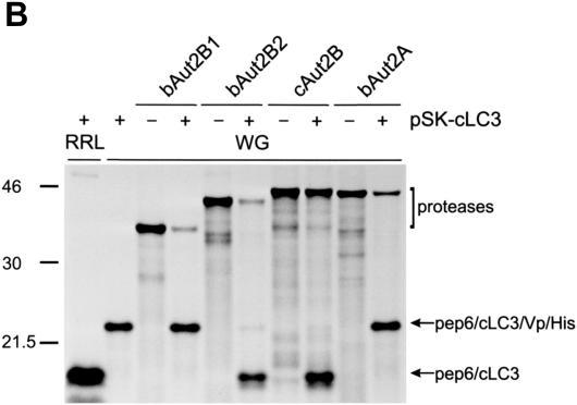
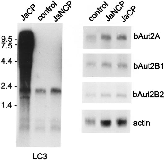
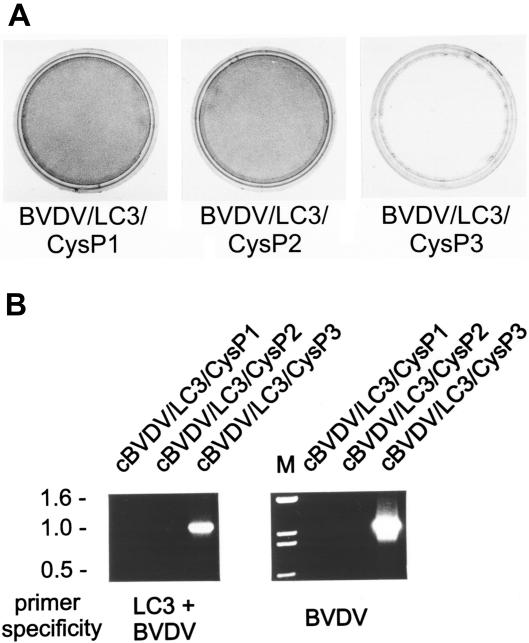
The genome of the cytopathogenic (cp) bovine viral diarrhea virus (BVDV) JaCP contains a cellular insertion coding for light chain 3 (LC3) of microtubule-associated proteins, the mammalian homologue of yeast Aut7p/Apg8p. The cellular insertion induces cp BVDV-specific processing of the viral polyprotein by a cellular cysteine protease homologous to the known yeast protease Aut2p/Apg4p. Three candidate bovine protease genes were identified on the basis of the sequence similarity of their products with the Saccharomyces cerevisiae enzyme. The search for a system for functional testing of these putative LC3-specific proteases revealed that the components involved in this processing have been highly conserved during evolution, so that the substrate derived from a mammalian virus is processed in cells of mammalian, avian, fish, and insect origin, as well as in rabbit reticulocyte lysate, but not in wheat germ extracts. Moreover, two of these proteases and a homologous protein from chickens were able to rescue the defect of a yeast AUT2 deletion mutant. In coexpression experiments with yeast and wheat germ extracts one of the bovine proteases and the corresponding enzyme from chickens were able to process the viral polyprotein containing LC3. Northern blots showed that bovine viral diarrhea virus infection of cells has no significant influence on the expression of either LC3 or its protease, bAut2B2. However, LC3-specific processing of the viral polyprotein containing the cellular insertion is essential for replication of the virus since mutants with changes in the LC3 insertion significantly affecting processing at the LC3/NS3 site were not viable.


Figures

FIG. 1.
FIG. 1.
Alignment of Aut2p/Apg4p-homologous sequences. (A) Amino acid sequence alignment of the bovine (bAut2A, bAut2B1, and bAut2B2) and chicken (cAut2B) Aut2p/Apg4p homologues with yeast Aut2p/Apg4 and two human homologues (HsApg4A and HsApg4B [19]). Sequence homology is indicated by gray boxes; a hyphen indicates that the respective sequence has a deletion of one amino acid at the position. The total lengths in amino acids of the aligned sequences are as follows: Aut2p, 506; HsApg4A, 398; bAut2A, 398; HsApg4B, 393; BAut2B1, 342; bAut2B2, 393; cAut2B, 393. (B) Dendritic diagram comparing the amino acid sequences in panel A. The diagram was generated with the Megalign program (Lasergene software; DNASTAR).
FIG. 1.
FIG. 1.
Alignment of Aut2p/Apg4p-homologous sequences. (A) Amino acid sequence alignment of the bovine (bAut2A, bAut2B1, and bAut2B2) and chicken (cAut2B) Aut2p/Apg4p homologues with yeast Aut2p/Apg4 and two human homologues (HsApg4A and HsApg4B [19]). Sequence homology is indicated by gray boxes; a hyphen indicates that the respective sequence has a deletion of one amino acid at the position. The total lengths in amino acids of the aligned sequences are as follows: Aut2p, 506; HsApg4A, 398; bAut2A, 398; HsApg4B, 393; BAut2B1, 342; bAut2B2, 393; cAut2B, 393. (B) Dendritic diagram comparing the amino acid sequences in panel A. The diagram was generated with the Megalign program (Lasergene software; DNASTAR).
FIG. 2.
FIG. 2.
Processing of a bovine LC3 fusion protein in cells of different origins. Shown is the expression of proteins containing LC3 sequences in cells of different origins. A Sindbis virus expression system was used to express a substrate composed of the LexA BD domain, the LC3 insertion as found in the BVDV isolate JaCP, and a fragment of the JaCP NS3 protein (plasmid, pSin-LC3; protein, BD/LC3/NS3) or the same protein with a deletion of the carboxy-terminal 18 amino acids of the LC3 insertion (plasmid, pSin-LC3Δ; protein, BD/LC3Δ/NS3). Protein extracts of infected cells were separated by SDS-PAGE and analyzed by Western blotting with a commercially available antiserum against the LexA-BD protein. The positions of molecular mass markers in kilodaltons are on the left. Arrows, indicated proteins.
FIG. 3.
FIG. 3.
Coexpression of candidate proteases and LC3 substrate in yeast. Shown is coexpression of a mammalian substrate protein containing LC3 with different candidate proteases. Plasmid pLex-B/LC3 (codes for protein BD/NS2/LC3/NS3) was expressed in aut2Δ yeast cells (strain YCV3) either with the pYES2 plasmid alone (left lane) or with pYES2 plasmids coding for one of the bovine candidate proteases (bAut2A, bAut2B1, or bAut2B2) or the chicken protease (cAut2B). The protein extracts were separated by SDS-PAGE and transferred to nitrocellulose membranes. The same blot was first analyzed with the antiserum against LexA-BD (α-BD; A) and then with a mixture of antisera directed against conserved regions within the A and B proteases (α-proteases; B). Note that in panel B the bands already detected in panel A are still visible. For unknown reasons the expression level of bAut2B2 seemed to be lower than that of bAut2A or cAut2B.
FIG. 4.
FIG. 4.
Rescue of yeast Aut2/Apg4 mutants by higher mammalian and avian proteases. Autophagy is impaired in yeast cells with inactivated protease Aut2p/Apg4p, as detectable in, e.g., a defect in maturation of pAPI and the inability to accumulate autophagic bodies in the vacuole during starvation. The different candidate proteases homologous to Aut2p/Apg4 were expressed in aut2-deficient yeast cells and tested with regard to trans complementation of the defects concerning pAPI maturation (A) and accumulation of autophagic bodies in the vacuole (B). (A) aut2Δ yeast cells expressing, from left to right, the yeast AUT2 gene, an empty pYES2 vector, and the indicated candidate mammalian proteinases. The cells were grown in selective galactose medium to the stationary phase (0 h starved) and further incubated in 1% potassium acetate for 4 h (4 h starved). Crude cell extracts were then subjected to SDS-PAGE, electroblotted on polyvinylidene difluoride membranes, and probed with antibodies against pAPI. mAPI, mature API. (B) aut2Δ yeast cells expressing the candidate mammalian proteinases from a pYES2 vector were checked for their ability to accumulate autophagic bodies in their vacuoles when starved for nitrogen in the presence of the proteinase B inhibitor PMSF. The cells were grown in selective galactose medium to the stationary phase and further starved for 4 h in nitrogen-free SD(-N) medium containing 1 mM PMSF. As controls cells expressing the yeast AUT2 and cells carrying an empty pYES2 vector are included. The cells were visualized by light microscopy with Nomarski optics. Bar: 10 μm. The vacuole is easily seen as a round structure in the center of the cell. Autophagic bodies look like granules within the vacuole.
FIG. 5.
FIG. 5.
In vitro translation of LC3 substrates and proteases in WG. Shown is coexpression of mammalian substrates and candidate proteases by in vitro translation. Fusion proteins composed of part of BVDV NS2 (pep6) and LC3-derived sequences with different carboxy-terminal extensions were expressed by in vitro translation using either the RRL or the WG system as indicated. (A) The LC3 sequence was either the authentic insertion identified in the BVDV JaCP polyprotein followed by part of BVDV NS3 (plasmid, pSK-LC3; protein, pep6/LC3/NS3) or an equivalent protein with the last 18 amino acids of LC3 deleted (plasmid, pSK-LC3Δ; protein, pep6/LC3Δ18/NS3). (B) The full-length bovine LC3 sequence, including the natural carboxy-terminal extension of five amino acids, followed by two tag sequences (VP5-six-His; plasmid pSK-cLC3) was expressed. The substrate was cotranslated with the different indicated proteases.
FIG. 5.
FIG. 5.
In vitro translation of LC3 substrates and proteases in WG. Shown is coexpression of mammalian substrates and candidate proteases by in vitro translation. Fusion proteins composed of part of BVDV NS2 (pep6) and LC3-derived sequences with different carboxy-terminal extensions were expressed by in vitro translation using either the RRL or the WG system as indicated. (A) The LC3 sequence was either the authentic insertion identified in the BVDV JaCP polyprotein followed by part of BVDV NS3 (plasmid, pSK-LC3; protein, pep6/LC3/NS3) or an equivalent protein with the last 18 amino acids of LC3 deleted (plasmid, pSK-LC3Δ; protein, pep6/LC3Δ18/NS3). (B) The full-length bovine LC3 sequence, including the natural carboxy-terminal extension of five amino acids, followed by two tag sequences (VP5-six-His; plasmid pSK-cLC3) was expressed. The substrate was cotranslated with the different indicated proteases.
FIG. 6.
FIG. 6.
Northern blot with an LC3 probe and different protease probes. RNA of MDBK cells (control) and MDBK cells infected with the indicated viruses was analyzed in a Northern blot with an LC3 probe (left) or probes specific for the different (putative) protease genes identified in this report. A chicken actin probe served as a control. The probes used for hybridization are indicated below or on the right of the gels. On the left, size marker bands for the left gel are indicated. For MDBK cells, hybridization with an LC3 probe leads to bands of about 2.0, 0.65, and 0.5 kb. Because of the smear resulting from the partially degraded viral RNA, the LC3 cellular signal could not be quantified for JaCP-infected cells.
FIG. 7.
FIG. 7.
Transfection of full-length BVDV RNA with mutated LC3 insertion. MDBK cells infected with a noncp helper virus (BVDV NCP1 [31]) were transfected with RNA transcribed in vitro from BVDV full-length plasmids (30) with mutated LC3 insertions and no duplication of viral sequences. As indicated, the mutations affected the LC3 sequence upstream of the LC3/NS3 cleavage site at position P1, P2, or P3. (A) Crystal violet staining of cells infected with cell lysate prepared after RNA transfection and three blind passages. (B) Agarose gel with samples obtained by RT-PCR with primers Ol-JasInsII and Ol-BVD30RII (specific for the combination of LC3 and BVDV NS3 coding sequences) (left) or Ol-BVD21.1 and Ol-BVD30RII (specific for a BVDV NCP7/CP7 NS2-3 fragment). Note that the primers do not amplify the sequences of the helper virus BVDV NCP1 used for infection of the cells prior to RNA transfection.
FIG. 8.
FIG. 8.
Transfection of full-length BVDV RNA containing duplicated viral sequences and mutated LC3 insertions. MDBK cells were transfected with RNA transcribed in vitro from plasmid pA/B-JaCP/dp (29) with mutated LC3 insertions. The parental plasmid mimics the BVDV JaCP genome with two duplications of viral sequences flanking the LC3 insertion. It was shown before that such an arrangement leads to a viable cp virus when the wild-type LC3 insertion is present (29). As indicated the mutations affected the LC3 sequence upstream of the LC3/NS3 cleavage site at position P1, P2, or P3. (A) Crystal violet staining of transfected cells (conducted about 72 h posttransfection). (B) Northern blot with a BVDV NCP1 probe (31) and RNAs of cells infected with the viruses cJaCP/CysP3 or cJaCP/CysP1 recovered from the above-described plasmids or the BVDV isolate JaCP or JaNCP. The “bands” visible in addition to the viral genome represent gel artifacts due to compression by the cellular 28S and 18S rRNAs. (C) Agarose gel with samples obtained by RT-PCR with primers Ol-JasInsII and Ol-BVD30RII (specific for the combination of LC3 and BVDV NS3 coding sequences) (left) or Ol-BVD21.1 and Ol-BVD30 RII (specific for a BVDV NCP7/CP7 NS2-3 fragment). The bands of about 0.7 kb in the right part correspond to an NS2-3 fragment without the LC3 insertion that is present in the 5′ terminal half of a genome with duplicated viral sequences in addition to the second copy of the gene that contains the LC3 insertion. For cJaCP/CysP1 the amplicon is derived from the noncp genome that was generated by deletion of the duplicated sequence including the LC3 insertion.

Similar articles

Cited by

References

    1. Ausubel, F. M., R. Brent, R. E. Kingston, and D. D. Moore (ed.). 1987. Current protocols in molecular biology. Greene Publishing Associates, New York, N.Y.
    1. Baroth, M., M. Orlich, H. J. Thiel, and P. Becher. 2000. Insertion of cellular NEDD8 coding sequences in a pestivirus. Virology 278:456-466. - PubMed
    1. Barth, H., and M. Thumm. 2001. A genomic screen identifies AUT8 as a novel gene essential for autophagy in the yeast Saccharomyces cerevisiae. Gene 274:151-156. - PubMed
    1. Becher, P., A. D. Shannon, N. Tautz, and H.-J. Thiel. 1994. Molecular characterization of border disease virus, a pestivirus from sheep. Virology 198:542-551. - PubMed
    1. Becher, P., H. J. Thiel, M. Collins, J. Brownlie, and M. Orlich. 2002. Cellular sequences in pestivirus genomes encoding gamma-aminobutyric acid (A) receptor-associated protein and Golgi-associated ATPase enhancer of 16 kilodaltons. J. Virol. 76:13069-13076. - PMC - PubMed

Publication types