. 2004 Jun;78(11):6043-7.
doi: 10.1128/JVI.78.11.6043-6047.2004.

Recombinant Sendai virus expressing the G glycoprotein of respiratory syncytial virus (RSV) elicits immune protection against RSV

Affiliations

Recombinant Sendai virus expressing the G glycoprotein of respiratory syncytial virus (RSV) elicits immune protection against RSV

Toru Takimoto et al. J Virol. 2004 Jun.

Abstract

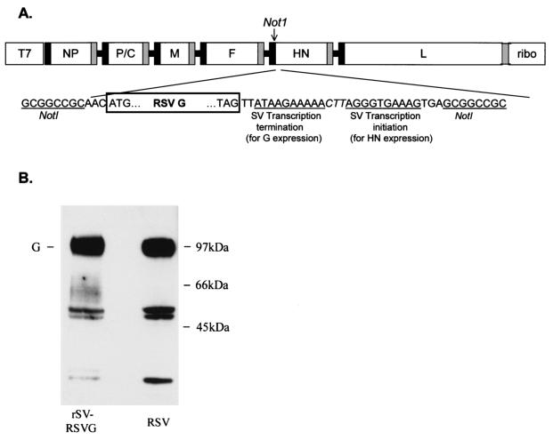
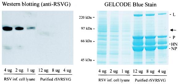
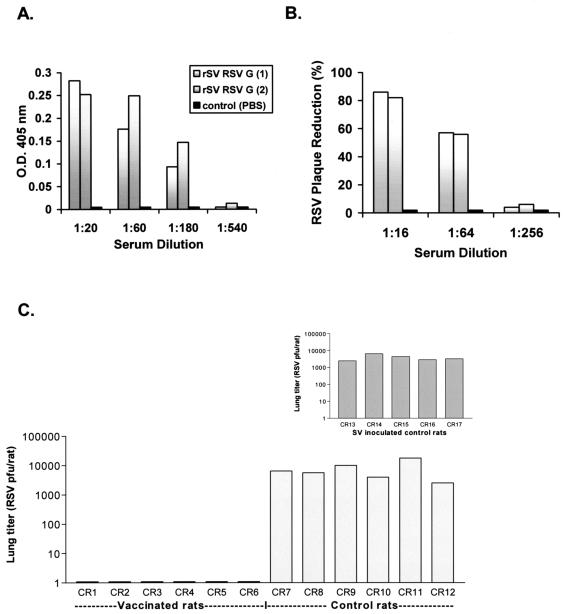
Although RSV causes serious pediatric respiratory disease, an effective vaccine does not exist. To capture the strengths of a live virus vaccine, we have used the murine parainfluenza virus type 1 (Sendai virus [SV]) as a xenogeneic vector to deliver the G glycoprotein of RSV. It was previously shown (J. L. Hurwitz, K. F. Soike, M. Y. Sangster, A. Portner, R. E. Sealy, D. H. Dawson, and C. Coleclough, Vaccine 15:533-540, 1997) that intranasal SV protected African green monkeys from challenge with the related human parainfluenza virus type 1 (hPIV1), and SV has advanced to clinical trials as a vaccine for hPIV1 (K. S. Slobod, J. L. Shenep, J. Lujan-Zilbermann, K. Allison, B. Brown, R. A. Scroggs, A. Portner, C. Coleclough, and J. L. Hurwitz, Vaccine, in press). Recombinant SV expressing RSV G glycoprotein was prepared by using reverse genetics, and intranasal inoculation of cotton rats elicited RSV-specific antibody and elicited protection from RSV challenge. RSV G-recombinant SV is thus a promising live virus vaccine candidate for RSV.


Figures

FIG. 1.
FIG. 1.
Design of pSV(+)RSVG and expression of RSV G target gene. (A) A unique NotI restriction enzyme site was created in the noncoding region of the HN gene to insert the RSV G glycoprotein gene. The NotI site was introduced into a subcloned ClaI-EcoRI fragment of pSeV(+) in pTF1 (21) by using a QuikChange Site-Directed Mutagenesis kit (Stratagene, La Jolla, Calif.). This modified fragment was then substituted for the wild-type fragment in pSeV(+) to create pSV(+)N. RSV G gene was cloned by using a forward primer which included a NotI site and a reverse primer which included an SV transcription termination signal and another transcription initiation signal (separated by an intergenic linker sequence [CTT]), followed by the NotI site. Thus, RSV G transcription initiated from the upstream SV HN transcription initiation sequence and terminated by using the new termination sequence. SV HN transcription initiated by using the newly introduced transcription initiation sequence. T7, T7 promoter; ribo, hepatitis delta virus ribozyme sequence. Black and gray boxes represent transcription initiation and termination sequences, respectively, of the nucleoprotein (NP), polymerase (P), matrix (M), fusion (F), hemagglutinin-neuraminidase (HN), and large (L) protein. (B) Western blot examination of lysates of HEp-2 cells (approximately 106 cells) infected with rSV RSVG (left lane) or wild-type RSV (right lane). Cells were lysed with 0.2 ml of TNE buffer (10 mM Tris [pH 7.4], 150 mM NaCl, 0.5% NP-40, and 1 mM EDTA) and were clarified (15,000 × g, 10 min). Supernatants were subjected to sodium dodecyl sulfate-polyacrylamide gel electrophoresis in nonreducing conditions, transferred to Immobilon membrane (Millipore, Danvers, Mass.), and developed with RSV G-specific monoclonal antibody (clone 63-10F; Chemicon International Inc., Temecula, Calif.). Fully glycosylated RSV G protein (both N- and O-linked glycosylation) runs at approximately 90 kDa (G). Middle band likely represents partially glycosylated G protein, and the lower band represents unglycosylated G.
FIG. 2.
FIG. 2.
rSV RSVG particles do not contain G protein. rSV RSVG viral particles were first purified from infected cell supernatant (HEp-2 cells; MOI 5; 72 h at 34°C) by sucrose gradient centrifugation and then resolved by sodium dodecyl sulfate-polyacrylamide gel electrophoresis (SDS-PAGE). RSV-infected HEp2 cell lysates (control) were also run on SDS-PAGE. (Left) Gels were transferred to a nitrocellulose sheet and were reacted with a monoclonal antibody directed against RSV G protein. The quantity of lysate run in each lane is indicated. RSV G detection assay (SuperSignal West Pico Chemiluminescent kit; Pierce Biotechnology, Rockland, Ill.) has a sensitivity of 1 pg. In contrast to the RSV G protein detected from all control preparations (RSV infected cell lysate), RSV G protein could not be detected from purified rSV RSVG, even with 12 μg of viral proteins. (Right) SDS-PAGE gels were also stained with GelCode Blue Stain Reagent (Pierce Biotechnology) to identify all proteins present in purified rSV RSVG particles. Arrow indicates the location of RSV G protein in the gel. SV protein abbreviations are the same as those described in the legend to Fig. 1.
FIG. 3.
FIG. 3.
rSV RSVG-primed cotton rats generate RSV-specific antibody. (A) Serial serum dilutions from immunized (gray bars) and control rats receiving PBS (black bars) were examined by enzyme-linked immunosorbent assay. RSV G-transfected 293T cells served as the source of antigen. 293T cells grown in polylysine-coated 24-well plates were transfected with pCAGGS (0.5 μg; in LipofectAMINE) for 24 h. After transfection, wells were washed and reacted with serial dilutions of cotton rat test serum (in PBS-0.1% bovine serum albumin [BSA]) for 30 min at room temperature (RT), washed again, and then reacted with rabbit anti-cotton rat IgG (1:3,000 in PBS-0.1% BSA; Virion Systems, Rockville, Md.) for 30 min at RT. Wells were washed and incubated with anti-rabbit immunoglobulin G-horseradish peroxidase conjugate (1:3,000 in PBS-0.1% BSA) for 30 min at RT. Wells were washed again and reacted with 2,2′-azinobis (3-ethylbenzothiazoline-6-sulfonic acid) substrate and measured by spectrophotometer. Results are reported as the absolute optical density at 405 nm (O.D. 405 nm) at each serum dilution. (B) RSV neutralizing activity was tested by plaque assay. Serum samples were mixed with RSV (100 to 500 PFU/well; 1 h at RT), and virus-serum mixtures were inoculated to HEp-2 cell monolayers (80 to 90% confluent) on 6-well plates, incubated for 1 h (37°C, 5% CO2), and then overlayed with medium containing methylcellulose. Plates were incubated for 7 days (37°C, 5% CO2), after which the methylcellulose was removed, the cells were fixed (formalin phosphate), and the plates were stained (hematoxylin and eosin) for plaque enumeration. Results are reported as the percent plaque reduction (y axis) observed at each serum dilution (x axis). (C) To measure protection from RSV challenge, sets of vaccinated (CR1 to CR6) and control (PBS recipients CR7 to CR12 and SV-inoculated CR13 to CR17, inset chart) were challenged with intranasal RSV (106 PFU) approximately 4 weeks after priming. Three days postchallenge animals were sacrificed, lungs were harvested, and lung tissue was cut into large fragments, mixed with PBS (1 ml), and processed with a mechanical Dounce homogenizer (PowerGen125 PCR Tissue Homogenizing kit; Fisher Scientific) over ice. Homogenates were then centrifuged and supernatants were collected and cryopreserved for virus quantitation (supernatant volume ranged between 4.0 to 6.5 ml/rat). RSV burden in lung supernatants was determined by plaque assay (see above) of serial dilutions of supernatants. Virus titers were determined by estimating the plaque number per volume plated, and the total virus burden per rat was calculated based on the total volume of supernatant obtained (reported along the y axis).

References

    1. Anderson, L. J., R. A. Parker, and R. L. Strikas. 1990. Association between respiratory syncytial virus outbreaks and lower respiratory tract deaths of infants and young children. J. Infect. Dis. 161:640-646. - PubMed
    1. Bousse, T., T. Matrosovich, A. Portner, A. Kato, Y. Nagai, and T. Takimoto. 2002. The long noncoding region of the human parainfluenza virus type 1 gene contributes to the read-through transcription at the M-F gene junction. J. Virol. 76:8244-8251. - PMC - PubMed
    1. Connors, M., A. B. Kulkarni, C.-Y. Firestone, K. L. Holmes, H. C. Morse, A. V. Sotnikov, and B. R. Murphy. 1992. Pulmonary histopathology induced by respiratory syncytial virus (RSV) challenge of formalin-inactivated RSV-immunized BALB/c mice is abrogated by depletion of CD4+ T cells. J. Virol. 66:7444-7451. - PMC - PubMed
    1. Crowe, J. E., Jr., P. L. Collins, W. T. London, R. M. Chanock, and B. R. Murphy. 1993. A comparison in chimpanzees of the immunogenicity and efficacy of live attenuated respiratory syncytial virus (RSV) temperature-sensitive mutant vaccines and vaccinia virus recombinants that express the surface glycoproteins of RSV. Vaccine 11:1395-1404. - PubMed
    1. Fulginiti, V. A., J. J. Eller, O. F. Sieber, J. W. Joyner, M. Minamitani, and G. Meiklejohn. 1969. I. A field trial of two inactivated respiratory virus vaccines; an aqueous trivalent parainfluenza virus vaccine and an alum-precipitated respiratory syncytial virus vaccine. Am. J. Epidemiol. 89:435-448. - PubMed

Publication types

Substances

LinkOut - more resources