

doi: 10.1128/JVI.78.11.6073-6076.2004.
Human APOBEC3F is another host factor that blocks human immunodeficiency virus type 1 replication
Affiliations
- PMID: 15141007
- PMCID: PMC415831
- DOI: 10.1128/JVI.78.11.6073-6076.2004
Item in Clipboard
Human APOBEC3F is another host factor that blocks human immunodeficiency virus type 1 replication
J Virol.
2004 Jun.
Abstract
Recently, APOBEC3G has been identified as a host factor that blocks retroviral replication. It introduces G to A hypermutations in newly synthesized minus strand viral cDNA at the step of reverse transcription in target cells. Here, we identified the human APOBEC3F protein as another host factor that blocks human immunodeficiency virus type 1 (HIV-1) replication. Similar to APOBEC3G, APOBEC3F also induced G to A hypermutations in HIV genomic DNA, and the viral Vif protein counteracted its activity. Thus, APOBEC family members might have evolved as a general defense mechanism of the body against retroviruses, retrotransposons, and other mobile genetic elements.
Figures
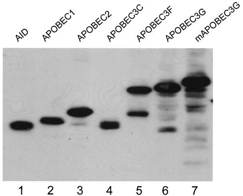
Expression of APOBEC proteins. Human APOBEC proteins AID, APOBEC1, APOBEC2, APOBEC3C, APOBEC3F, APOBEC3G, and mAPOBEC3G were cloned into pcDNA3 with a V5 tag. These proteins were expressed in 293T cells and detected with a monoclonal anti-V5 antibody.
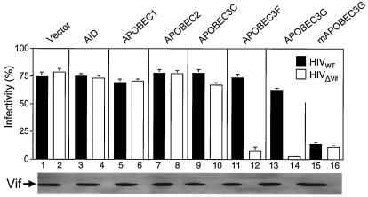
Function of APOBEC proteins in viral replication. APOBEC proteins were coexpressed with wild-type or Vif-defective HIV in 293T cells. After normalization by p24Gag enzyme-linked immunosorbent assay, equal amounts of viruses were used to infect GHOST-R3/X4/R5 cells. The expression of Vif in cell lysates from wild-type or Vif-defective viruses was demonstrated by Western blotting (bottom).
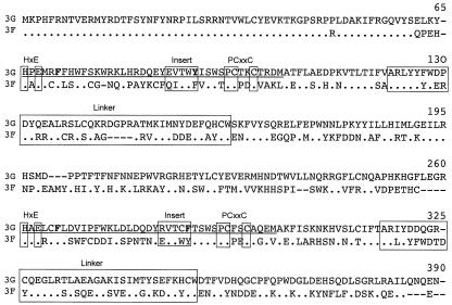
Sequence alignment between human APOBEC3G (3G) and APOBEC3F (3F). Two zinc-finger domains are underlined; two active sites for cytidine deaminase (C/HXE, PCXXC), two linker peptides, and two inserts are boxed, where X represents any amino acid residues; critical aromatic residues (F/Y) for RNA binding are presented in bold. For APOBEC3F, only residues different from APOBEC3G are presented. Dots indicate identity, and dashes represent deleted residues.
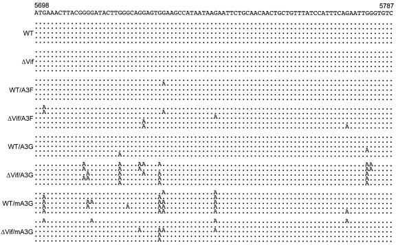
APOBEC3F induces G to A mutations in viral genome. Wild-type (WT) and Vif-defective viruses were produced in the presence and absence of APOBEC3G (A3G), APOBEC3F (A3F), and mAPOBEC3G (mA3G). These viruses were used to infect GHOST-R3/X4/R5 cells. Six hours later, cellular DNAs were extracted from these infected cells, and viral DNAs were amplified by PCR. After cloning into TA-cloning vector, these viral DNAs were then sequenced. The pNL4-3 sequence from nucleotides 5698 to 5787 is presented at the top. Five sequences from each infection are presented, and only the mutated nucleotides are shown. Dots indicate sequence identity.
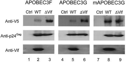
Levels of APOBEC proteins in viral particles. 293T cells were cotransfected with wild-type (WT) or Vif-defective proviruses and different APOBEC expression vectors. Viral particles were collected 48 h later and purified by ultracentrifugation through a 20% sucrose cushion. Equal amounts of viral proteins were separated by sodium dodecyl sulfate-polyacrylamide gel electrophoresis, and the expressions of APOBEC proteins, p24Gag, and Vif were determined by Western blotting.
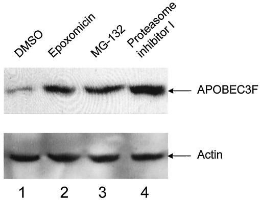
The degradation of APOBEC3F by Vif is blocked by proteasomal inhibitors in cells. 293T cells were cotransfected with Vif and APOBEC3F expression vectors. Twenty hours later, cells were treated with proteasomal inhibitors epoxomicin, MG-132, and proteasome inhibitor I at 10 μM for 12 h. Levels of APOBEC3F were then analyzed by Western blotting. DMSO, dimethyl sulfoxide.
References
-
- Bour, S., and K. Strebel. 2000. HIV accessory proteins: multifunctional components of a complex system. Adv. Pharmacol. 48:75-120. - PubMed
-
- Conticello, S. G., R. S. Harris, and M. S. Neuberger. 2003. The Vif protein of HIV triggers degradation of the human antiretroviral DNA deaminase APOBEC3G. Curr. Biol. 13:2009-2013. - PubMed
-
- Fisher, A. G., B. Ensoli, L. Ivanoff, M. Chamberlain, S. Petteway, L. Ratner, R. C. Gallo, and F. Wong-Staal. 1987. The sor gene of HIV-1 is required for efficient virus transmission in vitro. Science 237:888-893. - PubMed
Publication types
MeSH terms
Substances
LinkOut - more resources
Full Text Sources
Other Literature Sources
Molecular Biology Databases
