. 2004 May 11;32(8):2556-65.
doi: 10.1093/nar/gkh578. Print 2004.

Preferential binding to branched DNA strands and strand-annealing activity of the human Rad51B, Rad51C, Rad51D and Xrcc2 protein complex

Affiliations

Preferential binding to branched DNA strands and strand-annealing activity of the human Rad51B, Rad51C, Rad51D and Xrcc2 protein complex

Hiroshi Yokoyama et al. Nucleic Acids Res. .

Abstract

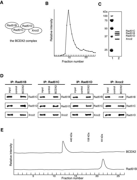
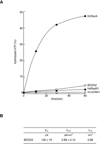
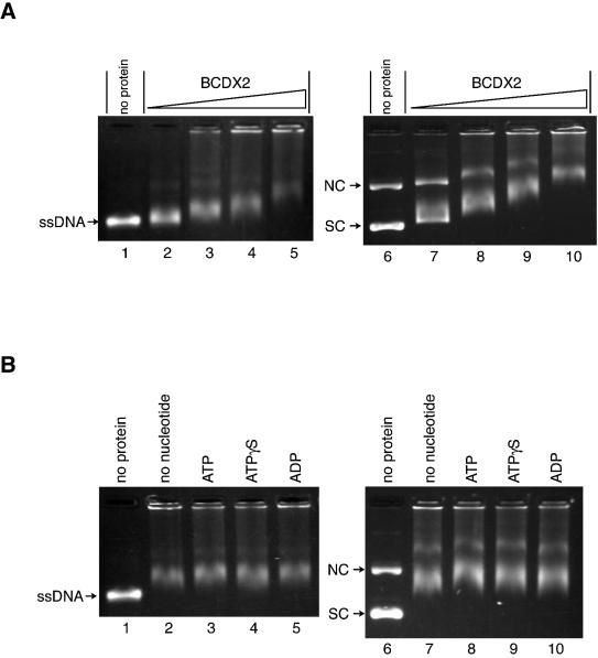
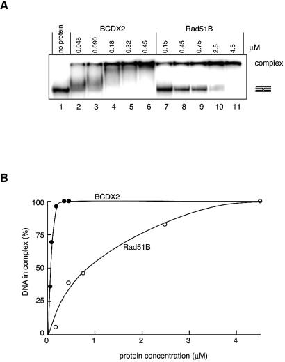
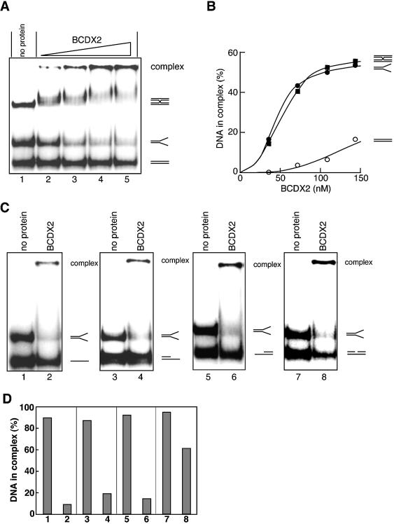
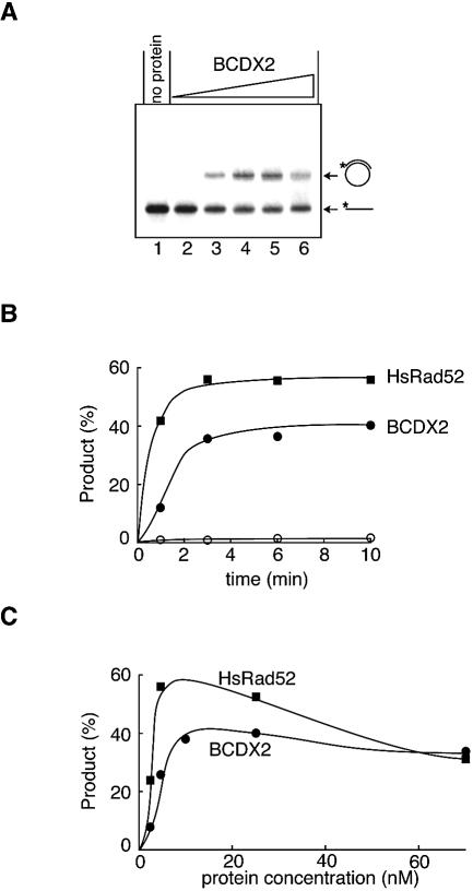
The Rad51B, Rad51C, Rad51D and Xrcc2 proteins are Rad51 paralogs, and form a complex (BCDX2 complex) in mammalian cells. Mutant cells defective in any one of the Rad51-paralog genes exhibit spontaneous genomic instability and extreme sensitivity to DNA-damaging agents, due to inefficient recombinational repair. Therefore, the Rad51 paralogs play important roles in the maintenance of genomic integrity through recombinational repair. In the present study, we examined the DNA-binding preference of the human BCDX2 complex. Competitive DNA-binding assays using seven types of DNA substrates, single-stranded DNA (ssDNA), double-stranded DNA, 5'- and 3'-tailed duplexes, nicked duplex DNA, Y-shaped DNA and a synthetic Holliday junction, revealed that the BCDX2 complex preferentially bound to the two DNA substrates with branched structures (the Y-shaped DNA and the synthetic Holliday junction). Furthermore, the BCDX2 complex catalyzed the strand-annealing reaction between a long linear ssDNA (1.2 kb in length) and its complementary circular ssDNA. These properties of the BCDX2 complex may be important for its roles in the maintenance of chromosomal integrity.


Figures

Figure 1
Figure 1
Purification of the BCDX2 complex. (A) Schematic representation of protein interactions within the BCDX2 complex. These protein interactions were previously identified by two-hybrid and biochemical analyses. (B) Elution profile of the Rad51B, Rad51C, Rad51D and Xrcc2 proteins in DEAE column chromatography. (C) The peak fraction (2 µg, lane 2) from the DEAE column was analyzed by 12% SDS–PAGE, and was stained with Coomassie Brilliant Blue. Lane 1 shows the molecular mass markers. (D) Immunoprecipitation (IP) experiments of the co-purified Rad51B, Rad51C, Rad51D and Xrcc2 proteins. The co-purified proteins were captured using anti-Rad51B, Rad51C, Rad51D or Xrcc2 antibody-conjugated beads, and the precipitates were analyzed by immunoblotting. As negative-control experiments, the co-purified proteins were incubated with normal IgG-conjugated beads. (E) Elution profiles of the co-purified Rad51B, Rad51C, Rad51D, and Xrcc2 proteins (upper) and the Rad51B protein (lower) in Superdex 200 HR gel filtration column chromatography.
Figure 2
Figure 2
ATPase activity of the BCDX2 complex. (A) Comparison of the ATPase activities among the BCDX2 complex, HsRad51 and EcRecA proteins. The ATPase activities of the three proteins were measured in the presence of 1 mM ATP and 75 µM ssDNA, and the percentages of hydrolyzed ATP at the indicated time points are presented. The open squares and the open circles indicate the experiments with the BCDX2 complex and without protein, respectively, and the closed squares and the closed circles indicate the experiments with HsRad51 and EcRecA, respectively. (B) Table of Km, Vmax and kcat values for the ATPase activity of the BCDX2 complex in the presence of ssDNA.
Figure 3
Figure 3
DNA-binding activities of the BCDX2 complex. (A) ssDNA and dsDNA binding by the BCDX2 complex. pGsat4 ssDNA (20 µM, lanes 1–5) or φX174 superhelical dsDNA (10 µM, lanes 6–10) was incubated with the BCDX2 complex at 30°C for 10 min. The concentrations of the BCDX2 complex used in the DNA-binding experiments were 0.12 µM (lanes 2 and 7), 0.35 µM (lanes 3 and 8), 0.69 µM (lanes 4 and 9) and 1.2 µM (lanes 5 and 10). The samples were analyzed by 1% agarose gel electrophoresis in 0.5× TBE buffer, and were stained with ethidium bromide (A and B). NC and SC indicate nicked circular and superhelical dsDNA, respectively (A and B). Lanes 1 and 5 indicate the negative control experiments without protein. (B) Nucleotide co-factor requirements for ssDNA and dsDNA binding. pGsat4 ssDNA (20 µM, lanes 1–5) or φX174 superhelical dsDNA (10 µM, lanes 6–10) was incubated with the BCDX2 complex (0.69 µM) at 30°C for 10 min in the presence of 1 mM ATP (lanes 3 and 8), 1 mM ATPγS (lanes 4 and 9) or 1 mM ADP (lanes 5 and 10). Lanes 1 and 6 indicate the negative control experiments without protein, and lanes 2 and 7 indicate the negative control experiments without nucleotide co-factor.
Figure 4
Figure 4
Holliday junction binding by the BCDX2 complex. (A) The 32P-labeled synthetic Holliday junction (0.33 µM) was incubated with the indicated amounts of the BCDX2 complex (lanes 2–6) or the Rad51B protein (lanes 7–11) at 30°C for 10 min. The samples were separated by electrophoresis on a 10% non-denaturing polyacrylamide gel in 0.5× TBE buffer, and were analyzed by the BAS2500 image analyzer. (B) Graphic representation of the amounts of the synthetic Holliday junction incorporated into the complex shown in (A). The closed and open circles indicate the experiments with the BCDX2 complex and Rad51B, respectively.
Figure 5
Figure 5
Preferential binding of the BCDX2 complex to branched DNA strands. (A) Increasing amounts of the BCDX2 complex were incubated with the DNA mixture, containing the three 32P-labeled DNA substrates (0.33 µM each), the synthetic Holliday junction, the Y-shaped DNA and the double-stranded oligonucleotide, at 30°C for 10 min. The samples were separated by electrophoresis on a 10% non-denaturing polyacrylamide gel in 0.5× TBE buffer, and were analyzed by the BAS2500 image analyzer. The concentrations of the BCDX2 complex used in the DNA-binding experiments were 36, 72, 110 and 140 nM (lanes 2–5). (B) Graphic representation of the amounts of each DNA incorporated into the complex shown in (A). The squares, closed circles and open circles indicate the synthetic Holliday junction, the Y-shaped DNA, and the double-stranded oligonucleotide, respectively. (C) The Y-shaped DNA (0.33 µM) was separately mixed with 0.33 µM of the four DNA substrates, a single-stranded oligonucleotide (lanes 1 and 2), a 5′-tailed duplex (lanes 3 and 4), a 3′-tailed duplex (lanes 5 and 6) or a nicked duplex (lanes 7 and 8), and was incubated with the BCDX2 complex (110 nM) at 30°C for 10 min. The samples were separated by electrophoresis on a 10% non-denaturing polyacrylamide gel in 0.5× TBE buffer, and were analyzed by the BAS2500 image analyzer. Lanes 1, 3, 5 and 7 indicate negative control experiments without protein. (D) Graphic representation of the amounts of each DNA incorporated into the complex shown in (C). Lanes 1 and 2 indicate the competition experiment with the Y-shaped DNA and the single-stranded oligonucleotide. Lanes 3 and 4 indicate the competition experiment with the Y-shaped DNA and the 5′-tailed duplex. Lanes 5 and 6 indicate the competition experiment with the Y-shaped DNA and the 3′-tailed duplex. Lanes 7 and 8 indicate the competition experiment with the Y-shaped DNA and the nicked duplex. Lanes 1, 3, 5 and 7 indicate the amounts of the Y-shaped DNA incorporated into the complex. Lanes 2, 3, 4, 6 and 8 indicate the amount of the single-stranded oligonucleotide, the 5′-tailed duplex, the 3′-tailed duplex, and the nicked duplex incorporated into the complex, respectively.
Figure 6
Figure 6
Strand-annealing activities of the BCDX2 complex. (A) Increasing amounts of the BCDX2 complex were incubated with the 32P-labeled linear ssDNA (1.2 kb, 35 nM) at 30°C for 5 min, and the strand-annealing reactions were started by the addition of the circular ssDNA (3.2 kb, 400 nM) and 1 mM ATP. After an incubation at 37°C for 10 min, the reactions were stopped by the addition of unlabeled linear ssDNA and 0.5% SDS, and the products were deproteinized with Proteinase K. The samples were analyzed by 0.8% agarose gel electrophoresis in TAE buffer, and were visualized by the BAS2500 image analyzer. The concentrations of the BCDX2 complex used in the strand-annealing experiments were 2, 4, 10, 25 and 70 nM (lanes 2–6). Lane 1 indicates a negative control experiment without protein. (B and C) Comparison of the strand-annealing activities between the BCDX2 complex and HsRad52, as functions of time (B) and protein concentration (C). (B) The closed circles and squares indicate the reactions of the BCDX2 complex (25 nM) and HsRad52 (25 nM), respectively, and the open circles indicate the negative control experiments without protein. (C) The circles and squares indicate the experiments with the increasing amounts of the BCDX2 complex and HsRad52, respectively.

Similar articles

Cited by

References

    1. Whitaker S.J. (1992) DNA damage by drugs and radiation: what is important and how is it measured? Eur. J. Cancer, 28, 273–276. - PubMed
    1. Cox M.M., Goodman,M.F., Kreuzer,K.N., Sherratt,D.J., Sandler,S.J. and Marians,K.J. (2000) The importance of repairing stalled replication forks. Nature, 404, 37–41. - PubMed
    1. Ward J.F. (1994) The complexity of DNA damage: relevance to biological consequences. Int. J. Radiat. Biol., 66, 427–432. - PubMed
    1. Caldecott K.W. (2001) Mammalian DNA single-strand break repair: an X-ra(y)ted affair. Bioessays, 23, 447–455. - PubMed
    1. van den Bosch M., Lohman,P.H. and Pastink,A. (2002) DNA double-strand break repair by homologous recombination. Biol. Chem., 383, 873–892. - PubMed

Publication types