. 2004 May 25;101(21):8138-43.
doi: 10.1073/pnas.0305683101. Epub 2004 May 12.

Leptin modulates beta cell expression of IL-1 receptor antagonist and release of IL-1beta in human islets

Affiliations

Leptin modulates beta cell expression of IL-1 receptor antagonist and release of IL-1beta in human islets

Kathrin Maedler et al. Proc Natl Acad Sci U S A. .

Abstract

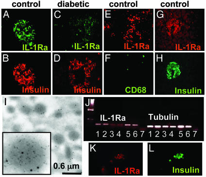
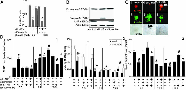
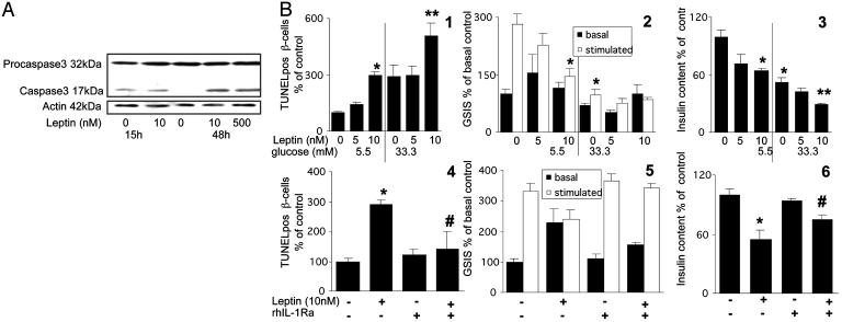
High concentrations of glucose induce beta cell production of IL-1beta, leading to impaired beta cell function and apoptosis in human pancreatic islets. IL-1 receptor antagonist (IL-1Ra) is a naturally occurring antagonist of IL-1beta and protects cultured human islets from glucotoxicity. Therefore, the balance of IL-1beta and IL-1Ra may play a crucial role in the pathogenesis of diabetes. In the present study, we observed expression of IL-1Ra in human pancreatic beta cells of nondiabetic individuals, which was decreased in tissue sections of type 2 diabetic patients. In vitro, chronic exposure of human islets to leptin, a hormone secreted by adipocytes, decreased beta cell production of IL-1Ra and induced IL-1beta release from the islet preparation, leading to impaired beta cell function, caspase-3 activation, and apoptosis. Exogenous addition of IL-1Ra protected cultured human islets from the deleterious effects of leptin. Antagonizing IL-1Ra by introduction of small interfering RNA to IL-1Ra into human islets led to caspase-3 activation, DNA fragmentation, and impaired beta cell function. Moreover, siIL-1Ra enhanced glucose-induced beta cell apoptosis. These findings demonstrate expression of IL-1Ra in the human beta cell, providing localized protection against leptin- and glucose-induced islet IL-1beta.


Figures

Fig. 1.
Fig. 1.
IL-1Ra is expressed by human islets and down-regulated in type 2 diabetes. Double immunostaining for IL-1Ra in green (A and C) or red (E) and insulin in red (B and D) or CD68 in green (F) in tissue sections of pancreases from a nondiabetic patient without (A, B, E, and F) and from a patient with type 2 diabetes (C and D). In situ hybridization for IL-1Ra mRNA in red (G) double immunostained for insulin in green (H) in tissue sections of pancreas from a nondiabetic patient. (I) Electron microscopy of secretory granules within a β cell in cultured human islets double gold-immunolabeled for insulin (small particles) and IL-1Ra (large particles). (J) RT-PCR analysis of IL-1Ra expression by purified human β cells (lanes 1 and 2), cultured human islets (lanes 3 and 4), human blood monocytes (lanes 5 and 6), and water (negative control; lane 7). One of four experiments from four donors is shown. Double immunostaining for IL-1Ra in red (K) and insulin in green (L) in purified human β cells.
Fig. 2.
Fig. 2.
Leptin induces phosphorylation of ERK1/2 and JAK2, decreases β cell production of IL-1Ra, and induces IL-1β release in human islets. (A) Double immunostaining for IL-1Ra in green (1, 3, and 5) and insulin in red (2, 4, and 6) in sections of cultured human islets exposed for 48 h to 5.5 mM glucose alone (1 and 2), 33.3 mM glucose (3 and 4), or 5.5 mM glucose and 10 nM leptin (5 and 6). (B) Immunoblotting of IL-1Ra and actin. Human islets cultured with and without 10 or 500 nM leptin were analyzed after 15 or 48 h of incubation. One of three experiments from three donors is shown. (C) Secretion of IL-1Ra (1 and 4) and RT-PCR detection and quantification of IL-1Ra mRNA expression (2 and 5) and secretion of IL-1β (3 and 6). Supernatants and total RNA were obtained from human islets (1–3) and human blood monocytes (4–6) cultured for 15 and 48 h in the presence of medium alone or 10 or 500 nM leptin. Data were collected from three tubes per treatment of five separate experiments from five islets donors. Results are means ± SE of percentage relative to control incubations (100%, in absolute values IL-1Ra release from islets: 152.0 ± 18.3 and 594.7 ± 24.5 pg/20islets per 2 ml after 15 and 48 h of culture, respectively, and from monocytes: 48.4 ± 1.7 and 96.4 ± 1.8 ng/ml after 15 and 48 h of culture, respectively. In absolute values for IL-1β release from islets: 0.28 ± 0.07 and 0.79 ± 0.13 pg/20 islets per 2 ml after 15 and 48 h of culture, respectively, and from monocytes: 332.0 ± 42.4 and 456.4 ± 88.6 pg/ml after 15 and 48 h of culture, respectively). *, P < 0.01 compared to controls. (D) Triple immunostaining for IL-1β in red (1), insulin in green (2), and DNA fragmentation by TUNEL assay in black (3). Human islets were cultured for 4 days at 5.5 mM glucose with 10 nM leptin. The arrows mark a β cell stained positive for IL-1β, insulin, and the TUNEL reaction. (E) Effects of 10 nM leptin on the time course of ERK1/2, JAK2, and STAT3 phosphorylation in cultured human islets. Lysates were subjected to Western blotting for activated ERK 1/2 (pERK1/2), JAK 2 (pJAK2), and STAT 3 (pSTAT3). One of three experiments from three donors is shown. (F) RT-PCR analysis for expression of the human long (hOB-Rl) and short forms (hOB-Rs) of the leptin receptor and tubulin (control) by cultured human islets.
Fig. 3.
Fig. 3.
Antagonizing IL-1Ra by siIL-1Ra induces β cell apoptosis and impairs β cell function. (A) RT-PCR detection and quantification of IL-1Ra mRNA expression. Total RNA was isolated from human islets cultured for 48 h in 5.5 or 33.3 mM glucose alone or after transfection with 50 nM siIL-1Ra or with 50 nM of a scrambled RNA sequence (siScramble) with or without addition of 500 ng/ml exogenous rh IL-1Ra. Results are presented as means ± SE of five independent experiments from five donors. *, P < 0.001 compared to control islets at the same glucose concentration. (B) Immunoblotting of procaspase 3, activated caspase 3, IL-1Ra, and actin. Human islets were cultured for 48 h in medium containing 5.5 mM glucose alone or after transfection with 50 nM siIL-1Ra or with 50 nM siScramble. The antibodies were blotted on the same membrane after stripping. One of four experiments from four donors is shown. (C and D) Human islets were cultured for 4 days in 5.5, 11.1, and 33.3 mM glucose alone or after transfection with 50 nM siIL-1Ra or 50 nM siScramble with or without addition of 500 ng/ml exogenous rh IL-1Ra. (C) Triple immunostaining for IL-1Ra in red (1, 4, and 7), insulin in green (2, 5, and 8), and DNA fragmentation by TUNEL assay in black (3, 6, and 9) in islets exposed to 5.5 mM glucose alone (1–3), with siIL-1Ra (4–6), or with siIL-1Ra and rh IL-1Ra (7–9). The arrows mark β cell nuclei stained positive for the TUNEL reaction. (D)(1) Results are means ± SE of percentage of TUNEL-positive β cells relative to control incubations at 5.5 mM glucose alone (100%, in absolute value: 0.33 ± 0.03% TUNEL-positive β cells). The mean number of islets scored from each donor was 44 (range 24–80) for each treatment condition. Islets were isolated from five organ donors. (2) Basal and glucose-stimulated insulin secretion (GSIS) during successive 1-h incubations at 3.3 (basal) and 16.7 (stimulated) mM glucose after the 4-day culture period at different glucose concentrations. (3) Insulin content. Results are means ± SE relative to 5.5 mM glucose alone (100%, in absolute values: 0.53 ± 0.08 and 7.9 ± 0.7 pmol/islet for basal insulin secretion and cell content, respectively). Data presented are means of three experiments from three separate donors. In each experiment, the data were collected from three plates per treatment. #, P < 0.05 compared with control islets at same glucose concentration; *, P < 0.05 compared with islets at 5.5 mM glucose alone; +, P < 0.05 compared to siIL-1Ra transfected islets alone at same glucose concentration.
Fig. 4.
Fig. 4.
Leptin induces β cell apoptosis and impairs β cell function via IL-1β signaling. (A) Immunoblotting of procaspase 3, activated caspase 3, and actin. Human islets were cultured for 15 and 48 h in 5.5 mM glucose alone or with 10 or 500 nM leptin. One of four experiments from four donors is shown. (B) Human islets were cultured on extracellular matrix-coated dishes for 4 days at 5.5 and 33.3 mM glucose with or without 5 and 10 nM leptin or with addition of 500 ng/ml exogenous rh IL-1Ra. (1 and 4) Results are means ± SE of percentage of TUNEL-positive β cells relative to 5.5 mM glucose alone [100%, in absolute value: 0.35 ± 0.04% (1) or 0.37 ± 0.06% (4) TUNEL-positive β cells]. The mean number of islets scored from each donor was 30 (range 21–45) for each treatment condition. Islets were isolated from five organ donors. (2 and 5) Basal and glucose-stimulated insulin secretion (GSIS) denote the amount secreted during successive 1-h incubations at 3.3 (basal) and 16.7 (stimulated) mM glucose after the 4-day culture period. (3 and 6) Insulin content. Results are means ± SE relative to control incubations at 5.5 mM glucose alone [100%, in absolute values: 0.70 ± 0.11 and 10.9 ± 0.7 pmol per islet (2 and 3) and 0.43 ± 0.10 and 7.5 ± 0.7 pmol per islet (5 and 6) for basal insulin secretion and cell content, respectively]. Data presented are means of three experiments from three separate donors. In each experiment, the data were collected from three plates per treatment. *, P < 0.01 compared to islets at 5.5 mM glucose alone; **, P < 0.01 compared to islets at 33.3 mM glucose alone; P < 0.05 compared to leptin-treated islets alone.

References

    1. Bonner-Weir, S. (2000) J. Mol. Endocrinol. 24, 297–302. - PubMed
    1. Butler, A. E., Janson, J., Bonner-Weir, S., Ritzel, R., Rizza, R. A. & Butler, P. C. (2003) Diabetes 52, 102–110. - PubMed
    1. Cerasi, E. (1995) Diabetologia 38, 992–997. - PubMed
    1. Donath, M. Y., Gross, D. J., Cerasi, E. & Kaiser, N. (1999) Diabetes 48, 738–744. - PubMed
    1. Leroith, D. (2002) Am. J. Med. 113 Suppl 6a, 3s–11s. - PubMed

Publication types

MeSH terms