Comparative Study
. 2004 Jun 2;23(11):2185-95.
doi: 10.1038/sj.emboj.7600212. Epub 2004 May 13.

Structural basis for the selective inhibition of JNK1 by the scaffolding protein JIP1 and SP600125

Affiliations
Comparative Study

Structural basis for the selective inhibition of JNK1 by the scaffolding protein JIP1 and SP600125

Yong-Seok Heo et al. EMBO J. .

Abstract

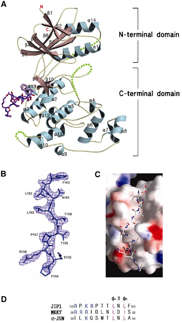
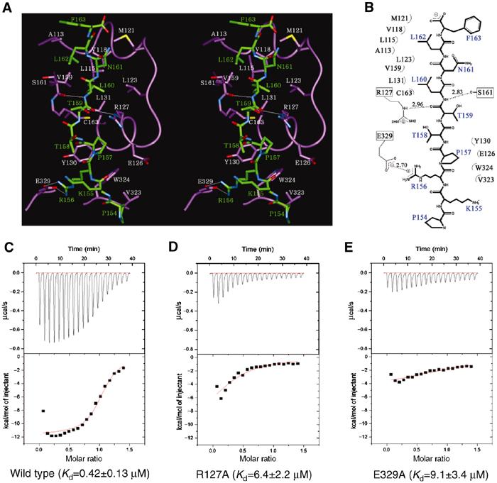
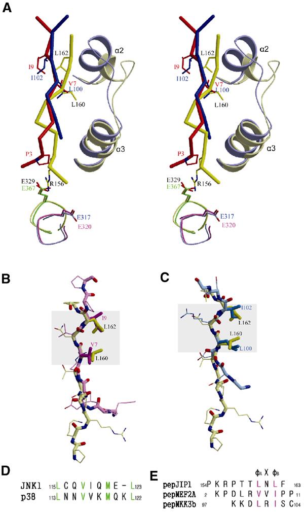
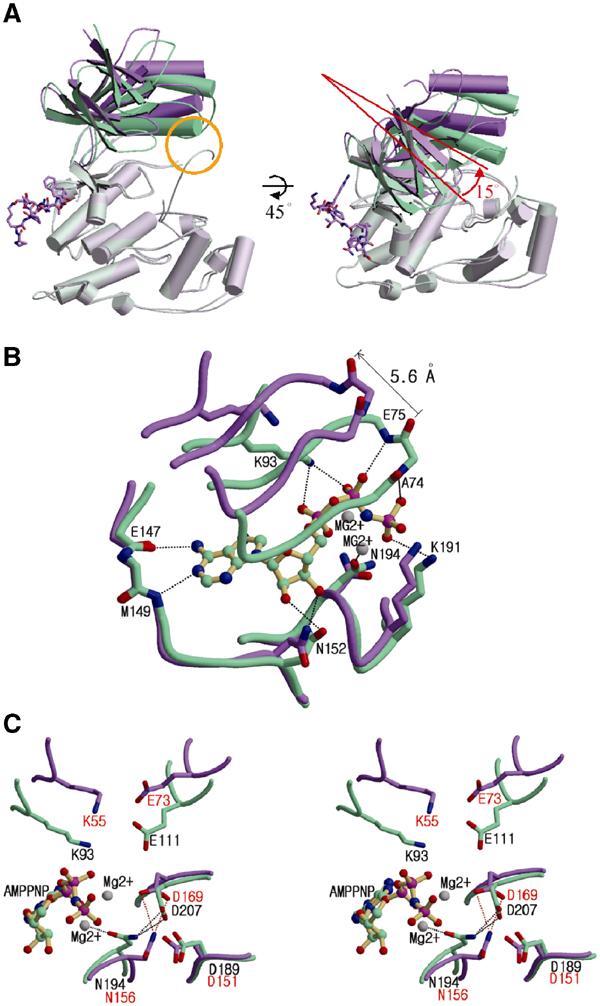
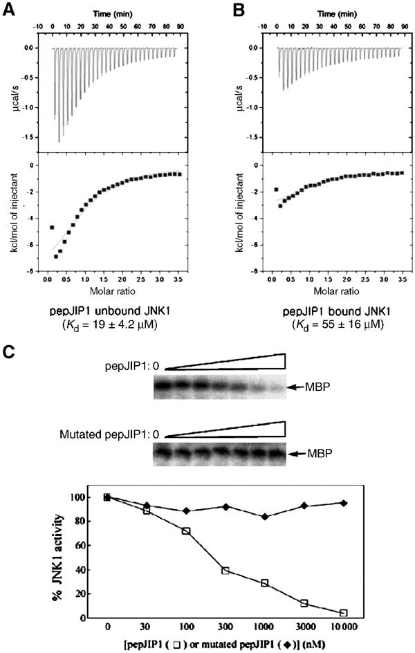
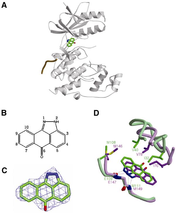
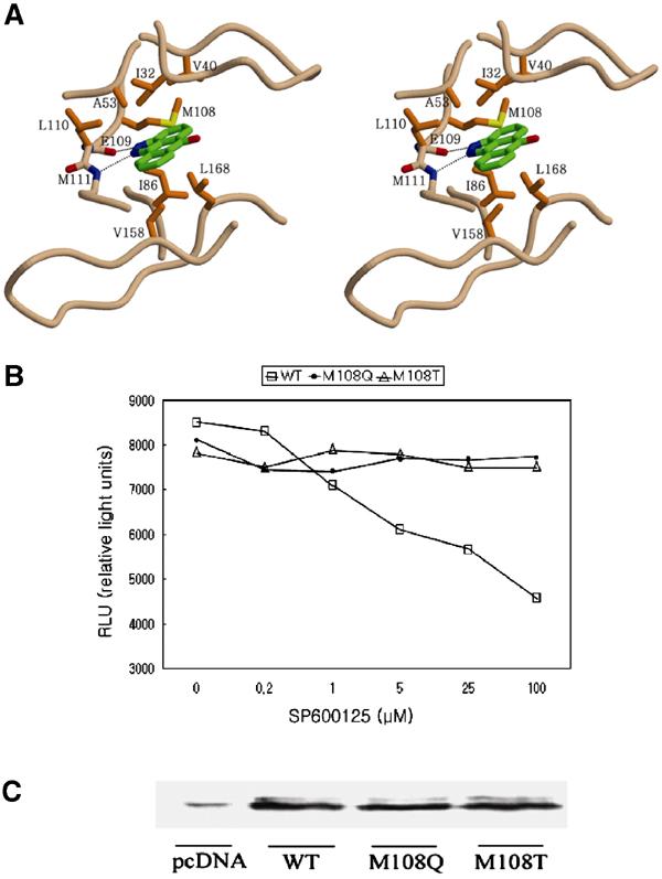
The c-jun N-terminal kinase (JNK) signaling pathway is regulated by JNK-interacting protein-1 (JIP1), which is a scaffolding protein assembling the components of the JNK cascade. Overexpression of JIP1 deactivates the JNK pathway selectively by cytoplasmic retention of JNK and thereby inhibits gene expression mediated by JNK, which occurs in the nucleus. Here, we report the crystal structure of human JNK1 complexed with pepJIP1, the peptide fragment of JIP1, revealing its selectivity for JNK1 over other MAPKs and the allosteric inhibition mechanism. The van der Waals contacts by the three residues (Pro157, Leu160, and Leu162) of pepJIP1 and the hydrogen bonding between Glu329 of JNK1 and Arg156 of pepJIP1 are critical for the selective binding. Binding of the peptide also induces a hinge motion between the N- and C-terminal domains of JNK1 and distorts the ATP-binding cleft, reducing the affinity of the kinase for ATP. In addition, we also determined the ternary complex structure of pepJIP1-bound JNK1 complexed with SP600125, an ATP-competitive inhibitor of JNK, providing the basis for the JNK specificity of the compound.


Figures

Figure 1
Figure 1
Overall structure of JNK1 complexed with pepJIP1 at a resolution of 2.35 Å. (A) JNK1 is shown in a ribbon model and the disordered regions of JNK1 (residues 173–189, 282–286, and 337–348) are expressed as dotted lines. The bound pepJIP1 is shown in a stick model of atomic color. The regions for the N- and C-terminal domains are indicated. (B) The sigma-A-weighted 2FoFc electron density map calculated with a final refined model without pepJIP1 at a level of 1.2σ. The peptide used in the experiment is RPKRPTTLNLF, but the starting residue, Arg153, is not shown in the density. (C) Surface representation of JNK1 in complex with pepJIP1, colored by electrostatic potential. (D) The docking site sequences of the scaffolding protein (JIP1), the upstream kinase (MKK7), and the substrate transcription factor (c-jun), which are components of the JNK signaling regulated by JIP1 scaffolding protein. The basic residues and φ residues are shown in blue and violet letters, respectively.
Figure 2
Figure 2
Interactions between JNK1 and pepJIP1. (A) Stereoview of the interactions between JNK1 (violet) and pepJIP1 (green). The residues of JNK1 involved in the interactions are shown in white labels and those of pepJIP1 in green labels. The hydrogen bonds in the interactions are shown as thin white lines. (B) Schematic expression of the interactions. (C) The binding affinity of pepJIP1 to wild-type JNK1 was measured by ITC. The upper panel shows the raw data, a trace of power with time. The lower panel shows the integrated heats from each injection, and the line through the measured points shows the best-fit model for a single binding site. (D, E) The respective Kd values of pepJIP1 to JNK1 mutants, R127A and E329A, were derived from the ITC data.
Figure 3
Figure 3
Structural comparison of the docking site peptides bound in the docking grooves of MAPKs. (A) Stereoview of the overlaid structures of pepJIP1 (saturated yellow) bound in JNK1 (whitened yellow) and pepMEF2A (saturated red) or pepMKK3b (saturated blue) bound in p38 MAPK (whitened blue) when the docking grooves of JNK1 and p38 are superimposed. The residues of pepJIP1, pepMEF2A, and pepMKK7 are labeled black, red, and blue, respectively. And the structure of E327 of JNK1 is compared with those of the corresponding glutamates of JNK3 (green), p38 (blue), and Erk2 (pink). (B) The structural deviation between pepJIP1 (yellow) and pepMEF2A (pink) when their φA-X-φB motifs are superimposed. (C) The structural comparison between pepJIP1 (yellow) and pepMKK3b (blue). In (B, C), the conformational conservations of the φA-X-φB motifs are highlighted by gray rectangles. (D) The differences of the sequences in the α2 helices of the docking grooves between JNK1 and p38 MAPK. The residues participating in hydrophobic interactions with the docking site peptides are shown in green letters. (E) The sequences of docking site peptides represented in (A–C). The φ residues are shown in violet letters.
Figure 4
Figure 4
Distortion of the ATP-binding site caused by interdomain rearrangement upon pepJIP1 binding. (A) Structural comparison between JNK3 (green) and pepJIP1-bound JNK1 (violet) when the C-terminal domains of the kinases are superimposed. The conformational differences of the N-terminal domains can be easily distinguished when the conventional view of kinases is rotated by 45° along the horizontal axis. The yellow circle indicates the interaction between the α1 helix and the phosphorylation loop in JNK3, but not existing in JNK1 complexed with pepJIP1. (B) Comparison of ATP-binding sites between the JNK1–pepJIP1 (violet) and JNK3–AMPPNP (green) complexes. The AMPPNP bound in JNK3 is shown in a ball-and-stick model. The residues of JNK3 involved in the hydrogen bonding with AMPPNP are labeled. The side chains of the residues in the glycine-rich loop including E75 and A74 of JNK3 are omitted for clarity because the backbone amide groups only are involved in the hydrogen bonds with the phosphate groups of AMPPNP. (C) The structural comparison of the residues crucial for the catalytic activity between the JNK1–pepJIP1 (violet) and JNK3–AMPPNP (green) complexes. The residues in JNK1 and JNK3 are labeled red and black, respectively. In (B, C), hydrogen bonds are indicated by dashed lines.
Figure 5
Figure 5
The inhibited phosphorylation of MBP, the docking site-independent substrate, due to the reduced ATP binding affinity to JNK1 by pepJIP1 binding. (A, B) The binding affinities of ATP to JNK1 were measured by ITC when pepJIP1 was unbound (A) and bound (B) to JNK1. (C) Dose-dependent inhibition of the kinase activity of JNK1 by pepJIP1 using MBP as substrates. The mutated pepJIP1 used for control experiment has the sequence of RPKAATTANAF.
Figure 6
Figure 6
The ternary complex JNK1–pepJIP1–SP600125. (A) SP600125 is bound at the hydrophobic pocket of the ATP-binding site. pepJIP1 is depicted as a single coil of orange color. (B) The numbering scheme of the compound SP600125. (C) The FoFc map calculated with a final refined model without SP600125 at the 3σ contour level. The refined atomic model of SP600125 is superimposed on the map. (D) Comparison of the positions of SP600125 when it is bound to JNK1 complexed with pepJIP1 (green) and free JNK3 (violet). The residues related with this shift are labeled and hydrogen bonds are represented by dashed lines.
Figure 7
Figure 7
The importance of the hydrophobic residues of JNK1 for binding of SP600125. (A) Stereoview of the binding mode of SP600125 in the JNK1–pepJIP1 complex. The residues important for inhibitor binding are represented and labeled. The side chains of E109 and M111 are omitted for clarity. (B) Cell-based assay of the JNK1 wild type and mutants using SP600125 as an inhibitor. The kinase activity was measured in HeLa cells using the PathDetect® c-jun trans-reporting systems (Stratagene). RLU is proportional to the kinase activity. (C) The physical amounts of the JNK1 wild type and mutants used in cell-based assay were measured by Western blot analysis. The weak band labeled as pcDNA means the amount of JNK1 isoforms existing in HeLa cells when the pcDNA3.1A expression vector only was transfected, where the cDNA of JNK1 was not inserted.

Similar articles

Cited by

References

    1. Bain J, McLauchlan H, Elliott M, Cohen P (2003) The specificities of protein kinase inhibitors: an update. Biochem J 371: 199–204 - PMC - PubMed
    1. Bardwell AJ, Flatauer LJ, Matsukuma K, Thorner J, Bardwell L (2001) A conserved docking site in MEKs mediates high-affinity binding to MAP kinases and cooperates with a scaffold protein to enhance signal transmission. J Biol Chem 276: 10374–10386 - PMC - PubMed
    1. Barr RK, Kendrick TS, Bogoyevitch MA (2002) Identification of the critical features of a small peptide inhibitor of JNK activity. J Biol Chem 277: 10987–10997 - PubMed
    1. Bennett BL, Sasaki DT, Murray BW, O'Leary EC, Sakata ST, Xu W, Leisten JC, Motiwala A, Pierce S, Satoh Y, Bhagwat SS, Manning AM, Anderson DW (2001) SP600125, an anthrapyrazolone inhibitor of Jun N-terminal kinase. Proc Natl Acad Sci USA 98: 13681–13686 - PMC - PubMed
    1. Bonny C, Oberson A, Negri S, Sauser C, Schorderet DF (2001) Cell-permeable peptide inhibitors of JNK: novel blockers of beta-cell death. Diabetes 50: 77–82 - PubMed

Publication types

MeSH terms