. 2004 Jun;24(11):4685-95.
doi: 10.1128/MCB.24.11.4685-4695.2004.

A direct binding site for Grb2 contributes to transformation and leukemogenesis by the Tel-Abl (ETV6-Abl) tyrosine kinase

Affiliations

A direct binding site for Grb2 contributes to transformation and leukemogenesis by the Tel-Abl (ETV6-Abl) tyrosine kinase

Ryan P Million et al. Mol Cell Biol. 2004 Jun.

Abstract

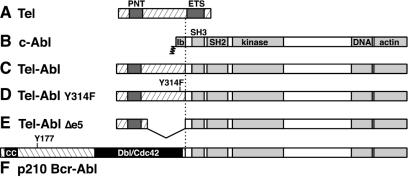
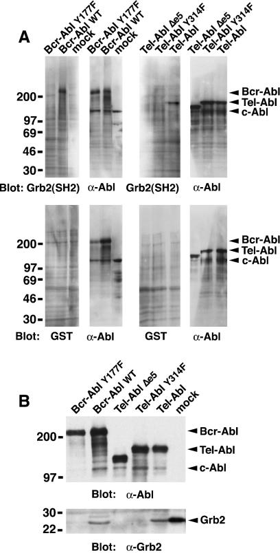
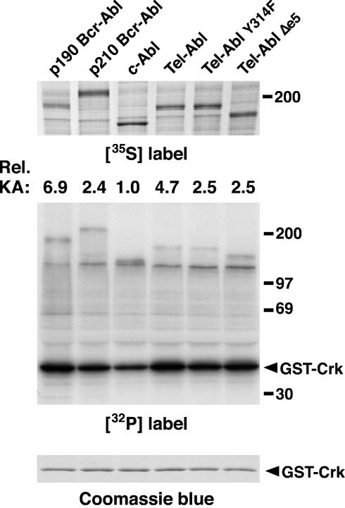
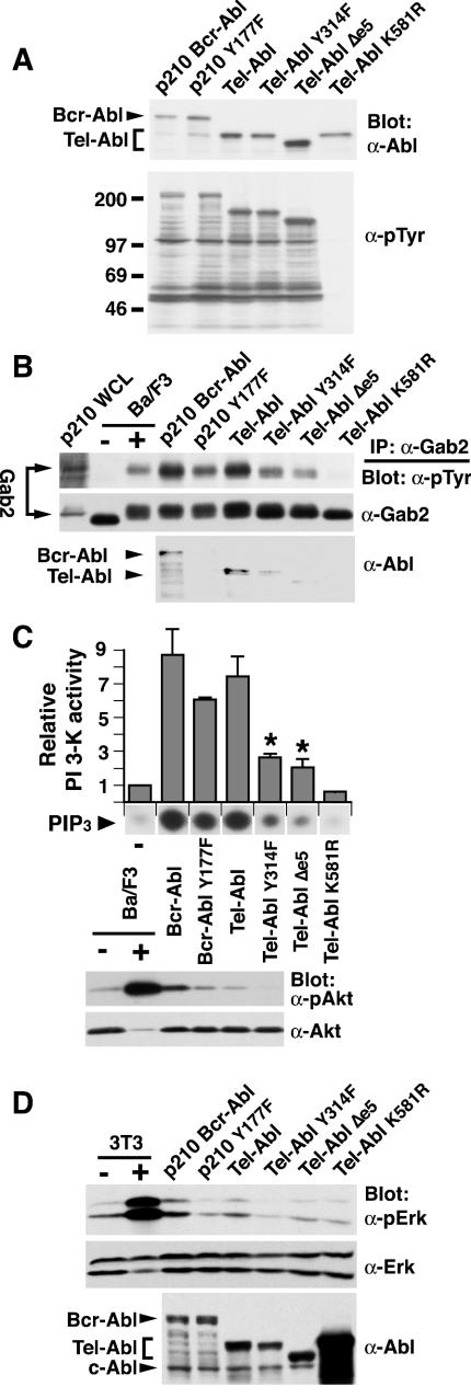
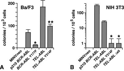
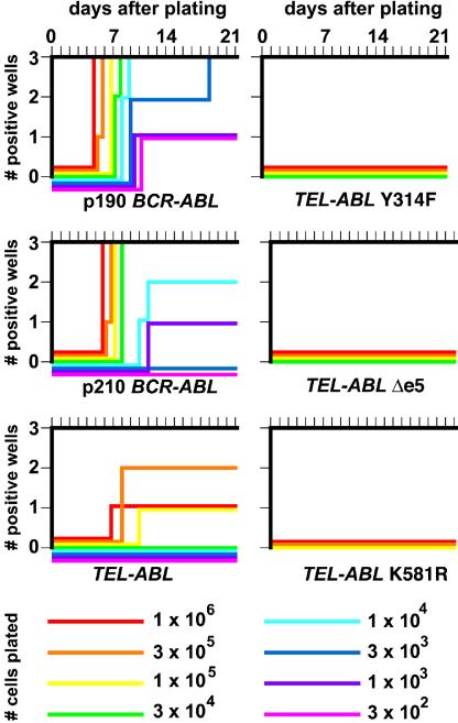
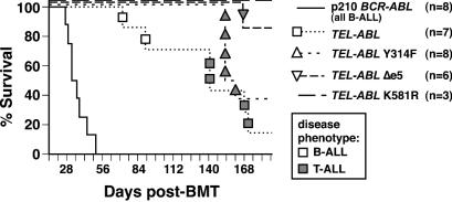
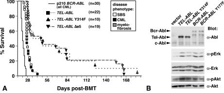
A direct binding site for the Grb2 adapter protein is required for the induction of fatal chronic myeloid leukemia (CML)-like disease in mice by Bcr-Abl. Here, we demonstrate direct binding of Grb2 to the Tel-Abl (ETV6-Abl) fusion protein, the product of complex (9;12) chromosomal translocations in human leukemia, via tyrosine 314 encoded by TEL exon 5. A Tel-Abl point mutant (Y314F) and a splice variant without TEL exon 5 sequences (Deltae5) lacked Grb2 interaction and exhibited decreased binding and phosphorylation of the scaffolding protein Gab2 and impaired activation of phosphatidylinositol 3-kinase, Akt, and extracellular signal-regulated kinase/mitogen-activated protein kinase in hematopoietic cells. Tel-Abl Y314F and Deltae5 were unable to transform fibroblasts to anchorage-independent growth and were defective for B-lymphoid transformation in vitro and lymphoid leukemogenesis in vivo. Previously, we demonstrated that full-length Tel-Abl induced two distinct myeloproliferative diseases in mice: CML-like leukemia similar to that induced by Bcr-Abl and a novel syndrome of small-bowel myeloid infiltration endotoxemia and hepatic and renal failure. Lack of the Grb2 binding site had no effect on development of small bowel syndrome but significantly attenuated the induction of CML-like disease by Tel-Abl. These results suggest that direct binding of Grb2 is a common mechanism contributing to leukemogenesis by oncogenic Abl fusion proteins.


Figures

FIG. 1.
FIG. 1.
Structure of Tel, Abl, and chimeric Tel-Abl and Bcr-Abl proteins. (A) Full-length Tel (ETV6) protein of 452 amino acids, with the positions of the PNT homology domain and ETS DNA-binding domain indicated by shaded boxes. (B) Full-length type Ib c-Abl protein of 1,142 amino acids, with the NH2-terminal myristoylation site, SH3 and SH2 domains, tyrosine kinase catalytic domain, and COOH-terminal DNA and actin binding domains indicated by shaded boxes. (C) Tel-Abl fusion protein, consisting of sequences encoded by TEL exons 1 to 5 (amino acids 1 to 336) fused to the 1,104 COOH-terminal amino acids of c-Abl. (D) Tel-Abl Y314F mutant, containing a point mutation of Tyr314 to Phe. (E) Tel-Abl Δexon5 (Δe5), consisting of sequences encoded by TEL exons 1 to 4 (amino acids 1 to 154) fused to the 1,104 COOH-terminal amino acids of c-Abl (43). (F) p210 Bcr-Abl fusion protein, consisting of Bcr amino acids 1 to 927 fused to the 1,104 COOH-terminal amino acids of c-Abl, with the NH2-terminal coiled-coil (CC) domain, Tyr177 Grb2 binding site, and region of homology to Db1/Cdc42 indicated.
FIG. 2.
FIG. 2.
Tyrosine 314 of Tel is a direct binding site for the SH2 domain of Grb2. (A) Far-Western blot. The indicated Abl proteins were expressed by transient transfection in 293T cells, and whole-cell extracts were fractionated by sodium dodecyl sulfate-polyacrylamide gel electrophoresis (SDS-PAGE), transferred to nitrocellulose filters, and hybridized with a GST-Grb2(SH2) fusion protein (first and third panels, top row) or with GST alone (first and third panels, bottom row). Bound GST protein was detected by anti-GST antibodies and enhanced chemiluminescence. Filters were subsequently stripped and rehybridized with anti-Abl antibody (second and fourth panels of each row). Molecular mass standards are on the left, and the positions of the Bcr-Abl, Tel-Abl, and c-Abl proteins are indicated by arrowheads on the right. (B) Coimmunoprecipitation. The indicated Abl proteins were expressed by transient transfection of 293T cells, immunoprecipitated with anti-Grb2 (mock-transfected cells only [right lane]) or anti-Abl (all other samples) antibody, fractionated by SDS-PAGE, transferred to nitrocellulose, and hybridized with anti-Abl (top panel) or anti-Grb2 (bottom panel) antibody. Molecular mass standards are on the left, and the positions of the Bcr-Abl, Tel-Abl, c-Abl, and Grb2 proteins are indicated by arrowheads on the right.
FIG. 3.
FIG. 3.
In vitro tyrosine kinase activity of Tel-Abl proteins. The indicated Abl proteins were expressed by transfection of 293T cells and labeled in vivo with l-35[S]methionine. The lysates were immunoprecipitated with anti-Abl antibodies, and immune complexes were incubated with [γ-32P]ATP and GST-Crk substrate. Results are representative of two independent experiments. (Top) 35S label, indicating relative levels of expression of the different Abl proteins. (Middle) 32P label. The position of the GST-Crk substrate is indicated by the arrowhead. The kinase activity (KA) of the different Abl proteins relative to c-Abl after correction for levels of expression is shown at the top. (Bottom) Coomassie blue stain demonstrating equal amounts of the GST-Crk substrate in all reaction mixtures.
FIG.4.
FIG.4.
The Grb2 binding site contributes to activation of PI 3-kinase and MAPK by Tel-Abl. (A) Western blot of whole-cell lysates of Ba/F3 cells expressing the indicated Abl fusion proteins. (Top) Anti-Abl antibody; (bottom) antiphosphotyrosine antibody (α-pTyr). The positions of the Bcr-Abl, Tel-Abl, and c-Abl proteins and molecular mass markers are indicated. (B) Gab2 phosphorylation and complex formation. Lysates from Ba/F3 cells expressing the indicated Abl fusion proteins were immunoprecipitated (IP) with anti-Gab2 antibodies and blotted with antiphosphotyrosine (top), anti-Gab2 (middle), or anti-Abl (bottom) antibodies. As a control, lysates from parental Ba/F3 cells that were starved of IL-3 and serum (−) and then stimulated for 5 min with IL-3 (+) were included. Whole-cell lysate from p210-expressing Ba/F3 cells was included to demonstrate the position of Gab2 (arrows on the left). (C) PI 3-kinase and Akt activation. (Top). The PI 3-kinase activity in antiphosphotyrosine immunoprecipitates from Ba/F3 cells expressing the indicated Abl fusion proteins was determined and is expressed as mean fold increased activity relative to serum- and IL-3-starved parental Ba/F3 cells; error bars indicate standard error. The relative levels of incorporation of 32P into PIP3 product in a representative experiment are shown below the histogram. The difference in PI 3-kinase activity induced by Tel-Abl WT and either Tel-Abl Y314F or Tel-Abl Δe5 (asterisks) was statistically significant (P = 0.05; unpaired t test), while the difference between PI 3-kinase activity induced by p210 Bcr-Abl and p210 Y177F was only of borderline significance (P = 0.10; unpaired t test). (Bottom) Whole-cell lysates from the indicated cells were blotted with phospho-Akt-specific antibodies (pAkt) and subsequently with pan-Akt antibodies (Akt). (D) ERK/MAPK activation. (Top and middle) 4A2+ fibroblasts expressing the indicated Bcr-Abl and Tel-Abl proteins after retroviral transduction were deprived of serum for 4 h, and lysates were prepared, fractionated by SDS-PAGE, and blotted with anti-phospho-ERK (top) or anti-ERK (middle) antibodies. As a control, lysates from parental 4A2+ cells without (−) or with (+) stimulation with 10% serum and 20 ng of platelet-derived growth factor per ml were included. (Bottom) Expression of Abl fusion proteins in these cells, with the positions of Bcr-Abl, Tel-Abl, and c-Abl indicated by the arrowheads. The kinase-defective Tel-Abl K581R protein was expressed at significantly higher levels than the other proteins, consistent with a lack of cytostatic or toxic effect.
FIG. 5.
FIG. 5.
The Grb2 binding site contributes to transformation of Ba/F3 cells and fibroblasts by Tel-Abl. Ba/F3 (A) and NIH 3T3 (B) cells were transduced with retrovirus lacking any insert (MINV neo) or containing p210 BCR-ABL, TEL-ABL, TEL-ABL Y314F, or TEL-ABL Δexon 5 (Δe5), and colony formation in soft agar was assessed as described in Materials and Methods. Error bars indicate standard deviation. (A) The difference between BCR-ABL WT and BCR-ABL Y177F (*) and between TEL-ABL and TEL-ABL Y314F (**) is statistically significant (P = 0.005 or P = 0.02, respectively; unpaired t test). (B) The difference between TEL-ABL and either TEL-ABL Y314F or TEL-ABL Δe5 (asterisks) is statistically significant (P = 0.01; unpaired t test).
FIG. 6.
FIG. 6.
The Grb2 binding site is required for transformation of primary bone marrow B-lymphoid cells by Tel-Abl in vitro. Primary bone marrow from non-5-FU-treated BALB/c donor mice was transduced with MSCV neo retrovirus expressing p190 or p210 BCR-ABL, TEL-ABL, TEL-ABL Y314F, or TEL-ABL Δexon5 (Δe5); transduction efficiency was equivalent based on determination of the proviral copy number in bone marrow cells by Southern blotting (data not shown). Serial dilutions of transduced marrow were plated in triplicate on syngeneic stromal layers derived from untransduced marrow and cultured for 3 weeks as described in Materials and Methods. The plating density is indicated by the line color, the number of cultures that reached confluence (defined as >106 nonadherent cells) is indicated on the ordinate, and the time to reach confluence is shown on the abscissa.
FIG. 7.
FIG. 7.
Induction of B-lymphoid leukemia in mice by Tel-Abl requires the Grb2 binding site. A Kaplan-Meier-style survival curve for recipients of bone marrow from non-5-FU-treated donors transduced with MSCV neo retrovirus expressing p210 BCR-ABL, TEL-ABL, TEL-ABL Y314F, and TEL-ABL Δexon5 (Δe5) is shown. The number of recipients in each arm is shown in parentheses; all recipients of p210 BCR-ABL-transduced marrow developed B-ALL. The symbols indicate individual TEL-ABL recipient mice, with the disease phenotype of each designated by the shading (open, B-ALL; shaded, T-ALL). The difference in the survival of recipients of p210 BCR-ABL-transduced marrow and any of the recipients of TEL-ABL-transduced marrow was highly significant (P < 0.001; Mantel-Cox test). The difference in survival between recipients of marrow transduced with full-length TEL-ABL and TEL-ABL Δe5 was significant (P = 0.016; Mantel-Cox test), while the difference between TEL-ABL and TEL-ABL Y314F or between the two TEL-ABL mutants was not significant. BMT, bone marrow transplantation.
FIG. 8.
FIG. 8.
Decreased myeloproliferative disease and activation of MAPK and Akt by Tel-Abl Grb2 binding mutants. (A) Loss of the Grb2 binding site attenuates the induction of CML-like disease by Tel-Abl but has no effect on development of small-bowel syndrome. A Kaplan-Meier-style survival curve for recipients of bone marrow from 5-FU-treated donors transduced with MSCV neo retrovirus expressing p210 BCR-ABL, TEL-ABL, TEL-ABL Y314F, or TEL-ABL Δexon5 (Δe5) is shown. The number of recipients in each arm is shown in parentheses. The survival curve for BCR-ABL is a composite of curves for previously reported mice (29) and additional mice transplanted concurrently with the TEL-ABL recipients; all recipients in this arm developed CML-like disease. The symbols indicate individual TEL-ABL recipient mice, with the disease phenotype of each designated by the shading (solid, CML-like disease; open, small-bowel necrosis with acute fatty liver [SBS] [38]); shaded, splenic and marrow fibrosis. Mice with prominent histopathological features of both disease processes are indicated by dually shaded symbols. The TEL-ABL (exons 1 to 5) recipients were from transplants conducted concurrently with the TEL-ABL mutants and have been reported previously (38). The difference in survival between recipients of TEL-ABL-transduced marrow and recipients of marrow transduced with either TEL-ABL mutant was highly statistically significant (P < 0.001; Mantel-Cox test), while there was no significant difference between the survival of recipients of marrow transduced by the two TEL-ABL mutants. BMT, bone marrow transplantation. (B) Impaired ERK/MAPK and Akt activation in primary myeloid progenitors by Tel-Abl Y314F. Lysates from primary bone marrow mononuclear cells transduced with empty MINVneo vector or MINVneo expressing the indicated Abl fusion protein were blotted with anti-Abl, anti-phospho-ERK (α-pErk), anti-pan-ERK (α-Erk), anti-phospho-Akt (α-pAkt), or anti-pan-Akt (α-Akt) antibodies, as indicated. The results are representative of two independent experiments.

Similar articles

Cited by

References

    1. Andreasson, P., B. Johansson, M. Carlsson, I. Jarlsfelt, T. Fioretos, F. Mitelman, and M. Hoglund. 1997. BCR/ABL-negative chronic myeloid leukemia with ETV6/ABL fusion. Genes Chromosomes Cancer 20:299-304. - PubMed
    1. Brasher, B. B., S. Roumiantsev, and R. A. Van Etten. 2001. Mutational analysis of the regulatory function of the c-Abl Src homology 3 domain. Oncogene 20:7744-7752. - PubMed
    1. Brunel, V., D. Sainty, N. Carbuccia, M. Mozzicolacci, F. Fernandez, J. Simonetti, J. Gabert, P. Dubreuil, M. Lafage-Pochitaloff, and F. Birg. 1996. A TEL/ABL fusion gene on chromosome 12p13 in a case of Ph−, BCR− atypical CML. Leukemia 10:2003.
    1. Carroll, M., S. Ohno-Jones, S. Tamura, E. Buchdunger, J. Zimmermann, N. B. Lydon, D. G. Gilliland, and B. J. Druker. 1997. CGP 57148, a tyrosine kinase inhibitor, inhibits the growth of cells expressing BCR-ABL, TEL-ABL, and TEL-PDGFR fusion proteins. Blood 90:4947-4952. - PubMed
    1. Cortez, D., L. Kadlec, and A. M. Pendergast. 1995. Structural and signaling requirements for BCR-ABL-mediated transformation and inhibition of apoptosis. Mol. Cell. Biol. 15:5531-5541. - PMC - PubMed

Publication types

MeSH terms