. 2004 Jun;24(11):4743-56.
doi: 10.1128/MCB.24.11.4743-4756.2004.

Glucocorticoids and tumor necrosis factor alpha cooperatively regulate toll-like receptor 2 gene expression

Affiliations

Glucocorticoids and tumor necrosis factor alpha cooperatively regulate toll-like receptor 2 gene expression

Marcela A Hermoso et al. Mol Cell Biol. 2004 Jun.

Abstract

Tumor necrosis factor alpha (TNF-alpha) and glucocorticoids are widely recognized as mutually antagonistic regulators of adaptive immunity and inflammation. Surprisingly, we show here that they cooperatively regulate components of innate immunity. The Toll-like receptor 2 (TLR2) gene encodes a transmembrane receptor critical for triggering innate immunity. Although TLR2 mRNA and protein are induced by inflammatory molecules such as TNF-alpha, we show that TLR2 is also induced by the anti-inflammatory glucocorticoids in cells where they also regulate MKP-1 mRNA and protein levels. TNF-alpha and glucocorticoids cooperate to regulate the TLR2 promoter, through the involvement of a 3' NF-kappaB site, a STAT-binding element, and a 3' glucocorticoid response element (GRE). Molecular studies show that the IkappaBalpha superrepressor or a STAT dominant negative element prevented TNF-alpha and dexamethasone stimulation of TLR2 promoter. Similarly, an AF-1 deletion mutant of glucocorticoid receptor or ablation of a putative GRE notably reduced the cooperative regulation of TLR2. Using chromatin immunoprecipitation assays, we demonstrate that all three transcription factors interact with both endogenous and transfected TLR2 promoters after stimulation by TNF-alpha and dexamethasone. Together, these studies define novel signaling mechanism for these three transcription factors, with a profound impact on discrimination of innate and adaptive immune responses.


Figures
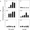
FIG. 1.
FIG. 1.
TNF-α and Dex induce TLR2 mRNA in A549 cells. (A) The expression of IL-8 mRNA in TNF-α-treated and untreated human lung A549 cells was measured by real-time quantitative PCR. IL-8 mRNA was used to confirm the pro- and anti-inflammatory effect of TNF-α and Dex, respectively. (B) TNF-α or Dex significantly increases TLR2 mRNA levels. TLR2 mRNA levels after TNF-α or Dex treatment were determined by real-time quantitative reverse transcription-PCR. (C) Cyclophilin B (cyclo) was used as a control for the amount of RNA used in each reaction and for each treatment. All sample analyses were carried out in duplicate after 8 h of treatment. Values are the mean ± standard error (SE) of three experiments. *, P < 0.05 for pair comparison analysis (Tukey-Kramer test).
FIG. 2.
FIG. 2.
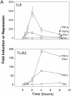
Glucocorticoids synergistically enhance TNF-α-induced TLR2 expression. (A) Glucocorticoids enhance TNF-α-induced TLR2 mRNA production. (Upper panel). IL-8 was profoundly up-regulated by TNF-α treatment and down-regulated by Dex treatment. When the two agents were added together, the TNF-α-induced increase in the amount of IL-8 mRNA was partially counteracted as determined by real-time PCR. A549 cells were harvested at six different time points (0, 0.5, 1, 2, 4, and 8 h), after addition of TNF-α and/or Dex. (Lower panel). Kinetics of the TNF-α and Dex treatments. The increase in the amount of TNF-α-induced TLR2 mRNA was sensitized by Dex. A549 cells were harvested at six different time points (0, 0.5, 1, 2, 4, and 8 h) after addition of TNF-α and/or Dex, and the TLR2 mRNA content was detected by real-time quantitative reverse transcription-PCR. (B) Mechanism of the cooperative effect of Dex on the TNF-α-induced TLR2 mRNA up-regulation. (Upper graph). The GR antagonist, RU486, counteracts the repressive effect of Dex on the TNF-α-induced up-regulation of the level of IL-8 mRNA. A549 cells were stimulated with each of the agents or the combination of all of them for 8 h, and the IL-8 mRNA levels were measured by real-time quantitative PCR in duplicate. RU486 counteracts the enhancing effect of Dex on TNF-α-induced up-regulation of TLR2 mRNA levels. A549 cells were stimulated with each of the agents or a combination of all of them for 8 h, and the TLR2 mRNA levels were measured by real-time quantitative PCR in duplicate. Values are the mean ± SE of three experiments. (C) TLR2 protein levels in A549 cells after TNF-α and Dex treatment. (Left) Major TLR2 protein expression was detected after TNF-α and Dex treatment. Western blot analysis was performed to confirm the cooperative effect between Dex and TNF-α. A549 cells were harvested after 6 h of treatment and subsequently labeled for 1 h with [35S]Cys-[35S]Met and then immunoprecipitated (IP) with anti-TLR2 antibody. (Right) Exogenous expression of TLR2-Flag comigrated with endogenously stimulated receptor with TNF-α and Dex. A549 cells were transiently transfected with the complete TLR2 cDNA tagged to a Flag epitope, labeled for 1 h with [35S]Cys-[35S]Met, and then immunoprecipitated with anti-TLR2 or anti-Flag antibodies. Cells were then harvested, and Western blot (IB) analysis was performed using the anti-TLR2 antibody. (Graph) TLR2 protein expression from cells treated with TNF-α and/or Dex was normalized to the control immunoprecipitated receptor (vehicle-treated cells). Analysis of the immunoreactive bands with the NIH-Image software reproduced the cooperative effect of Dex on the TNF-α-induced increase in the level of TLR2. Values are the mean ± SE of three experiments. *, P < 0.05 for pair comparison analysis (Tukey-Kramer test).
FIG. 2.
FIG. 2.
Glucocorticoids synergistically enhance TNF-α-induced TLR2 expression. (A) Glucocorticoids enhance TNF-α-induced TLR2 mRNA production. (Upper panel). IL-8 was profoundly up-regulated by TNF-α treatment and down-regulated by Dex treatment. When the two agents were added together, the TNF-α-induced increase in the amount of IL-8 mRNA was partially counteracted as determined by real-time PCR. A549 cells were harvested at six different time points (0, 0.5, 1, 2, 4, and 8 h), after addition of TNF-α and/or Dex. (Lower panel). Kinetics of the TNF-α and Dex treatments. The increase in the amount of TNF-α-induced TLR2 mRNA was sensitized by Dex. A549 cells were harvested at six different time points (0, 0.5, 1, 2, 4, and 8 h) after addition of TNF-α and/or Dex, and the TLR2 mRNA content was detected by real-time quantitative reverse transcription-PCR. (B) Mechanism of the cooperative effect of Dex on the TNF-α-induced TLR2 mRNA up-regulation. (Upper graph). The GR antagonist, RU486, counteracts the repressive effect of Dex on the TNF-α-induced up-regulation of the level of IL-8 mRNA. A549 cells were stimulated with each of the agents or the combination of all of them for 8 h, and the IL-8 mRNA levels were measured by real-time quantitative PCR in duplicate. RU486 counteracts the enhancing effect of Dex on TNF-α-induced up-regulation of TLR2 mRNA levels. A549 cells were stimulated with each of the agents or a combination of all of them for 8 h, and the TLR2 mRNA levels were measured by real-time quantitative PCR in duplicate. Values are the mean ± SE of three experiments. (C) TLR2 protein levels in A549 cells after TNF-α and Dex treatment. (Left) Major TLR2 protein expression was detected after TNF-α and Dex treatment. Western blot analysis was performed to confirm the cooperative effect between Dex and TNF-α. A549 cells were harvested after 6 h of treatment and subsequently labeled for 1 h with [35S]Cys-[35S]Met and then immunoprecipitated (IP) with anti-TLR2 antibody. (Right) Exogenous expression of TLR2-Flag comigrated with endogenously stimulated receptor with TNF-α and Dex. A549 cells were transiently transfected with the complete TLR2 cDNA tagged to a Flag epitope, labeled for 1 h with [35S]Cys-[35S]Met, and then immunoprecipitated with anti-TLR2 or anti-Flag antibodies. Cells were then harvested, and Western blot (IB) analysis was performed using the anti-TLR2 antibody. (Graph) TLR2 protein expression from cells treated with TNF-α and/or Dex was normalized to the control immunoprecipitated receptor (vehicle-treated cells). Analysis of the immunoreactive bands with the NIH-Image software reproduced the cooperative effect of Dex on the TNF-α-induced increase in the level of TLR2. Values are the mean ± SE of three experiments. *, P < 0.05 for pair comparison analysis (Tukey-Kramer test).
FIG. 3.
FIG. 3.
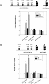
Glucocorticoids cooperate in the TNF-α-induced increase in TLR2 promoter activity. Functional analysis of 5′ deletion constructs of the TLR2 promoter is shown. (A) Scheme of the 5′ TLR2 promoter deletion luciferase constructs (top). Binding sites for different transcription factors are indicated. The number of each construct corresponds to its 5′ end. The luciferase activity of each of the 5′ deletion constructs of the TLR2 promoter transfected into A549 cells is shown in the graph. Cells were treated for 16 h with 10 ng of TNF-α per ml or 100 nM Dex or a combination and then harvested for luciferase activity determination. Bars represent the ratio between relative luciferase units (RLU) to protein content and normalized to the control (fold induction). All samples were analyzed in duplicate, and the values are the mean ± SE of three experiments. (B) Study of the role of GR in the activation of the TLR2 promoter. A diagram of the pGL3-297 TLR2 promoter construct used to study GR participation in the enhancement induced by 10 ng of TNF-α per ml plus 100 nM Dex is shown (top). The luciferase activity of the pGL3-297 TLR2 promoter construct transfected into A549 cells is shown in the graph. The transfected cells were left untreated or treated for 16 h with 10 ng of TNF-α per ml, 100 nM Dex, 1 μM RU486, or a combination of them, as indicated. Bars represent the ratio between relative luciferase units (RLU) and protein content and normalized to the control (fold induction). All samples were analyzed in duplicate, and the values are the mean ± SE of three experiments. *, P < 0.05 for pair comparison analysis (Tukey-Kramer test) to each control condition.
FIG. 4.
FIG. 4.
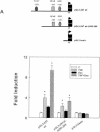
Role of the NF-κB transcription factor in TLR2 promoter activity in A549 cells. (A) The TLR2 promoter construct pGL3-1486 was used as a wild type to produce the NF-κB deletion construct (pGL3-1486/NFm). The X indicates that the NF-κB transcription factor site has been deleted. Bars represent the fold induction by TNF-α, Dex, or both after 16 h. (B) The shortened TLR2 promoter construct, pGL3-297wt TLR2, was used as a template to construct pGL3-297/NFm, in which the 3′ NF-κB consensus site was deleted, as indicated. The bar graph represents the luciferase activity of the wild type (wt), mutated constructs, and pure pGL3-basic vector (empty vector) normalized to untreated cells. All samples were analyzed in duplicate, and the values are the mean ± SE of three experiments. (C) A549 cells were transiently cotransfected with the pGL3-297wt TLR2 reporter construct and the IκBα superrepressor expression plasmid that was ramped from 0 to 5 μg. The fold induction of luciferase activities by TNF-α, Dex, or both is shown. The luciferase activity of the pGL3-297 TLR2 construct in the presence of different concentrations of the IκBα superrepressor expression plasmid is shown. All samples were analyzed in duplicate, and the values are the mean ± SE of three experiments. *, P < 0.05 for pair comparison analysis (Tukey-Kramer test) to each control condition.
FIG. 4.
FIG. 4.
Role of the NF-κB transcription factor in TLR2 promoter activity in A549 cells. (A) The TLR2 promoter construct pGL3-1486 was used as a wild type to produce the NF-κB deletion construct (pGL3-1486/NFm). The X indicates that the NF-κB transcription factor site has been deleted. Bars represent the fold induction by TNF-α, Dex, or both after 16 h. (B) The shortened TLR2 promoter construct, pGL3-297wt TLR2, was used as a template to construct pGL3-297/NFm, in which the 3′ NF-κB consensus site was deleted, as indicated. The bar graph represents the luciferase activity of the wild type (wt), mutated constructs, and pure pGL3-basic vector (empty vector) normalized to untreated cells. All samples were analyzed in duplicate, and the values are the mean ± SE of three experiments. (C) A549 cells were transiently cotransfected with the pGL3-297wt TLR2 reporter construct and the IκBα superrepressor expression plasmid that was ramped from 0 to 5 μg. The fold induction of luciferase activities by TNF-α, Dex, or both is shown. The luciferase activity of the pGL3-297 TLR2 construct in the presence of different concentrations of the IκBα superrepressor expression plasmid is shown. All samples were analyzed in duplicate, and the values are the mean ± SE of three experiments. *, P < 0.05 for pair comparison analysis (Tukey-Kramer test) to each control condition.
FIG. 5.
FIG. 5.
Analysis of the role of STAT transcription factor in the activation of the TLR2 promoter in A549 cells. (A) The TLR2 promoter construct pGL3-297 was used as the wild type to produce the deletion construct pGL3-297wt-Δ269-208, in which a specific deletion was conducted to remove the STAT-binding site, as indicated in Materials and Methods. The fold induction of luciferase by TNF-α, Dex, or both is shown. All samples were analyzed in duplicate, and the values are the mean ± SE of three experiments. Luciferase activities of the wild type (wt), mutated constructs, and pGL3-basic vector are shown. All samples were analyzed in duplicate, and the values are the mean ± SE of three experiments. (B) The pGL3-297wt TLR2 promoter construct was cotransfected with the STAT5b (Y699F) dominant negative expression plasmid, ramped from 0 to 5 μg. The fold induction of luciferase activities by TNF-α, Dex, or both is shown. Luciferase activities of all constructs and the pGL3-basic vector are shown. All samples were analyzed in duplicate, and the values are the mean ± SE of three experiments. (C) Interaction between NF-κB and STAT transcriptional factor binding sites during TLR2 gene transcription. A diagram of the pGL3-297 TLR2 promoter construct used as the wild type and the insertion and deletion constructs is shown (top). The shortened luciferase reporter construct pGL3-297 TLR2 was used as a template to construct pGL3-297wt-in300 and pGL3-297wt-in600 (containing a 300- and a 600-bp fragment, respectively, inserted between the NF-κB and STAT sites). The pGL3-297Δ285-269 was obtained after deletion of the DNA fragment between the NF-κB and STAT sites. The fold induction of luciferase activities by TNF-α, Dex, or both is shown. All samples were analyzed in duplicate, and the values are the mean ± SE of three experiments. *, P < 0.05 for pair comparison analysis (Tukey-Kramer test) to each control condition.
FIG. 5.
FIG. 5.
Analysis of the role of STAT transcription factor in the activation of the TLR2 promoter in A549 cells. (A) The TLR2 promoter construct pGL3-297 was used as the wild type to produce the deletion construct pGL3-297wt-Δ269-208, in which a specific deletion was conducted to remove the STAT-binding site, as indicated in Materials and Methods. The fold induction of luciferase by TNF-α, Dex, or both is shown. All samples were analyzed in duplicate, and the values are the mean ± SE of three experiments. Luciferase activities of the wild type (wt), mutated constructs, and pGL3-basic vector are shown. All samples were analyzed in duplicate, and the values are the mean ± SE of three experiments. (B) The pGL3-297wt TLR2 promoter construct was cotransfected with the STAT5b (Y699F) dominant negative expression plasmid, ramped from 0 to 5 μg. The fold induction of luciferase activities by TNF-α, Dex, or both is shown. Luciferase activities of all constructs and the pGL3-basic vector are shown. All samples were analyzed in duplicate, and the values are the mean ± SE of three experiments. (C) Interaction between NF-κB and STAT transcriptional factor binding sites during TLR2 gene transcription. A diagram of the pGL3-297 TLR2 promoter construct used as the wild type and the insertion and deletion constructs is shown (top). The shortened luciferase reporter construct pGL3-297 TLR2 was used as a template to construct pGL3-297wt-in300 and pGL3-297wt-in600 (containing a 300- and a 600-bp fragment, respectively, inserted between the NF-κB and STAT sites). The pGL3-297Δ285-269 was obtained after deletion of the DNA fragment between the NF-κB and STAT sites. The fold induction of luciferase activities by TNF-α, Dex, or both is shown. All samples were analyzed in duplicate, and the values are the mean ± SE of three experiments. *, P < 0.05 for pair comparison analysis (Tukey-Kramer test) to each control condition.
FIG. 6.
FIG. 6.
Study of the role of the GR and the TLR2 GRE-like element in the cooperative effect between TNF-α and Dex on TLR2 gene activation. (A) The pGL3-297 TLR2 reporter construct was cotransfected with the full-length GR cDNA (wtGR) expression plasmid or with the AF-1 deletion mutant of the glucocorticoid receptor (GRΔ77-262) expression plasmid. The fold induction of luciferase activities by TNF-α, Dex, or both is shown. All samples were analyzed in duplicate, and the values are the mean ± SE of three experiments. (B) The TLR2 promoter constructs pGL3-1486 and pGL3-297 were used as wild type to produce the mutant GRE construct (pGL3- 1486/GREm and pGL3-297/GREm). The X indicates that the GRE site has been mutated. The graph represents the luciferase activity induced by treatment by TNF-α, Dex, or both, after 16 h, of the wild-type and mutated constructs normalized to untreated cells. All samples were analyzed in duplicate, and the values are the mean ± SE of three experiments. *, P < 0.05 for pair comparison analysis (Tukey-Kramer test) to each control condition.
FIG. 6.
FIG. 6.
Study of the role of the GR and the TLR2 GRE-like element in the cooperative effect between TNF-α and Dex on TLR2 gene activation. (A) The pGL3-297 TLR2 reporter construct was cotransfected with the full-length GR cDNA (wtGR) expression plasmid or with the AF-1 deletion mutant of the glucocorticoid receptor (GRΔ77-262) expression plasmid. The fold induction of luciferase activities by TNF-α, Dex, or both is shown. All samples were analyzed in duplicate, and the values are the mean ± SE of three experiments. (B) The TLR2 promoter constructs pGL3-1486 and pGL3-297 were used as wild type to produce the mutant GRE construct (pGL3- 1486/GREm and pGL3-297/GREm). The X indicates that the GRE site has been mutated. The graph represents the luciferase activity induced by treatment by TNF-α, Dex, or both, after 16 h, of the wild-type and mutated constructs normalized to untreated cells. All samples were analyzed in duplicate, and the values are the mean ± SE of three experiments. *, P < 0.05 for pair comparison analysis (Tukey-Kramer test) to each control condition.
FIG. 7.
FIG. 7.
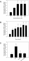
Dex induces MKP-1. (A) MKP-1 mRNA was measured by real-time quantitative reverse transcription-PCR in A549 cells after 8 h of treatment with the indicated concentrations of Dex. Asterisks denote a significant increase between untreated and Dex-treated cultures as determined by Tukey-Kramer pair comparison analysis (P < 0.05). (B) Kinetics of MKP-1 induction by Dex. The MKP-1 mRNA level was measured by real-time quantitative reverse transcription-PCR. A549 cells were treated with 100 nM Dex and harvested at the indicated time points (0, 1, 2, 4, 6, 8, and 24 h). Asterisks denote a significant increase between untreated and Dex-treated cultures as determined by Tukey-Kramer pair comparison analysis (P < 0.05). (C) The GR antagonist, RU486, counteracts the Dex-induced up-regulation of the MKP-1 mRNA level. A549 cells were stimulated with Dex for 8 h, and the MKP-1 mRNA levels were measured by real-time quantitative reverse transcription-PCR. RU486 counteracts the enhancing effect of Dex on MKP-1 mRNA levels. For each well, MKP-1 expression was normalized to cyclophilin B, and the fold induction for each experiment was determined by dividing the normalized expression from treated wells by that from control (untreated) wells. The asterisk denotes a statistically significant increase between the indicated Dex-treated wells and all other treatments as determined by Tukey-Kramer pair comparison analysis (P < 0.05). Values are the mean ± SE of three experiments. (D) MKP-1 protein levels in A549 cells after Dex treatment. MKP-1 protein expression was detected after Dex treatment. Western blot (IB) analysis was performed to confirm the Dex-induced effect. A549 cells were harvested after 8 h of treatment and immunoprecipitated (IP) with anti-MKP-1 antibody. The graph shows MKP-1 protein expression from cells treated with Dex and/or RU486, normalized to the control immunoprecipitated protein (vehicle treated cells). Analysis of the immunoreactive bands with NIH-Image software reproduced the Dex-induced increase in the level of MKP-1. Values are the mean ± SE of three experiments. *, P < 0.05 for pair comparison analysis (Tukey-Kramer test).
FIG. 7.
FIG. 7.
Dex induces MKP-1. (A) MKP-1 mRNA was measured by real-time quantitative reverse transcription-PCR in A549 cells after 8 h of treatment with the indicated concentrations of Dex. Asterisks denote a significant increase between untreated and Dex-treated cultures as determined by Tukey-Kramer pair comparison analysis (P < 0.05). (B) Kinetics of MKP-1 induction by Dex. The MKP-1 mRNA level was measured by real-time quantitative reverse transcription-PCR. A549 cells were treated with 100 nM Dex and harvested at the indicated time points (0, 1, 2, 4, 6, 8, and 24 h). Asterisks denote a significant increase between untreated and Dex-treated cultures as determined by Tukey-Kramer pair comparison analysis (P < 0.05). (C) The GR antagonist, RU486, counteracts the Dex-induced up-regulation of the MKP-1 mRNA level. A549 cells were stimulated with Dex for 8 h, and the MKP-1 mRNA levels were measured by real-time quantitative reverse transcription-PCR. RU486 counteracts the enhancing effect of Dex on MKP-1 mRNA levels. For each well, MKP-1 expression was normalized to cyclophilin B, and the fold induction for each experiment was determined by dividing the normalized expression from treated wells by that from control (untreated) wells. The asterisk denotes a statistically significant increase between the indicated Dex-treated wells and all other treatments as determined by Tukey-Kramer pair comparison analysis (P < 0.05). Values are the mean ± SE of three experiments. (D) MKP-1 protein levels in A549 cells after Dex treatment. MKP-1 protein expression was detected after Dex treatment. Western blot (IB) analysis was performed to confirm the Dex-induced effect. A549 cells were harvested after 8 h of treatment and immunoprecipitated (IP) with anti-MKP-1 antibody. The graph shows MKP-1 protein expression from cells treated with Dex and/or RU486, normalized to the control immunoprecipitated protein (vehicle treated cells). Analysis of the immunoreactive bands with NIH-Image software reproduced the Dex-induced increase in the level of MKP-1. Values are the mean ± SE of three experiments. *, P < 0.05 for pair comparison analysis (Tukey-Kramer test).
FIG. 8.
FIG. 8.
Study of transcription factor occupancy of the TLR2 promoter after TNF-α and Dex treatment. A549 cells were lysed and analyzed for the ChIP assay. After a 1-h treatment with TNF-α and/or Dex, cells were sonicated and immunoprecipitated with specific antibodies. Thereafter, the immunoprecipitated TLR2 promoter was identified by PCR. Endogenous GR, p65, and STAT5 binding to the endogenous TLR2 promoter was explored after treatment with TNF-α and/or Dex. The PCR amplification product from immunoprecipitated endogenous TLR2 promoter from A549 cells using the specific GR antibody Ab57 (anti-GR), the antibody against p65 (anti-p65), or the anti-STAT5 antibody (anti-STAT5) is shown.
FIG. 9.
FIG. 9.
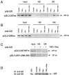
GR association with overexpressed TLR2 promoter mutants after TNF-α and Dex treatment. Transfected A549 cells were lysed and analyzed for the ChIP assay. After a 1-h treatment with TNF-α and/or Dex, cells were sonicated and immunoprecipitated with the specific anti-GR antibody, Ab57. Thereafter, the immunoprecipitated TLR2 promoter mutants was identified by PCR. (A) PCR amplification product from immunoprecipitated A549 cells transiently expressing the pGL3-297wt TLR2 promoter constructs after TNF-α and/or Dex treatment. (B) PCR Amplification products from immunoprecipitated A549 cells transiently expressing the pGL3-297GREm TLR2 promoter constructs after TNF-α and/or Dex treatment. (C) PCR amplicon from immunoprecipitated A549 cells transiently expressing the pGL3-297/NFm or the pGL3-297Δ285-269 TLR2 promoter mutants. (D) The immunoprecipitated (IP) GR content was similar for all the treatment conditions. A transiently transfected hGRα construct was used as the control (first lane). IB, western blotting.

Similar articles

Cited by

References

    1. Aliprantis, A. O., R. B. Yang, M. R. Mark, S. Suggett, B. Devaux, J. D. Radolf, G. R. Klimpel, P. Godowski, and A. Zychlinsky. 1999. Cell activation and apoptosis by bacterial lipoproteins through toll-like receptor-2. Science 285:736-739. - PubMed
    1. An, H., Y. Yu, M. Zhang, H. Xu, R. Qi, X. Yan, S. Liu, W. Wang, Z. Guo, J. Guo, Z. Qin, and X. Cao. 2002. Involvement of ERK, p38 and NF-kappaB signal transduction in regulation of TLR2, TLR4 and TLR9 gene expression induced by lipopolysaccharide in mouse dendritic cells. Immunology 106:38-45. - PMC - PubMed
    1. Chang, L., and M. Karin. 2001. Mammalian MAP kinase signalling cascades. Nature 410:37-40. - PubMed
    1. Chrousos, G. P. 1995. The hypothalamic-pituitary-adrenal axis and immune-mediated inflammation. N. Engl. J. Med. 332:1351-1362. - PubMed
    1. Chuang, T., and R. J. Ulevitch. 2001. Identification of hTLR10: a novel human Toll-like receptor preferentially expressed in immune cells. Biochim. Biophys. Acta 1518:157-161. - PubMed

MeSH terms