. 2004 Jun;24(11):4757-68.
doi: 10.1128/MCB.24.11.4757-4768.2004.

Wnt proteins induce dishevelled phosphorylation via an LRP5/6- independent mechanism, irrespective of their ability to stabilize beta-catenin

Affiliations

Wnt proteins induce dishevelled phosphorylation via an LRP5/6- independent mechanism, irrespective of their ability to stabilize beta-catenin

José M González-Sancho et al. Mol Cell Biol. 2004 Jun.

Abstract

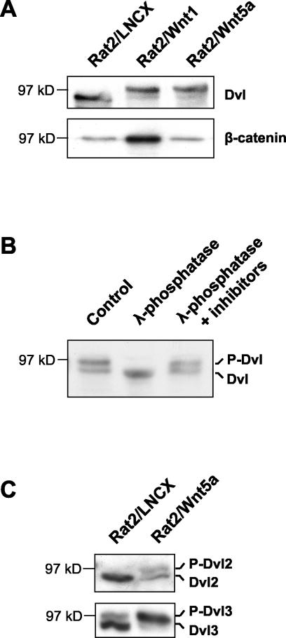
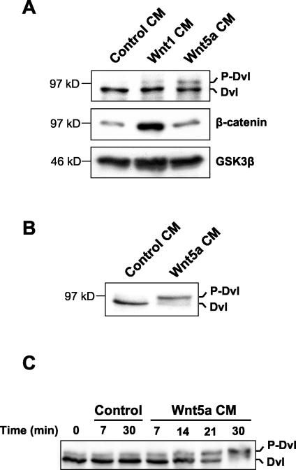
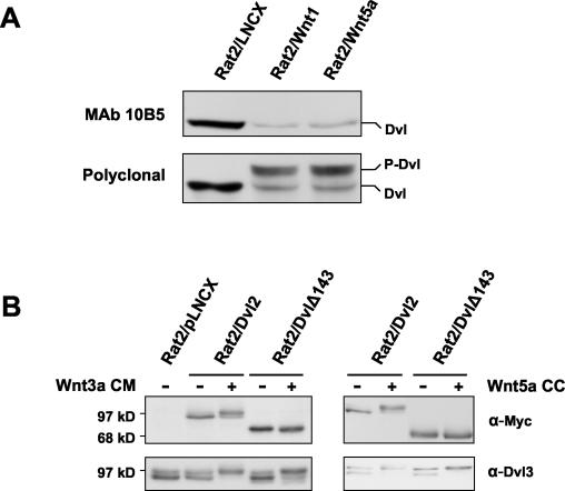
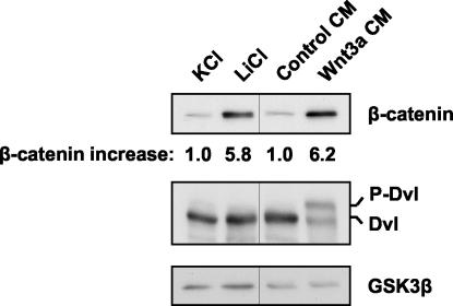
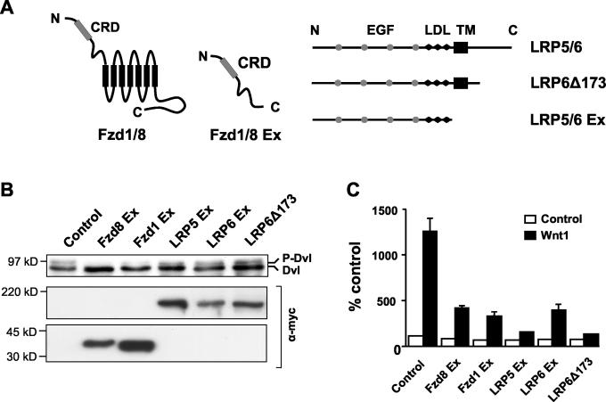
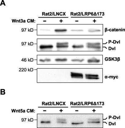
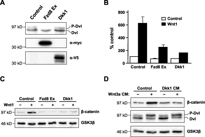
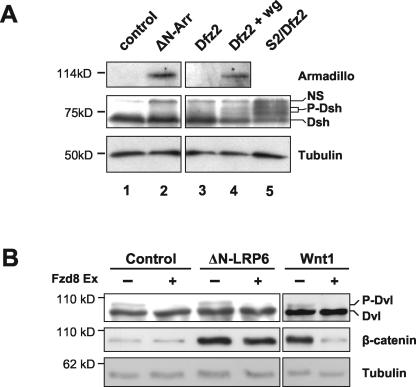
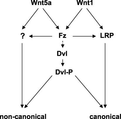
Wnt glycoproteins play essential roles in the development of metazoan organisms. Many Wnt proteins, such as Wnt1, activate the well-conserved canonical Wnt signaling pathway, which results in accumulation of beta-catenin in the cytosol and nucleus. Other Wnts, such as Wnt5a, activate signaling mechanisms which do not involve beta-catenin and are less well characterized. Dishevelled (Dvl) is a key component of Wnt/beta-catenin signaling and becomes phosphorylated upon activation of this pathway. In addition to Wnt1, we show that several Wnt proteins, including Wnt5a, trigger phosphorylation of mammalian Dvl proteins and that this occurs within 20 to 30 min. Unlike the effects of Wnt1, phosphorylation of Dvl in response to Wnt5a is not concomitant with beta-catenin stabilization, indicating that Dvl phosphorylation is not sufficient to activate canonical Wnt/beta-catenin signaling. Moreover, neither Dickkopf1, which inhibits Wnt/beta-catenin signaling by binding the Wnt coreceptors LRP5 and -6, nor dominant-negative LRP5/6 constructs could block Wnt-mediated Dvl phosphorylation. We conclude that Wnt-induced phosphorylation of Dvl is independent of LRP5/6 receptors and that canonical Wnts can elicit both LRP-dependent (to beta-catenin) and LRP-independent (to Dvl) signals. Our data also present Dvl phosphorylation as a general biochemical assay for Wnt protein function, including those Wnts that do not activate the Wnt/beta-catenin pathway.


Figures

FIG. 1.
FIG. 1.
Wnt5a causes Dvl phosphorylation but not β-catenin stabilization. (A) Stable expression of either Wnt1 or Wnt5a leads to Dvl phosphorylation, but only Wnt1 causes β-catenin stabilization. Whole-cell extracts (upper panel) and cytosolic extracts (lower panel) from Rat2 cells stably expressing the control vector LNCX, Wnt1, or Wnt5a were analyzed by Western blotting with antibodies against Dvl proteins or β-catenin, as indicated. (B) Western analysis of Dvl in Rat2/Wnt5a cell extracts either untreated (control), treated with λ-phosphatase, or treated with phosphatase in the presence of phosphatase inhibitors. The mobility shift upon phosphatase treatment confirms that the upper Dvl band in Rat2/Wnt5a cells is hyperphosphorylated. For simplicity, the two forms of Dvl protein are here referred to as unphosphorylated (Dvl) and phosphorylated Dishevelled (P-Dvl). (C) Wnt5a induces phosphorylation of both Dvl2 and Dvl3. Extracts from control Rat2/LNCX and from Rat2/Wnt5a cells were analyzed by Western blotting using Dvl2-specific and Dvl3-specific antibodies. Dvl2 and Dvl3 appear to comigrate on SDS-polyacrylamide gels.
FIG. 2.
FIG. 2.
CM from Wnt5a-expressing cells induces Dvl phosphorylation. (A) Rat2 fibroblasts were treated for 2 h with CM from Rat2 cells expressing control vector, Wnt1, or Wnt5a. Extracts were then analyzed as described for Fig. 1A for phosphorylation of Dvl and cytosolic β-catenin. Analysis of cytosolic GSK3β protein levels provided a loading control. (B) Treatment of 10T1/2 fibroblasts with Wnt5a CM also induces Dvl phosphorylation. (C) Kinetics of Dvl phosphorylation induced by Wnt5a. 10T1/2 fibroblasts were treated with either control or Wnt5a CM for the indicated times. Cells were then lysed, and extracts were analyzed by Western blotting with Dvl antibodies as described for panels A and B.
FIG. 3.
FIG. 3.
Canonical and noncanonical Wnts induce phosphorylation of a common region of Dvl2. (A) Phosphorylation of Dvl2 induced by either Wnt1 or Wnt5a masks the epitope for recognition by monoclonal antibody 10B5. Whole-cell extracts from Rat2 cells stably expressing the control vector LNCX, Wnt1, or Wnt5a were analyzed for Dvl2 phosphorylation by Western blotting using either monoclonal antibody 10B5 (upper panel) or a polyclonal Dvl2 antibody (lower panel). 10B5 recognizes an epitope within amino acids 594 to 736 of human Dvl2. (B) The Dvl2 derivative DvlΔ143, lacking amino acids 594 to 736, is not phosphorylated in response to signaling by Wnt3a or Wnt5a, but full-length Dvl2 shows a mobility shift. Rat2 cells stably expressing either Dvl2 or DvlΔ143 were either treated for 2 h with control or Wnt3a CM or were cocultured (CC) with control (−) or Wnt5a-producing Rat2 cells. Myc-tagged exogenous Dvl phosphorylation was analyzed by Western blotting using an anti-Myc antibody. Phosphorylation of endogenous Dvl3 was monitored as a positive control.
FIG. 4.
FIG. 4.
Lithium treatment stabilizes β-catenin but has no effect on Dvl phosphorylation. Rat2 cells were treated for 2 h with 40 mM KCl, 40 mM LiCl, control CM, or Wnt3a CM. Extracts were then analyzed as described for Fig. 1A for cytosolic β-catenin levels, Dvl phosphorylation, and cytosolic GSK3β as a loading control. The relative increases in β-catenin levels determined by densitometry are indicated.
FIG. 5.
FIG. 5.
DN Frizzled, but not DN LRP5 or -6, can inhibit Dvl phosphorylation. (A) Diagram of Fzd and LRP5 and -6 constructs used in this study. The extracellular cysteine-rich domain (CRD) and the seven transmembrane domains (black rectangles) are indicated for Fzd 1 and 8, as are the epidermal growth factor (EGF) repeat domains (grey circles), LDL receptor repeats (LDL; black lozenges), and transmembrane domain (TM; black rectangle) for LRP5/6. (B) 293T cells were transfected with vectors encoding the indicated Fzd or LRP5/6 DN constructs. Dvl phosphorylation was analyzed by Western blotting as described in the legend for Fig. 1A. Expression of the epitope-tagged Frizzled and LRP derivatives was confirmed with an anti-Myc antibody. Only Fzd8 Ex, and to a lesser extent Fzd1 Ex, inhibited Dvl phosphorylation. (C) Both DN Frizzled and LRP5 and -6 proteins can inhibit TCF/β-catenin-dependent transcription. 293T cells were transfected with vectors encoding the indicated Fzd or LRP5/6 DN constructs, in the absence (white bars) or presence (black bars) of a Wnt1-expressing construct, together with the TCF/β-catenin-responsive reporter pTOPflash. The cells were also transfected with a Renilla luciferase plasmid, pRL-TK, as an internal control. Luciferase activities were measured, and TOPflash values were normalized to Renilla values. Results shown are means + standard deviations of six replicates.
FIG. 6.
FIG. 6.
DN LRP does not inhibit Wnt3a- or Wnt5a-induced Dvl phosphorylation. (A) Rat2 cells stably expressing either LRP6Δ173 or the control vector LNCX were treated for 2 h with either control or Wnt3a CM. Cytosolic β-catenin levels and Dvl phosphorylation were analyzed by Western blotting as described in the legend for Fig. 1A. LRP6Δ173 blocked the Wnt-induced stabilization of β-catenin but did not block the phosphorylation of Dvl. GSK3β was used as a loading control, and expression of the Myc epitope-tagged LRP6Δ173 protein was confirmed using an anti-Myc antibody. (B) Rat2 cells stably expressing LRP6Δ173 or empty vector were treated for 2 h with either control or Wnt5a CM. Whole-cell lysates were analyzed for Dvl phosphorylation as described for panel A.
FIG. 7.
FIG. 7.
Dkk1 inhibits the Wnt/β-catenin pathway but does not block Dvl phosphorylation. (A) Dkk1 does not inhibit endogenous Dvl phosphorylation. 293T cells were transfected with vectors encoding either Dkk1 or Fzd8 Ex, and Dvl phosphorylation was analyzed by Western blotting. Expression of epitope-tagged Dkk1 and Fzd8 Ex was confirmed using anti-V5 and anti-Myc antibodies, respectively. (B) Dkk1 inhibits TCF/β-catenin-dependent transcription. 293T cells were transfected with vectors encoding Dkk1 or Fzd8 Ex, together with pTOPflash and pRL-TK. These cells were then cocultured with Rat2 cells stably expressing either Wnt1 (black bars) or empty vector (white bars). Luciferase assays were performed as described for Fig. 5C. Results shown are the mean + standard deviation of six replicates. (C) Dkk1 inhibits Wnt1-dependent β-catenin stabilization. 293T cells were transfected either with empty vector or vector encoding Wnt1, in the absence or presence of vectors encoding Dkk1 or Fzd8 Ex. Cytosolic β-catenin was analyzed by Western blotting as described before, and GSK3β was used as a loading control. (D) Dkk1 inhibits Wnt3a-mediated β-catenin stabilization but not Dvl phosphorylation. 10T1/2 cells were pretreated for 2 h with either control or Dkk1 CM, and this was then supplemented with control or Wnt3a CM for a further 2 h. Cell extracts were analyzed for β-catenin levels and Dvl phosphorylation as described above.
FIG. 8.
FIG. 8.
Activated LRP6/Arrow can stabilize β-catenin/Armadillo without an apparent change in Dvl/Dsh phosphorylation. (A) A truncated form of Drosophila Arrow (ΔN-Arr) activates signaling to Armadillo (Arm) in S2 cells with no evidence of Dishevelled (Dsh) phosphorylation. Drosophila S2 cells were transiently transfected with control vector (lane 1), ΔN-Arr (lane 2), Dfrizzled2 (Dfz2; lane 3), or Dfz2 plus Wingless (Wg; lane 4). Cellular levels of Arm and phosphorylation of Dsh were analyzed by Western blotting, and β-tubulin provided a loading control. Dishevelled is constitutively phosphorylated in S2 cells stably transfected with Dfz2 (lane 5), allowing identification of two bands corresponding to phosphorylated Dsh. These lie below a nonspecific background band (labeled NS) in each lane. While Wg induces Arm stabilization and Dsh phosphorylation (lane 4), ΔN-Arr induces Arm stabilization only (lane 2). (B) ΔN-LRP6 causes stabilization of β-catenin but does not induce Dvl phosphorylation. 293T cells were transiently transfected with ΔN-LRP6, Wnt1, or control vector, in the presence or absence of Fzd8 Ex. Whole-cell and cytosolic extracts were analyzed for Dvl phosphorylation and β-catenin levels, respectively. Tubulin levels provided a loading control. Fzd8 Ex abolishes basal Dvl phosphorylation in all cases but does not prevent β-catenin stabilization in ΔN-LRP6-transfected cells.
FIG. 9.
FIG. 9.
Model illustrating Dvl phosphorylation as a common response to Wnt proteins that activate distinct signaling pathways. Wnt signaling via the canonical β-catenin pathway requires both Fzd and LRP5/6, while noncanonical Wnt signaling requires Fzd and possibly additional receptors as yet uncharacterized. For simplicity, we have indicated that signals leading to phosphorylation of Dvl are derived from Fzd alone. We propose that Dvl phosphorylation is a common response to both the Wnt1 class and Wnt5a class of Wnt proteins and that it potentiates Wnt signaling via either the canonical or noncanonical pathways. Which of these pathways dominates in a given circumstance may depend on the involvement of Wnt coreceptor components in addition to Fzd and possibly on the subcellular distribution of Dvl. The reported association between different subcellular locations of Dvl protein and different signaling pathways suggests that this might be a key determinant (17).

Similar articles

Cited by

References

    1. Adler, P. N. 2002. Planar signaling and morphogenesis in Drosophila. Dev. Cell 2:525-535. - PubMed
    1. Amit, S., A. Hatzubai, Y. Birman, J. S. Andersen, E. Ben-Shushan, M. Mann, Y. Ben-Neriah, and I. Alkalay. 2002. Axin-mediated CKI phosphorylation of beta-catenin at Ser 45: a molecular switch for the Wnt pathway. Genes Dev. 16:1066-1076. - PMC - PubMed
    1. Axelrod, J. D. 2001. Unipolar membrane association of Dishevelled mediates Frizzled planar cell polarity signaling. Genes Dev. 15:1182-1187. - PMC - PubMed
    1. Axelrod, J. D., K. Matsuno, S. Artavanis-Tsakonas, and N. Perrimon. 1996. Interaction between Wingless and Notch signaling pathways mediated by Dishevelled. Science 271:1826-1832. - PubMed
    1. Axelrod, J. D., J. R. Miller, J. M. Shulman, R. T. Moon, and N. Perrimon. 1998. Differential recruitment of Dishevelled provides signaling specificity in the planar cell polarity and Wingless signaling pathways. Genes Dev. 12:2610-2622. - PMC - PubMed

Publication types

MeSH terms

Substances