. 2004 Jun;24(11):4769-80.
doi: 10.1128/MCB.24.11.4769-4780.2004.

The Rpd3-Sin3 histone deacetylase regulates replication timing and enables intra-S origin control in Saccharomyces cerevisiae

Affiliations

The Rpd3-Sin3 histone deacetylase regulates replication timing and enables intra-S origin control in Saccharomyces cerevisiae

Jennifer G Aparicio et al. Mol Cell Biol. 2004 Jun.

Abstract

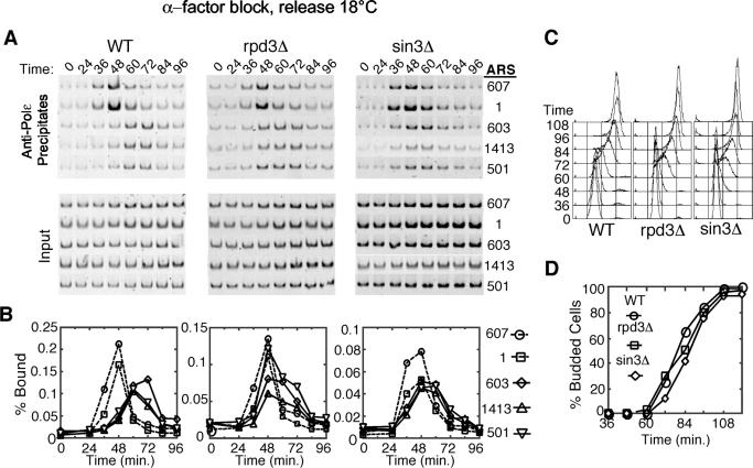
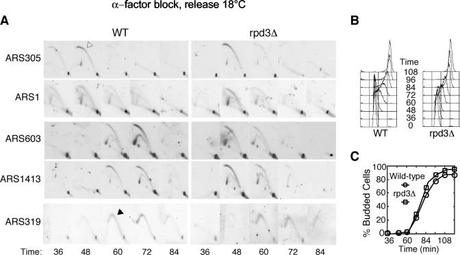
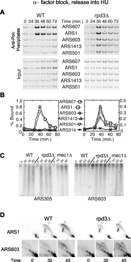
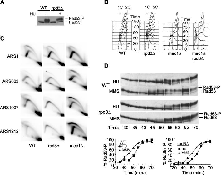
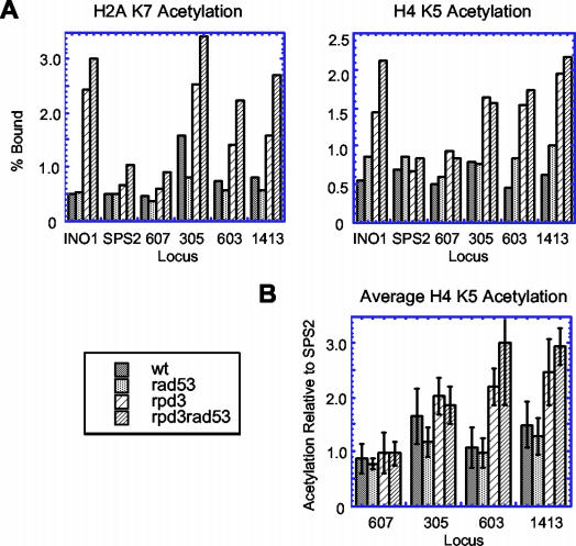
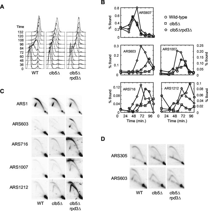
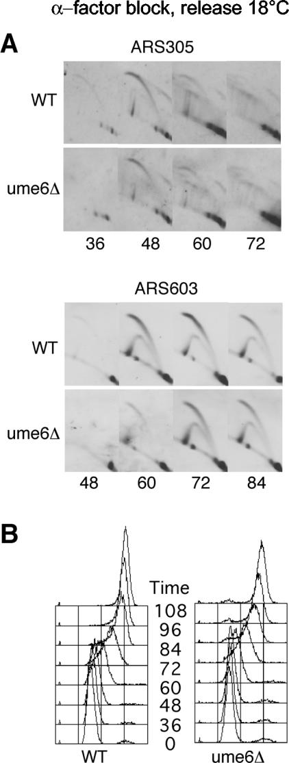
The replication of eukaryotic genomes follows a temporally staged program, in which late origin firing often occurs within domains of altered chromatin structure(s) and silenced genes. Histone deacetylation functions in gene silencing in some late-replicating regions, prompting an investigation of the role of histone deacetylation in replication timing control in Saccharomyces cerevisiae. Deletion of the histone deacetylase Rpd3 or its interacting partner Sin3 caused early activation of late origins at internal chromosomal loci but did not alter the initiation timing of early origins or a late-firing, telomere-proximal origin. By delaying initiation relative to the earliest origins, Rpd3 enables regulation of late origins by the intra-S replication checkpoint. RPD3 deletion suppresses the slow S phase of clb5Delta cells by enabling late origins to fire earlier, suggesting that Rpd3 modulates the initiation timing of many origins throughout the genome. Examination of factors such as Ume6 that function together with Rpd3 in transcriptional repression indicates that Rpd3 regulates origin initiation timing independently of its role in transcriptional repression. This supports growing evidence that for much of the S. cerevisiae genome transcription and replication timing are not linked.


Figures

FIG. 1.
FIG. 1.
Rpd3 and Sin3 delay activation of late-firing replication origins. Wild-type (OAy618), rpd3Δ (OAy781), and sin3Δ (JAy44) cells were synchronized in late G1 with α-factor at 23°C and released into S phase at 18°C. At the indicated intervals, cells were fixed for analysis. (A) DNA Polɛ association with early (ARS607 and ARS1) and late (ARS603, ARS1413, and ARS501) replication origins was analyzed by ChIP. Origin-specific PCR analysis of immunoprecipitated (Precipitates) and total (Input) DNA is shown. (B) Quantification of the data shown in panel A plotted as the percent bound. (C) DNA content of cells was analyzed by FACScan. (D) The percentage of budded cells was determined by microscopic analysis of at least 100 cells at each time point.
FIG. 2.
FIG. 2.
Rpd3 regulates internal late-firing origins. Wild-type (OAy618) and rpd3Δ (OAy781) cells were synchronized in late G1 at 23°C with α-factor and released into S phase at 18°C. At the indicated intervals, cells were fixed for analysis. (A) 2-D agarose gel electrophoresis analysis of ARS305, ARS1, ARS603, ARS1413, and ARS319. An open arrow indicates a bubble arc, and a Y arc is marked with a filled arrow. (B) DNA content was analyzed by FACScan. (C) The percentage of budded cells was determined by microscopic analysis of at least 100 cells at each time point.
FIG. 3.
FIG. 3.
Late origins escape checkpoint inhibition in rpd3Δ cells treated with HU. Wild-type (OAy618) and rpd3Δ (OAy781) cells (A and B) or wild-type (OAy470), rpd3Δ (JAy22), and mec1Δ (DGy159) cells (C and D) were synchronized in late G1 with α-factor at 23°C and released into S phase in the presence of 200 mM HU at 23°C. Samples were collected at the indicated intervals. DNA Polɛ association with origins was analyzed by ChIP (A) and quantified (B) as described in the Fig. 1 legend. (C) Nascent DNA strand analysis of ARS305 and ARS603. (D) Replication structures detected by 2-D gel analysis.
FIG. 4.
FIG. 4.
The checkpoint pathway is intact in rpd3Δ cells. (A) Wild-type (FHy20) and rpd3Δ (DGy140) cells were grown for 1 h at 30°C in the presence or absence of 200 mM HU. Protein extracts prepared by trichloroacetic acid precipitation were analyzed by Western blotting using anti-HA antibody (16B12) to detect Rad53-HA (33). (B) Wild-type (OAy470), rpd3Δ (JAy22), mec1Δ (DGy159), and mec1Δ rpd3Δ (DGy170) cells were synchronized in late G1 with α-factor at 23°C and released into S phase at 30°C in yeast extract-peptone-dextrose containing 0.033% MMS. At the indicated intervals, cells were fixed for DNA content analysis. (C) Cells were treated as for panel B and fixed for analysis at 25, 32, 39, 46, and 53 min after release from α-factor. Cells from all time points were pooled to prepare S-phase DNA for 2-D gel analysis. (D) Wild-type (FHy20) and rpd3Δ (DGy140) cells synchronized at 23°C with α-factor were released into S phase at 18°C, and samples were collected at the indicated times. Rad53 phosphorylation was analyzed by Western blotting as for panel A and was quantified (percent Rad53-P) by direct analysis of the chemiluminescent signal using a Bio-Rad ChemiDoc system and QuantityOne software. Percent phosphorylation was calculated as (Rad53-P)/(Rad53-P + Rad53), where Rad53-P includes all slower-migrating forms of Rad53.
FIG. 5.
FIG. 5.
Histone deacetylation by Rpd3 functions independently of Rad53. (A) ChIP analysis of histone acetylation in asynchronous cultures of wild-type (DGy166), rad53Δ (DGy164), rpd3Δ (JAy72), and rad53Δ rpd3Δ (JAy71) cells. (B) Average H4 K5 acetylation from three experiments as in panel A was calculated for each locus relative to that of SPS2. SPS2 serves as an internal control, as its acetylation is not affected by Rpd3 and enables comparison between different experiments in which the absolute levels of chromatin immunoprecipitated can vary significantly.
FIG. 6.
FIG. 6.
Advanced timing of late origin activation upon deletion of RPD3 restores normal S-phase kinetics in clb5Δ cells. Wild-type (OAy617), clb5Δ (DGy197), and rpd3Δ clb5Δ (DGy198) cells were synchronized in late G1 at 23°C with α-factor and released into S phase at 18°C. (A) At the indicated intervals, cells were fixed for DNA content analysis. (B) Association of Cdc45-HA with origin DNA was monitored by ChIP at the indicated times. (C) Wild-type (OAy470), clb5Δ (JAy30), and rpd3Δ clb5Δ (JAy24) cells were synchronized in late G1 at 23°C with α-factor and released at 23°C into medium containing 200 mM HU for 60 min. Replication structures were analyzed by 2-D agarose gel electrophoresis. (D) Replication structures from asynchronous cultures (30°C) of wild-type (OAy470), clb5Δ (JAy30), and rpd3Δ clb5Δ (DGy189) cells were analyzed by 2-D agarose gel electrophoresis.
FIG. 7.
FIG. 7.
Ume6 is not required for regulation of origin initiation timing. Wild-type (OAy618) and ume6Δ (JAy43) cells were analyzed for replication structures at ARS305 and ARS603 (A) or by DNA content analysis (B) as described in the legend for Fig. 2.

Similar articles

Cited by

References

    1. Aparicio, O. M. 1999. Characterization of proteins bound to chromatin by immunoprecipitation from whole-cell extracts, p. 21.3.1-21.3.12. In F. M. Ausubel, R. Brent, R. E. Kingston, D. D. Moore, J. G. Seidman, J. A. Smith, and K. Struhl (ed.), Current protocols in molecular biology, vol. 4. John Wiley and Sons, Inc., New York, N.Y.
    1. Aparicio, O. M., A. M. Stout, and S. P. Bell. 1999. Differential assembly of Cdc45p and DNA polymerases at early and late origins of DNA replication. Proc. Natl. Acad. Sci. USA 96:9130-9135. - PMC - PubMed
    1. Aparicio, O. M., D. M. Weinstein, and S. P. Bell. 1997. Components and dynamics of DNA replication complexes in S. cerevisiae: redistribution of MCM proteins and Cdc45p during S phase. Cell 91:59-69. - PubMed
    1. Bell, S. P. 2002. The origin recognition complex: from simple origins to complex functions. Genes Dev. 16:659-672. - PubMed
    1. Bernstein, B. E., J. K. Tong, and S. L. Schreiber. 2000. Genomewide studies of histone deacetylase function in yeast. Proc. Natl. Acad. Sci. USA 97:13708-13713. - PMC - PubMed

Publication types

MeSH terms

Substances