. 2004 Jun;24(11):4810-23.
doi: 10.1128/MCB.24.11.4810-4823.2004.

The homeodomain protein CDP regulates mammary-specific gene transcription and tumorigenesis

Affiliations

The homeodomain protein CDP regulates mammary-specific gene transcription and tumorigenesis

Quan Zhu et al. Mol Cell Biol. 2004 Jun.

Abstract

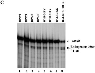
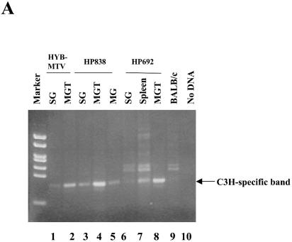
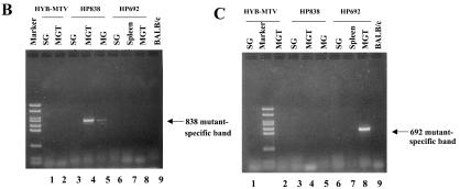
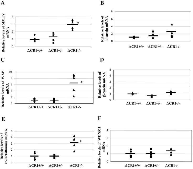
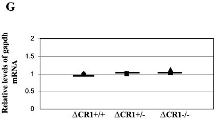
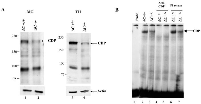
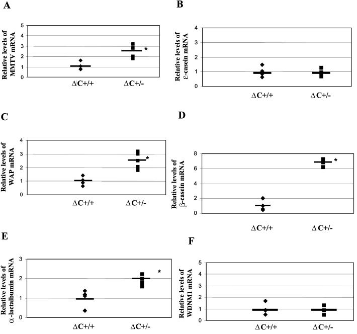
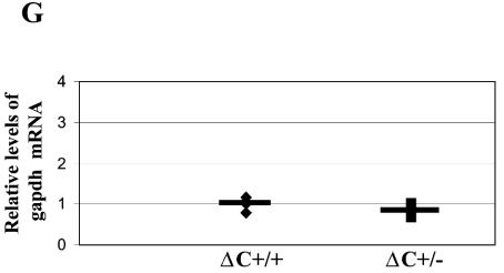
The CCAAT-displacement protein (CDP) has been implicated in developmental and cell-type-specific regulation of many cellular and viral genes. We previously have shown that CDP represses mouse mammary tumor virus (MMTV) transcription in tissue culture cells. Since CDP-binding activity for the MMTV long terminal repeat declines during mammary development, we tested whether binding mutations could alter viral expression. Infection of mice with MMTV proviruses containing CDP binding site mutations elevated viral RNA levels in virgin mammary glands and shortened mammary tumor latency. To determine if CDP has direct effects on MMTV transcription rather than viral spread, virgin mammary glands of homozygous CDP-mutant mice lacking one of three Cut repeat DNA-binding domains (DeltaCR1) were examined by reverse transcription-PCR. RNA levels of endogenous MMTV as well as alpha-lactalbumin and whey acidic protein (WAP) were elevated. Heterozygous mice with a different CDP mutation that eliminated the entire C terminus and the homeodomain (DeltaC mice) showed increased levels of MMTV, beta-casein, WAP, and alpha-lactalbumin RNA in virgin mammary glands compared to those from wild-type animals. No differences in amounts of WDNM1, epsilon-casein, or glyceraldehyde-3-phosphate dehydrogenase RNA were observed between the undifferentiated mammary tissues from wild-type and mutant mice, indicating the specificity of this effect. These data show independent contributions of different CDP domains to negative regulation of differentiation-specific genes in the mammary gland.


Figures

FIG. 1.
FIG. 1.
Experimental design for MMTV proviruses with CDP binding site mutations. (A) Scheme for analysis of CDP binding site mutants. The 5′ half of the hybrid infectious provirus is composed of the 5′ LTR and gag-pol genes from the endogenous Mtv-1 provirus, whereas the 3′ end is composed of the env and 3′ LTR from a C3H MMTV provirus. An inverted triangle represents the CDP binding site mutations within the U3 region of the LTR. Transfection of proviral DNA leads to integration and transcription from the 5′ LTR by RNA polymerase II followed by RNA packaging into virions. Subsequent infection will allow RT of viral RNA so that the 3′ LTR mutations are duplicated in the 5′ LTR of the provirus. BALB/c mice were injected with XC cells containing the stably transfected proviruses and analyzed for T-cell deletion and the appearance of tumors. (B) Positions of mutations introduced in the distal NRE (dNRE) and proximal NRE (pNRE) of MMTV proviruses.
FIG. 2.
FIG. 2.
MMTV proviruses with CDP binding site mutations show elevated viral RNA and protein levels in transfected cells. (A and B) Total RNA was harvested from XC cells without (A) or with treatment of 10−6 M DEX for 24 h (B). RNA samples (50 μg in lanes 4 to 7) from normal XC cells (lane 4), XC cells stably transfected with HYB-MTV (lane 5), HP692 (lane 6), or HP838 (lane 7) were subjected to an RPA using riboprobe specific for the C3H LTR (5 × 10⁵ cpm) and a gapdh riboprobe (5 × 10³ cpm) as an RNA loading control. Lane 1 shows RNA molecular weight standards; lanes 2 and 3 show the position of the gapdh and LTR probes, respectively. Lane 8 (panel A) shows RNA from HP838-transfected XC cells hybridized to C3H LTR riboprobe in the absence of the gapdh riboprobe. (C) CDP-binding mutants show elevated viral protein levels. After treatment with 10−6 M DEX for 24 h, XC cell lysates containing wild-type virus (HYB-MTV) (lanes 2 and 6) or mutant provirus with a CDP binding site mutation in the proximal NRE (HP838 and HP837) (lanes 4 and 7) or in the distal NRE (HP692 and HP691) (lanes 3 and 8) were harvested and incubated with protein A. Precleared lysates (40 μg each) were analyzed by Western blotting using MMTV CA-specific antibody. XC rat cells (lanes 1 and 5) lack endogenous MMTVs.
FIG. 2.
FIG. 2.
MMTV proviruses with CDP binding site mutations show elevated viral RNA and protein levels in transfected cells. (A and B) Total RNA was harvested from XC cells without (A) or with treatment of 10−6 M DEX for 24 h (B). RNA samples (50 μg in lanes 4 to 7) from normal XC cells (lane 4), XC cells stably transfected with HYB-MTV (lane 5), HP692 (lane 6), or HP838 (lane 7) were subjected to an RPA using riboprobe specific for the C3H LTR (5 × 10⁵ cpm) and a gapdh riboprobe (5 × 10³ cpm) as an RNA loading control. Lane 1 shows RNA molecular weight standards; lanes 2 and 3 show the position of the gapdh and LTR probes, respectively. Lane 8 (panel A) shows RNA from HP838-transfected XC cells hybridized to C3H LTR riboprobe in the absence of the gapdh riboprobe. (C) CDP-binding mutants show elevated viral protein levels. After treatment with 10−6 M DEX for 24 h, XC cell lysates containing wild-type virus (HYB-MTV) (lanes 2 and 6) or mutant provirus with a CDP binding site mutation in the proximal NRE (HP838 and HP837) (lanes 4 and 7) or in the distal NRE (HP692 and HP691) (lanes 3 and 8) were harvested and incubated with protein A. Precleared lysates (40 μg each) were analyzed by Western blotting using MMTV CA-specific antibody. XC rat cells (lanes 1 and 5) lack endogenous MMTVs.
FIG. 3.
FIG. 3.
Kinetics of infection and tumorigenesis by wild-type or CDP binding site mutants. (A) Deletion of C3H MMTV-specific Sag-reactive T cells in mice infected with wild-type or mutant viruses. BALB/c mice were injected intraperitoneally with XC cells that were stably transfected with wild-type MMTV proviruses (HYB-MTV) or CDP binding site mutants (HP692 and HP838), and lymphocytes were tested at 6, 16, and 26 weeks postinjection. Peripheral blood lymphocytes were stained with fluorescein isothiocyanate-conjugated antibodies to Vβ14 and phycoerythrin-conjugated antibodies to CD4. The lymphocyte population was determined by forward and side scatter using FACS analysis. Stars indicate values that were statistically different (P < 0.05) from those for wild-type virus-injected animals by Student's t tests and Tukey honestly significant difference mean comparison post-hoc tests at the same times after inoculation. (B) Mammary tumor development in mice infected by CDP binding site mutants. BALB/c weanling mice were injected intraperitoneally with XC cells that were stably transfected with wild-type hybrid MMTV proviruses (HYB-MTV) or CDP binding site mutants (HP692 and HP838) and monitored for tumor development. There was no detectable difference in the histology of mammary tumors induced by the wild-type virus compared to the mutants. Tumorigenesis results were analyzed by the Kaplan-Meier survival analysis technique using a log-rank test of significance. Symbols (+) represent animals that died for reasons other than mammary tumors and may represent more than one animal.
FIG. 4.
FIG. 4.
Expression of wild-type and mutant MMTVs in virgin mammary glands of inoculated mice. (A) Total RNA was extracted from virgin mammary glands 6 weeks after injection with virus-producing XC cells. RNA samples were subjected to an RPA using riboprobes specific for the C3H MMTV LTR (5 × 10⁵ cpm) and gapdh (1 × 10⁴ cpm) as an RNA loading control. Lane 9 contains 20 μg of mammary gland tumor (MGT) RNA from a BALB/c mouse that was infected by foster nursing on a C3H mother (BALB/cfC3H). Lane 10 contains 40 μg of BALB/c mammary gland (MG) RNA. Lanes 11 and 12 show the position of the LTR and gapdh probes, respectively. Lane 13 contains 40 μg of yeast tRNA. (B) Quantitation of relative levels of C3H riboprobe-protected bands from RPAs. RNA samples from individual HYB-MTV- (n = 3), HP692- (n = 5), and HP838- (n = 4) injected mice were analyzed by RPAs, and the intensities of C3H and gapdh riboprobe protected bands were quantitated using a phosphorimager and normalized to gapdh levels. The average for each group is indicated by a horizontal bar. Stars indicate values that were statistically different (P < 0.05) from those of wild-type virus-injected animals by Student's t test. (C) Total RNA was extracted from salivary glands 6 weeks after injection with virus-producing XC cells. RNA samples (70 μg in lanes 1 to 6) from HP692 (lanes 1 and 2), HP838 (lanes 3 and 4), and HYB-MTV (lanes 5 and 6) virus-injected mice were subjected to an RPA as described for panel A. Lane 7 contains 70 μg of salivary gland (SG) RNA from a BALB/c mouse. Lane 8 contains 35 μg of salivary gland RNA from a BALB/c mouse infected by foster nursing on a C3H mother (BALB/cfC3H).
FIG. 4.
FIG. 4.
Expression of wild-type and mutant MMTVs in virgin mammary glands of inoculated mice. (A) Total RNA was extracted from virgin mammary glands 6 weeks after injection with virus-producing XC cells. RNA samples were subjected to an RPA using riboprobes specific for the C3H MMTV LTR (5 × 10⁵ cpm) and gapdh (1 × 10⁴ cpm) as an RNA loading control. Lane 9 contains 20 μg of mammary gland tumor (MGT) RNA from a BALB/c mouse that was infected by foster nursing on a C3H mother (BALB/cfC3H). Lane 10 contains 40 μg of BALB/c mammary gland (MG) RNA. Lanes 11 and 12 show the position of the LTR and gapdh probes, respectively. Lane 13 contains 40 μg of yeast tRNA. (B) Quantitation of relative levels of C3H riboprobe-protected bands from RPAs. RNA samples from individual HYB-MTV- (n = 3), HP692- (n = 5), and HP838- (n = 4) injected mice were analyzed by RPAs, and the intensities of C3H and gapdh riboprobe protected bands were quantitated using a phosphorimager and normalized to gapdh levels. The average for each group is indicated by a horizontal bar. Stars indicate values that were statistically different (P < 0.05) from those of wild-type virus-injected animals by Student's t test. (C) Total RNA was extracted from salivary glands 6 weeks after injection with virus-producing XC cells. RNA samples (70 μg in lanes 1 to 6) from HP692 (lanes 1 and 2), HP838 (lanes 3 and 4), and HYB-MTV (lanes 5 and 6) virus-injected mice were subjected to an RPA as described for panel A. Lane 7 contains 70 μg of salivary gland (SG) RNA from a BALB/c mouse. Lane 8 contains 35 μg of salivary gland RNA from a BALB/c mouse infected by foster nursing on a C3H mother (BALB/cfC3H).
FIG. 5.
FIG. 5.
In vivo infection by CDP binding site mutants detected using PCR. Genomic DNA was extracted from different tissues in mice that were injected with XC transfectants of wild-type (HYB-MTV; lanes 1 and 2) or CDP binding site mutant proviruses (HP692, lanes 6 to 8; HP838, lanes 3 to 5). (A) PCR products detected with primers specific for C3H MMTV. The C3H-specific band was 393 bp. (B) PCR products detected with primers specific for the HP838 mutation. The HP838-specific band was 703 bp. (C) PCR products detected with primers specific for the HP692 mutation. The HP692-specific band was 561 bp. Reaction mixtures contained 100 ng of DNA, except for those shown in panel A, lanes 1 and 6 (200 ng) and lane 7 (400 ng). DNA from an uninjected BALB/c mouse was used as a negative control (lane 9). MG, mammary gland; MGT, mammary gland tumor; SG, salivary gland.
FIG. 5.
FIG. 5.
In vivo infection by CDP binding site mutants detected using PCR. Genomic DNA was extracted from different tissues in mice that were injected with XC transfectants of wild-type (HYB-MTV; lanes 1 and 2) or CDP binding site mutant proviruses (HP692, lanes 6 to 8; HP838, lanes 3 to 5). (A) PCR products detected with primers specific for C3H MMTV. The C3H-specific band was 393 bp. (B) PCR products detected with primers specific for the HP838 mutation. The HP838-specific band was 703 bp. (C) PCR products detected with primers specific for the HP692 mutation. The HP692-specific band was 561 bp. Reaction mixtures contained 100 ng of DNA, except for those shown in panel A, lanes 1 and 6 (200 ng) and lane 7 (400 ng). DNA from an uninjected BALB/c mouse was used as a negative control (lane 9). MG, mammary gland; MGT, mammary gland tumor; SG, salivary gland.
FIG. 6.
FIG. 6.
Increased RNA levels of endogenous MMTVs and mammary-specific genes in virgin mammary glands of ΔCR1 mice. RNA levels in virgin mammary glands of individual animals (each represented by a symbol) were detected by semiquantitative RT-PCR. Six wild-type (+/+), four ΔCR1 heterozygous (+/−), and five homozygous ΔCR1 (−/−) mice were tested. The average for each group of age-matched mice is indicated by a horizontal line. The average for the wild-type animals was assigned a value of 1, and the RNA levels of the heterozygous and homozygous ΔCR1 mice were assigned values relative to the wild-type levels after normalization for gapdh levels in individual samples. No significant differences in gapdh levels were observed between mammary glands from wild-type or mutant animals (G). RNA levels of the following genes are shown: endogenous MMTV (A), ɛ-casein (B), WAP (C), β-casein (D), α-lactalbumin (E), WDNM1 (F), and gapdh (G) genes. An asterisk indicates that the values for the wild-type and mutant mice were statistically different (P < 0.05) as determined by the two-tailed Student t test.
FIG. 6.
FIG. 6.
Increased RNA levels of endogenous MMTVs and mammary-specific genes in virgin mammary glands of ΔCR1 mice. RNA levels in virgin mammary glands of individual animals (each represented by a symbol) were detected by semiquantitative RT-PCR. Six wild-type (+/+), four ΔCR1 heterozygous (+/−), and five homozygous ΔCR1 (−/−) mice were tested. The average for each group of age-matched mice is indicated by a horizontal line. The average for the wild-type animals was assigned a value of 1, and the RNA levels of the heterozygous and homozygous ΔCR1 mice were assigned values relative to the wild-type levels after normalization for gapdh levels in individual samples. No significant differences in gapdh levels were observed between mammary glands from wild-type or mutant animals (G). RNA levels of the following genes are shown: endogenous MMTV (A), ɛ-casein (B), WAP (C), β-casein (D), α-lactalbumin (E), WDNM1 (F), and gapdh (G) genes. An asterisk indicates that the values for the wild-type and mutant mice were statistically different (P < 0.05) as determined by the two-tailed Student t test.
FIG. 7.
FIG. 7.
CDP protein levels and DNA-binding activity in ΔC mutant mice. (A) Western blotting of nuclear extracts from tissues of ΔC wild-type and heterozygous animals. Extracts from virgin mammary glands (MG; 50 μg; lanes 1 and 2) or thymi (TH; 25 μg; lanes 3 and 4) were analyzed on a 6% polyacrylamide gel containing sodium dodecyl sulfate followed by Western blotting and detection with CDP-specific antibody. The same blots were stripped and incubated with antiactin as a loading control (lower panel). (B) DNA-binding activity of nuclear extracts from virgin mammary glands of ΔC mice. Extracts (5 μg) from wild-type (lanes 2, 4, and 6) or heterozygous (lanes 3, 5, and 7) animals were incubated in the presence or absence of anti-CDP or preimmune (PI) serum as indicated prior to incubation with a labeled MMTV pNRE4 probe (ca. 3 fmol) (24). Complexes were separated on a 4% nondenaturing polyacrylamide gel prior to autoradiography. (C) CDP levels in ΔC-heterozygous animals during mammary gland development. Nuclear extracts (75 μg) of mammary glands from virgin (V-MG), pregnant (P-MG), or lactating (L-MG) mice were analyzed by Western blotting with CDP-specific antibody as described for panel A. The blot was stripped and incubated with actin-specific antibody to serve as a protein loading control (lower panel). The 150-kDa differentiation-specific form of CDP is indicated as CDP*.
FIG. 7.
FIG. 7.
CDP protein levels and DNA-binding activity in ΔC mutant mice. (A) Western blotting of nuclear extracts from tissues of ΔC wild-type and heterozygous animals. Extracts from virgin mammary glands (MG; 50 μg; lanes 1 and 2) or thymi (TH; 25 μg; lanes 3 and 4) were analyzed on a 6% polyacrylamide gel containing sodium dodecyl sulfate followed by Western blotting and detection with CDP-specific antibody. The same blots were stripped and incubated with antiactin as a loading control (lower panel). (B) DNA-binding activity of nuclear extracts from virgin mammary glands of ΔC mice. Extracts (5 μg) from wild-type (lanes 2, 4, and 6) or heterozygous (lanes 3, 5, and 7) animals were incubated in the presence or absence of anti-CDP or preimmune (PI) serum as indicated prior to incubation with a labeled MMTV pNRE4 probe (ca. 3 fmol) (24). Complexes were separated on a 4% nondenaturing polyacrylamide gel prior to autoradiography. (C) CDP levels in ΔC-heterozygous animals during mammary gland development. Nuclear extracts (75 μg) of mammary glands from virgin (V-MG), pregnant (P-MG), or lactating (L-MG) mice were analyzed by Western blotting with CDP-specific antibody as described for panel A. The blot was stripped and incubated with actin-specific antibody to serve as a protein loading control (lower panel). The 150-kDa differentiation-specific form of CDP is indicated as CDP*.
FIG. 8.
FIG. 8.
Increased RNA levels of endogenous MMTVs and mammary-specific genes in virgin mammary glands of ΔC mice. RNA levels in virgin mammary glands of individual animals (each represented by a symbol) were detected by semiquantitative RT-PCR. Four wild-type and six heterozygous ΔC mice were tested. The average for each group of age-matched mice is indicated by a horizontal line. The average for the wild-type animals was assigned a value of 1, and the RNA levels of the heterozygous ΔC mice were assigned values relative to the wild-type levels after normalization for gapdh levels in individual samples. No significant differences in gapdh levels were observed between mammary glands from wild-type or mutant animals (G). RNA levels of the following genes are shown: endogenous MMTV (A), ɛ-casein (B), WAP (C), β-casein (D), α-lactalbumin (E), WDNM1 (F), and gapdh (G) genes. An asterisk indicates that the values for the wild-type and mutant mice were statistically different (P < 0.05) as determined by the two-tailed Student t test.
FIG. 8.
FIG. 8.
Increased RNA levels of endogenous MMTVs and mammary-specific genes in virgin mammary glands of ΔC mice. RNA levels in virgin mammary glands of individual animals (each represented by a symbol) were detected by semiquantitative RT-PCR. Four wild-type and six heterozygous ΔC mice were tested. The average for each group of age-matched mice is indicated by a horizontal line. The average for the wild-type animals was assigned a value of 1, and the RNA levels of the heterozygous ΔC mice were assigned values relative to the wild-type levels after normalization for gapdh levels in individual samples. No significant differences in gapdh levels were observed between mammary glands from wild-type or mutant animals (G). RNA levels of the following genes are shown: endogenous MMTV (A), ɛ-casein (B), WAP (C), β-casein (D), α-lactalbumin (E), WDNM1 (F), and gapdh (G) genes. An asterisk indicates that the values for the wild-type and mutant mice were statistically different (P < 0.05) as determined by the two-tailed Student t test.

Similar articles

Cited by

References

    1. Ai, W., E. Toussaint, and A. Roman. 1999. CCAAT displacement protein binds to and negatively regulates human papillomavirus type 6 E6, E7, and E1 promoters. J. Virol. 73:4220-4229. - PMC - PubMed
    1. Aufiero, B., E. J. Neufeld, and S. H. Orkin. 1994. Sequence-specific DNA binding of individual cut repeats of the human CCAAT displacement/cut homeodomain protein. Proc. Natl. Acad. Sci. USA 91:7757-7761. - PMC - PubMed
    1. Banan, M., I. C. Rojas, W. H. Lee, H. L. King, J. V. Harriss, R. Kobayashi, C. F. Webb, and P. D. Gottlieb. 1997. Interaction of the nuclear matrix-associated region (MAR)-binding proteins, SATB1 and CDP/Cux, with a MAR element (L2a) in an upstream regulatory region of the mouse CD8α gene. J. Biol. Chem. 272:18440-18452. - PubMed
    1. Barberis, A., G. Superti-Furga, and M. Busslinger. 1987. Mutually exclusive interaction of the CCAAT-binding factor and of a displacement protein with overlapping sequences of a histone gene promoter. Cell 50:347-359. - PubMed
    1. Bodmer, R., S. Barbel, S. Sheperd, J. W. Jack, L. Y. Jan, and Y. N. Jan. 1987. Transformation of sensory organs by mutations of the cut locus of D. melanogaster. Cell 51:293-307. - PubMed

Publication types

LinkOut - more resources